Fatal Clostridium sordellii-mediated hemorrhagic and necrotizing gastroenteropathy in a dog: case report - BMC Veterinary Research
←
→
Page content transcription
If your browser does not render page correctly, please read the page content below
Capewell et al. BMC Veterinary Research    (2020) 16:152
https://doi.org/10.1186/s12917-020-02362-y

 CASE REPORT                                                                                                                                         Open Access

Fatal Clostridium sordellii-mediated
hemorrhagic and necrotizing
gastroenteropathy in a dog: case report
Paul Capewell* , Angie Rupp, Manuel Fuentes, Michael McDonald and William Weir

  Abstract
  Background: Canine hemorrhagic gastroenteritis (also canine gastrointestinal hemorrhagic syndrome) is commonly
  associated with Clostridium perfringens, although in some cases the etiology remains unclear. This report describes a
  fatal acute hemorrhagic and necrotizing gastroenteropathy in a dog associated with Clostridium sordellii, a bacterial
  species never before identified as the etiological agent of hemorrhagic and necrotizing gastroenteropathy in dogs.
  Case presentation: A fully vaccinated, eight-year-old, female neutered Labrador presented with a history of
  vomiting without diarrhea. Clinical examination revealed pink mucous membranes, adequate hydration,
  normothermia, and normocardia. The dog was discovered deceased the following day. Post-mortem examination
  showed moderate amounts of dark red, non-clotted fluid within the stomach that extended into the jejunum.
  Discoloration was noted in the gastric mucosa, liver, lungs, and kidneys, with small petechial hemorrhages present
  in the endocardium over the right heart base and thymic remnants. Histological analysis demonstrated that the
  gastric fundic mucosa, the pyloric region, small intestine, and large intestine exhibited superficial coagulative
  necrosis and were lined with a layer of short Gram-positive rods. Anaerobic culture of the gastric content revealed
  C. sordellii as the dominant bacterial species and neither Salmonella spp., Campylobacter spp., C. perfringens, nor C.
  difficile were isolated. Unexpectedly, whole genome sequencing of the C. sordellii isolate showed that it lacked the
  main plasmid-encoded virulence factors typical of the species, indicating that the genetic determinants of
  pathogenicity of this strain must be chromosomally encoded. Further phylogenetic analysis revealed it to be
  genetically similar to C. sordellii isolates associated with gastroenteric disease in livestock, indicating that the
  infection may have been acquired from the environment.
  Conclusions: This case demonstrates that C. sordellii can associate with a canine hemorrhagic and necrotizing
  gastroenteropathy in the absence of C. perfringens and illustrates the benefits of using bacterial whole genome
  sequencing to support pathological investigations in veterinary diagnostics. These data also update the molecular
  phylogeny of C. sordellii, indicating a possible pathogenic clade in the environment that is distinct from currently
  identified clades.
  Keywords: Bacterial toxins, Clostridium sordellii, Clostridium perfringens, Dog diseases, Genomics, Hemorrhagic
  gastroenteropathy, Hemorrhagic canine gastroenteritis

* Correspondence: paul.capewell@glasgow.ac.uk
College of Medical, Veterinary and Life Sciences, Institute of Biodiversity
Animal Health and Comparative Medicine, University of Glasgow, Urquhart
Building, 464 Bearsden Road, Glasgow G61 1QH, UK

                                         © The Author(s). 2020 Open Access This article is licensed under a Creative Commons Attribution 4.0 International License,
                                         which permits use, sharing, adaptation, distribution and reproduction in any medium or format, as long as you give
                                         appropriate credit to the original author(s) and the source, provide a link to the Creative Commons licence, and indicate if
                                         changes were made. The images or other third party material in this article are included in the article's Creative Commons
                                         licence, unless indicated otherwise in a credit line to the material. If material is not included in the article's Creative Commons
                                         licence and your intended use is not permitted by statutory regulation or exceeds the permitted use, you will need to obtain
                                         permission directly from the copyright holder. To view a copy of this licence, visit http://creativecommons.org/licenses/by/4.0/.
                                         The Creative Commons Public Domain Dedication waiver (http://creativecommons.org/publicdomain/zero/1.0/) applies to the
                                         data made available in this article, unless otherwise stated in a credit line to the data.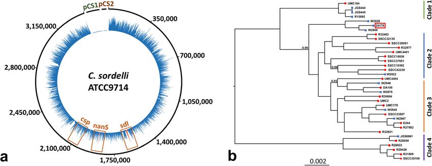
Capewell et al. BMC Veterinary Research   (2020) 16:152                                                         Page 2 of 5

Background                                                    Case presentation
Canine hemorrhagic gastroenteritis (also canine gastro-       In the present case, a fully vaccinated, eight-year-old, fe-
intestinal hemorrhagic syndrome or acute hemorrhagic          male neutered Labrador presented with a 24-h history of
diarrhea syndrome) is clinically characterized by (per)       vomiting without diarrhea. Until this episode, the dog
acute hemorrhagic diarrhea, frequently accompanied by         had been completely healthy, with a single vomiting epi-
vomiting and hemoconcentration. The disease is often          sode reported approximately 3 months prior. It was
associated with the presence of Clostridium perfringens,      noted that the dog was a known scavenger. Upon clinical
an aerobic, spore-forming, rod-shaped Gram-positive           examination, the dog presented with pink mucous mem-
bacterium [1]. C. perfringens can produce several major       branes, adequate hydration, normothermia, and normo-
toxins (Cpa, Cpb, Etx, and Iap/Iab) that are used to type     cardia. The abdomen lacked any signs of bloating or
infection (types A–E). However, the description of            dilation. The following morning the dog was found de-
several new toxin genes (netB, cpe, netE, and netF) has       ceased. Gross examination conducted 7 h post-mortem
recently expanded the range of toxinotypes [2]. In par-       revealed moderate amounts of dark red, non-clotted
ticular, isolates expressing both Cpe and NetF (termed        fluid within the stomach. The fluid extended caudally
type F) have been linked to acute cases of hemorrhagic        into the first two-thirds of the jejunum, whereas the
diarrhea in canines and equines [1, 3–6]. This includes       remaining small intestine and large intestine were devoid
studies that identified the toxinotype in half of dogs pre-   of content. The gastric mucosa was diffusely discolored
senting with acute hemorrhagic diarrhea [1, 6]. However,      dark red, with the fundic mucosa containing additional
these reports contain several animals with hemorrhagic        irregular patches of more intense reddening that were
gastroenteritis and an uncharacterized clostridial infec-     interpreted as hemorrhages (Fig. 1a). The mucosa of the
tion, suggesting further pathogenic strains or species        remaining gastrointestinal system was mildly reddened
may contribute to disease.                                    and this was interpreted as evidence of congestion. Liver,
   Clostridium sordellii is an emerging pathogen of           lungs, and kidneys were dark red, with small petechial
humans and animals that is commonly found in soil and         hemorrhages present in the endocardium overlying the
sewage [7]. While many strains are non-pathogenic,            right heart base and thymic remnants.
some are virulent, particularly those expressing lethal         Upon histological examination, the superficial third of
toxin (TscL) and hemorrhagic toxic (TcsH) encoded on          the gastric fundic mucosa exhibited coagulative necrosis
separate plasmids, pCS1 and pCS2 [7]. Humans are              (Fig. 1b and c), and in a multifocal to coalescing distri-
commonly infected at sites of soft tissue trauma and typ-     bution, the mucosal surface was lined by a thin layer of
ically exhibit gas gangrene, edema, hypotension, absence      short Gram-positive rods (length: 6 μm; thickness: 1 μm;
of fever, tachycardia, intense leucocytosis, and hemocon-     Fig. 1d), with small groups of rods also extending
centration. Mortality is usually due to hypotension and       multifocally along the gastric pits into the necrotic
multiorgan failure, likely mediated by capillary leak syn-    layers. Inflammatory cells were not evident in any of the
drome, septic processes, and toxic shock [8]. In contrast     mural layers, although small multifocal hemorrhages
to this, cattle and sheep are more commonly infected or-      were present in the mucosa. The proximal mucosa of
ally and exhibit gastrointestinal disease and sudden          the pyloric region, small intestine, and large intestine
death [9–12], whereas equines exhibit fatal internal          exhibited similar changes to those observed in the
omphalitis and atypical myopathy [13, 14]. Infection with     fundic region with these lesions mildly tapering to-
C. sordellii has also been linked to incidences of necrotic   wards a more multifocal to coalescing distribution in
enteritis in chickens [9].                                    the large intestines and regions of more intense bac-
   TscL has been shown to be leucocidal, leading to le-       terial infiltrates and pronounced mucosal necrosis
sions characterized by a profound absence or mild in-         commonly associated with one another. Additionally,
flammatory responses [15]. TcsH is less commonly              multifocal     intra-alveolar    and     intra-bronchiolar
found and is hypothesized to cause alteration to the          hemorrhages were evident in the lungs and the ad-
cytoskeleton, resulting in capillary leakage [10]. How-       renal cortex exhibited acute, multifocal to coalescing
ever, it is important to note that the majority of C. sor-    hemorrhages. Anaerobic culture of gastric content
dellii strains do not possess TscL nor TscH but are still     using horse blood agar revealed a profuse growth of a
pathogenic, albeit less so than those possessing these        clostridial species that was subsequently identified as
toxins [7, 16]. These strains do not contain any              C. sordellii using the Analytic Profile Index (API)
recognizable plasmids and additional chromosomally-           method (bioMérieux) and designated as strain 24,178.
encoded virulence factors have been suggested, including      Aerobic culture using sheep blood agar revealed a
sordellilysin (Sdl), neuraminidase (NanS), and phospho-       profuse growth of Cellulomonas/Microbacterium, also
lipase C (Csp), for which the exact mechanisms of             identified by API. Salmonella spp., Campylobacter
pathophysiology remain to be established [16].                spp., C. perfringens, and C. difficile were not isolated.
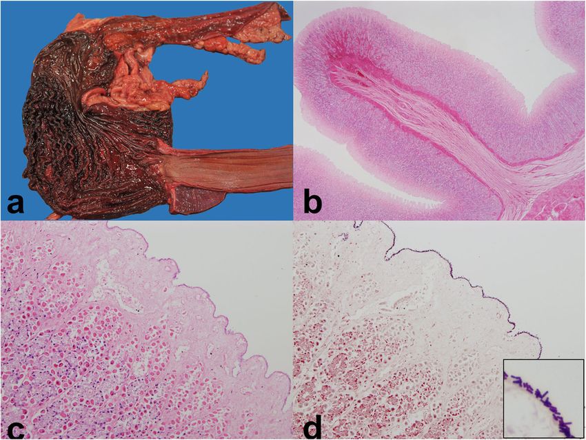
Capewell et al. BMC Veterinary Research      (2020) 16:152                                                                              Page 3 of 5

 Fig. 1 Gross and histological appearance of a canine stomach following infection with C. sordellii. The gastric mucosa was diffusely discolored
 dark red and the fundic region also exhibited irregular patches of more intense reddening (interpreted as hemorrhages) a. Hematoxylin and
 eosin stained tissue showed that the most proximal aspects of the fundic mucosa were necrotic and lacked appreciable inflammatory infiltrates
 (b-c). Small mucosal hemorrhages were present, and subsequent Gram-stain revealed that the mucosa was covered by a thin layer of small,
 Gram-positive rods (inset shows an enlarged view) (d)

  A whole genome sequencing approach was utilized to                       mutation. These mutations have not previously been de-
fully characterize C. sordellii strain 24,178. To this end,                scribed and it is unknown what impact, if any, these
purified DNA was prepared from culture using a                             would have on virulence. Finally, a core C. sordellii gen-
QIAamp DNA Mini Kit (Qiagen GmbH, Hilden,                                  ome was created using 35 publicly available sequences
Germany) and an aliquot containing 24 ng/ul was com-                       and a previously established method [7]. This resulted in
mercially sequenced using the Illumina HiSeq platform                      1157 shared genes that were aligned using Mauve [20]
(MicrobesNG, Birmingham). This resulted in 378,245                         to construct a phylogenetic tree with FastTree2 [21].
read pairs aligned to a reference C. sordellii strain                      Similar to previous work [7], the tree was rooted with C.
ATCC9714 that possesses both pCS1 and pCS2 plas-                           difficile strain R20291. The tree topology suggested that
mids. However, despite an average read depth of 18.1                       the isolated C. sordellii strain is closely related to two
across the genome, no reads were found to align to ei-                     livestock C. sordellii strains (W3026 and W2948), pos-
ther plasmid, indicating that the tscL and tscH toxin                      sibly indicating that the dog contracted the infection via
genes were not present in the isolate under investigation                  environmental ingestion. Neither of the closely related
(Fig. 2a). Sequence reads were assembled de novo using                     veterinary strains possess the plasmid-encoded toxins.
VelvetOptimiser for Velvet [17] and BLASTN in order                        While the four clades of C. sordellii were largely recon-
to identify sequences similar to tscL and tscH in the                      structed in this analysis, the additional data provided by
resulting assembly and this also resulted in no hits, again                our analysis show that the new strain 24,178 and the re-
confirming that the major toxin genes were not present.                    lated veterinary strains are placed within a distinct group
Further in silico plasmid detection using HyAsP [18]                       between clade 1 and clades 2 and 3. This may suggest
and plasmidSPAdes [19] detected no evidence of novel                       these strains are a novel environmental clade more
plasmids, whilst reciprocal BLASTN searches of the 123                     closely related to the virulent clade 1 strains despite not
de novo contigs assembled by Velvet against ATCC9714                       possessing TcsL nor TcsH.
also did not detect any regions that were unique to the
isolate. The virulence associated genes sordellilysin (sdl),               Discussion and conclusions
neuraminidase (nanS), and phospholipase C (csp) were                       This case report demonstrates that C. sordellii can cause
all found to be present (Fig. 2b). The predicted sordelli-                 a fatal hemorrhagic and necrotizing gastroenteropathy in
lysin amino acid sequence was identical to that found in                   dogs and should be considered as a potential causative
other strains, while neuraminidase possessed a Leu397Ile                   agent for such syndromes alongside C. perfringens. The
mutation and phospholipase C featured an Asp480Glu                         close relatedness of this isolate to strains previously
Capewell et al. BMC Veterinary Research      (2020) 16:152                                                                                       Page 4 of 5

 Fig. 2 Genomic analysis of C. sordellii isolate. A schematic showing the depth of sequencing reads from strain 24,178 (blue) that mapped to the
 genome of C. sordellii reference strain ATCC9714 and the two toxin-encoding plasmids (pCS1 and pCS2). The numbers indicate base pair position.
 No reads aligned to pCS1 or pCS2 (highlighted in green and brown), although reads were found that mapped to the putative virulence factors
 highlighted in red; sordellilysin (sdl), neuraminidase (nanS), and phospholipase C (csp) (a). A cladogram of publicly available genomes for human
 (red circles) and veterinary isolates (blue squares) indicates that strain 24,178 (red box) is closely related to two livestock samples (W3026 and
 W2948). There is strong node support to suggest that this cluster does not sit within any of the four classical clades (support values are shown at
 the corresponding node if above 0.8 and the scale bar denotes nucleotide changes per position) (b)

recovered from diseased livestock suggests that the in-                     unlikely. Our findings therefore highlight the need for
fection of this dog may have occurred via ingestion from                    further genomic studies to quantify the importance of
the environment. C. sordelli infections associating with                    chromosomally encoded virulence factors in this patho-
severe necrotizing gastroenteric disease have previously                    gen. Genetic characterization beyond the straightforward
been described for several livestock species, including                     presence or absence of plasmid-encoded virulence fac-
chickens [11], sheep [9], and cattle [12]. Fatal infections                 tors may allow the pathogenic potential of an isolate to
have also been reported for equines [13, 14]. Therefore,                    be estimated and an array of genetic polymorphisms as-
a history of scavenging or contact with livestock may                       sociated with subtle pathogenic differences to be identi-
represent a risk factor in such cases. It is notable that                   fied. An important aspect of this retrospective case study
the strain of C. sordellii under investigation caused se-                   was the application of whole-genome sequencing to fully
vere disease despite lacking the classical tcsL and tcsH                    characterize the pathogen involved. As sequencing tech-
virulence factors sometimes found in this species. Inter-                   nology becomes a more readily accessible tool to sup-
estingly, the genetic basis for virulence associated with                   port diagnostic veterinary clinical pathology, the use of
companion animal hemorrhagic gastroenteritis caused                         predictive models designed to gauge isolate virulence
by C. perfringens has been the subject of recent investi-                   may lead to more tailored treatment for canine
gations and this has resulted in the identification of the                  hemorrhagic canine gastroenteritis and other Clostrid-
NetF toxin in isolates from canine [1, 4–6] and equine                      ium-mediated infections.
cases [4, 5]. This pore-forming cytotoxin co-locates with
the cpe enterotoxin gene on a plasmid [4]. Since its dis-                   Abbreviations
                                                                            Cpa: Clostridium perfringens alpha toxin; Cpb: Clostridium perfringens beta
covery, large-scale studies have indicated a significant as-                toxin; Etx: Clostridium perfringens epsilon toxin; Iap/Iab: Clostridium perfringens
sociation with dogs with acute hemorrhagic diarrhea                         iota toxin; Cpe: Clostridium perfringens enterotoxin; NetB: Clostridium
syndrome [1, 3–6] and a causal link has been proposed                       perfringens necrotic enteritis toxin B; NetE: Clostridium perfringens necrotic
                                                                            enteritis toxin E; NetF: Clostridium perfringens necrotic enteritis toxin F;
[1]. It is striking that we did not find any plasmid-                       TcsL: Clostridium sordellii lethal toxin; TcsH: Clostridium sordellii hemorrhagic
encoded virulence factors in our strain of C. sordellii.                    toxic; pCS1: Clostridium sordellii plasmid 1; pCS2: Clostridium sordellii plasmid
While it is possible that the toxin-encoding plasmids                       2; Sdl: Sordellilysin; NanS: Neuraminidase S; Csp: Clostridium sordellii
                                                                            Phospholipase C; API: Analytic Profile Index
were lost during culture adaption of isolate 24,178, as
has been described for other isolates [7], the short-term
                                                                            Acknowledgements
growth in vitro and the close relationship between iso-                     We would like to thank the VDS histopathology team for technical support
lates 24,178, W3026, and W2948 suggest that this is                         with tissue processing for the histological assessments.
Capewell et al. BMC Veterinary Research (2020) 16:152 Page 5 of 5
Authors’ contributions Proceedings of the Fifth Anaerobe Discussion Group Symposium Held at
PC analyzed the genomic data and contributed to the writing of the Churchill College, University of Cambridge, July 23–25, 1987. New York:
manuscript. AR performed the gross and histological examination and Wiley; 1988. p. 61.
contributed to the writing of the manuscript. MF performed the 13. Ortega J, Daft B, Assis R, Kinde H, Anthenill L, Odani J, Uzal FA. Infection of
microbiological work. MM co-ordinated the genomic sequencing effort and internal umbilical remnant in foals by Clostridium sordellii. Vet Pathol. 2007;
contributed to the writing of the manuscript. WW contributed to the gen- 44(3):269–75.
omic analysis and the writing of the manuscript. All authors read and ap- 14. Unger-Torroledo L, Straub R, Lehmann AD, Graber F, Stahl C, Frey J, Gerber
proved the final manuscript. V, Hoppeler H, Baum O. Lethal toxin of Clostridium sordellii is associated with
fatal equine atypical myopathy. Vet Microbiol. 2010;144(3–4):487–92.
Funding 15. Popoff MR. Clostridium difficile and Clostridium sordellii toxins, proinflammatory
Not applicable. versus anti-inflammatory response. Toxicon. 2018;149:54–64.
16. Voth DE, Martinez OV, Ballard JD. Variations in lethal toxin and
Availability of data and materials cholesterol-dependent cytolysin production correspond to differences in
The datasets supporting the conclusions of this article are available in the cytotoxicity among strains of Clostridium sordellii. FEMS Microbiol Lett.
NCBI repository BioProject number PRJNA607435 http://www.ncbi.nlm.nih. 2006;259(2):295–302.
gov/bioproject/607435. 17. Zerbino DR, Birney E. Velvet: algorithms for de novo short read assembly
using de Bruijn graphs. Genome Res. 2008;18(5):821–9.
Ethics approval and consent to participate 18. Müller R, Chauve C. HyAsP, a greedy tool for plasmids identification.
Consent was obtained from the owner to participate in the examination. Bioinformatics. 2019.
19. Antipov D, Hartwick N, Shen M, Raiko M, Lapidus A, Pevzner P.
plasmidSPAdes: assembling plasmids from whole genome sequencing data.
Consent for publication
bioRxiv, vol. 048942; 2016.
Written consent was obtained from the owner to participate for the
20. Darling AE, Mau B, Perna NT. Progressivemauve: multiple genome
publication of this case report and the accompanying images.
alignment with gene gain, loss and rearrangement. PLoS One. 2010;5(6):
e11147.
Competing interests
21. Price MN, Dehal PS, Arkin AP. FastTree 2–approximately maximum-
The authors declare that they have no competing interests.
likelihood trees for large alignments. PLoS One. 2010;5(3):e9490.
Received: 19 February 2020 Accepted: 10 May 2020
Publisher’s Note
Springer Nature remains neutral with regard to jurisdictional claims in
References published maps and institutional affiliations.
1. Leipig-Rudolph M, Busch K, Prescott JF, Mehdizadeh Gohari I, Leutenegger
CM, Hermanns W, Wolf G, Hartmann K, Verspohl J, Unterer S. Intestinal
lesions in dogs with acute hemorrhagic diarrhea syndrome associated with
netF-positive Clostridium perfringens type a. J Vet Diagn Investig. 2018;30(4):
495–503.
2. Rood JI, Adams V, Lacey J, Lyras D, McClane BA, Melville SB, Moore RJ,
Popoff MR, Sarker MR, Songer JG, et al. Expansion of the Clostridium
perfringens toxin-based typing scheme. Anaerobe. 2018;53:5–10.
3. Diniz AN, Coura FM, Rupnik M, Adams V, Stent TL, Rood JI, de Oliveira CA,
Jr., Lobato FCF, Silva ROS: The incidence of Clostridioides difficile and
Clostridium perfringens netF-positive strains in diarrheic dogs. Anaerobe
2018;49:58–62.
4. Mehdizadeh Gohari I, Parreira VR, Nowell VJ, Nicholson VM, Oliphant K,
Prescott JF. A novel pore-forming toxin in type a Clostridium perfringens is
associated with both fatal canine hemorrhagic gastroenteritis and fatal foal
necrotizing enterocolitis. PLoS One. 2015;10(4):e0122684.
5. Mehdizadeh Gohari I, Unterer S, Whitehead AE, Prescott JF. NetF-producing
Clostridium perfringens and its associated diseases in dogs and foals. J Vet
Diagn Investig. 2020;32(2):230–8.
6. Sindern N, Suchodolski JS, Leutenegger CM, Mehdizadeh Gohari I, Prescott
JF, Proksch AL, Mueller RS, Busch K, Unterer S. Prevalence of Clostridium
perfringens netE and netF toxin genes in the feces of dogs with acute
hemorrhagic diarrhea syndrome. J Vet Intern Med. 2019;33(1):100–5.
7. Couchman EC, Browne HP, Dunn M, Lawley TD, Songer JG, Hall V, Petrovska
L, Vidor C, Awad M, Lyras D. Clostridium sordellii genome analysis reveals
plasmid localized toxin genes encoded within pathogenicity loci. BMC
Genomics. 2015;16(1):392.
8. Aldape M, Bryant A, Stevens D. Clostridium sordellii infection: epidemiology,
clinical findings, and current perspectives on diagnosis and treatment. Clin
Infect Dis. 2006;43(11):1436–46.
9. Lewis C, Naylor R. Sudden death in sheep associated with Clostridium
sordellii. Vet Rec. 1998;142(16):417–21.
10. Craven R, Lacy DB. Clostridium sordellii lethal-toxin autoprocessing and
membrane localization activities drive GTPase glucosylation profiles in
endothelial cells. mSphere. 2016;1(1):e00012–5.
11. Rimoldi G, Uzal F, Chin RP, Palombo EA, Awad M, Lyras D, Shivaprasad HL.
Necrotic enteritis in chickens associated with Clostridium sordellii. Avian Dis.
2015;59(3):447–51.
12. Taylor DJ, Estrada AE, Al-Mashat RR. Toxigenic Clostridium Sordellii and
Clostridium perfringens Type A Infections in Animals. In: Anaerobes Today:You can also read