Structure and mode of action of clostridial glucosylating toxins: the ABCD model
←
→
Page content transcription
If your browser does not render page correctly, please read the page content below
Review

Structure and mode of action of
clostridial glucosylating toxins: the
ABCD model
Thomas Jank and Klaus Aktories
Institut für Experimentelle und Klinische Pharmakologie und Toxikologie der Albert-Ludwigs-Universität Freiburg,
Otto-Krayer-Haus, Albertstrasse 25, D-79104 Freiburg, Germany

Toxins A and B, which are the major virulence factors of                                  Toxins with multimodular structure
antibiotic-associated diarrhea and pseudomembranous                                       Toxin A consists of 2710 amino acid residues with a
colitis caused by Clostridium difficile, are the prototypes                               molecular mass of 308 kDa and toxin B comprises 2366
of the family of clostridial glucosylating toxins. The                                    residues with a mass of 269.6 kDa. These toxins are there-
toxins inactivate Rho and Ras proteins by glucosylation.                                  fore also known as large clostridial cytotoxins [10]. On the
Recent findings on the autocatalytic processing of the                                    basis of their amino acid sequences, a tripartite structure for
toxins and analysis of the crystal structures of their                                    the toxins had been suggested [10,12,13], with a biologically
domains have made a revision of the current model of                                      active N-terminal domain, a middle translocation section
their actions on the eukaryotic target cells necessary.                                   characterized by a small hydrophobic stretch (prediction of
                                                                                          transmembrane regions), and a C-terminal receptor-bind-
Introduction                                                                              ing domain. This prediction of the structure–function
Antibiotic-associated diarrhea and pseudomembranous                                       relationship was in line with the model of AB-toxins such
colitis induced by Clostridium difficile have emerged as                                  as diphtheria toxin, consisting of a biologically active
important nosocomial infections. During the past decade,                                  domain and a binding or translocation domain [14]. The
these diseases seem to have become more serious and more                                  binding domain can be separated into subdomains (e.g. for
frequently refractory to therapy [1]. The occurrence of                                   receptor binding and translocation) resulting in a tripartite
hypervirulent strains of C. difficile such as PCR ribotype                                structure. However, recent studies indicate that a multi-
027/PFGE type NAP1 [2] with attributable mortality rates                                  modular structure more accurately describes the structure–
of up to 16.7% is concerning [3]. The major virulence factors                             function relationship of the clostridial glucosylating toxins
of C. difficile are two protein toxins, named toxin A and toxin                           (Figure 1). These toxins have a biologically active domain
B (also designated TcdA and TcdB), which have been recog-                                 and a binding and translocation domain but in addition they
nized for their important role in disease for the past 30 years.                          have an autocatalytic, self-cutting protease domain for toxin
In addition, some strains of C. difficile produce a binary                                processing. Thus, the AB-toxin model can be extended to an
ADP-ribosylating toxin (C. difficile transferase, CDT) that                               ABCD model (A, biological activity; B, binding; C, cutting; D,
modifies G-actin [4]. Notably, hypervirulent strains are                                  delivery).
characterized by production of 10- to 20-fold larger amounts
of toxins A and B, the resistance to fluoroquinolones and                                 The C terminus binds to target cell membranes
production of the actin-ADP-ribosylating toxin [2].                                       The receptor-binding domain located at the C terminus of
                                                                                          the clostridial glucosylating toxins consists of repeating
Structure–function relationships of clostridial                                           polypeptide units [12,15–17]. Recently, two C-terminal
glucosylating toxins                                                                      fragments (terminal 127 and 255 residues) of toxin A
C. difficile toxin A and toxin B are the prototypes of the                                (toxinotype VI) were crystallized [18,19]. These studies
family of clostridial glucosylating toxins [5,6]. Other mem-                              gave new insights into the overall structure of the C
bers of this toxin family are the hemorrhagic and the lethal                              terminus of the toxin (Figure 1). Toxin A folds in a sole-
toxins from Clostridium sordellii and the a-toxin from                                    noid-like structure with 32 small repeats consisting of 15–
Clostridium novyi. Moreover, several isoforms of toxin A                                  21 residues and seven large repeats of 30 residues. Each
and B have been described, adding to this family of cyto-                                 repeat forms a single b-hairpin that is rotated by 1208 to
toxins [7–9]. All of these toxins share sequence identities                               each other, thereby forming a screw-like superfold
ranging from 36% to 90% and have molecular masses                                         (Figure 1). Solenoid structures are frequently found in
between 250 and 308 kDa [10,11]. Because toxins A and                                     bacterial virulence proteins [20]. They increase the sur-
B are of major clinical importance and because most data                                  face area of proteins and enable protein–protein or
obtained are from these toxins, this review focuses on                                    protein–carbohydrate interactions. Some years ago, Kri-
recent progress in the understanding of the structure–                                    van and Tucker showed binding of the trisaccharide
function relationship of these toxins.                                                    Gala1–3Galb1–4GlcNAc to toxin A [21,22]. This was con-
                                                                                          firmed and structurally explained by co-crystallization of
      Corresponding author: Aktories, K. (klaus.aktories@pharmakol.uni-freiburg.de).      toxin A with an artificial trisaccharide containing the
222                                                                           0966-842X/$ – see front matter ß 2008 Elsevier Ltd. All rights reserved. doi:10.1016/j.tim.2008.01.011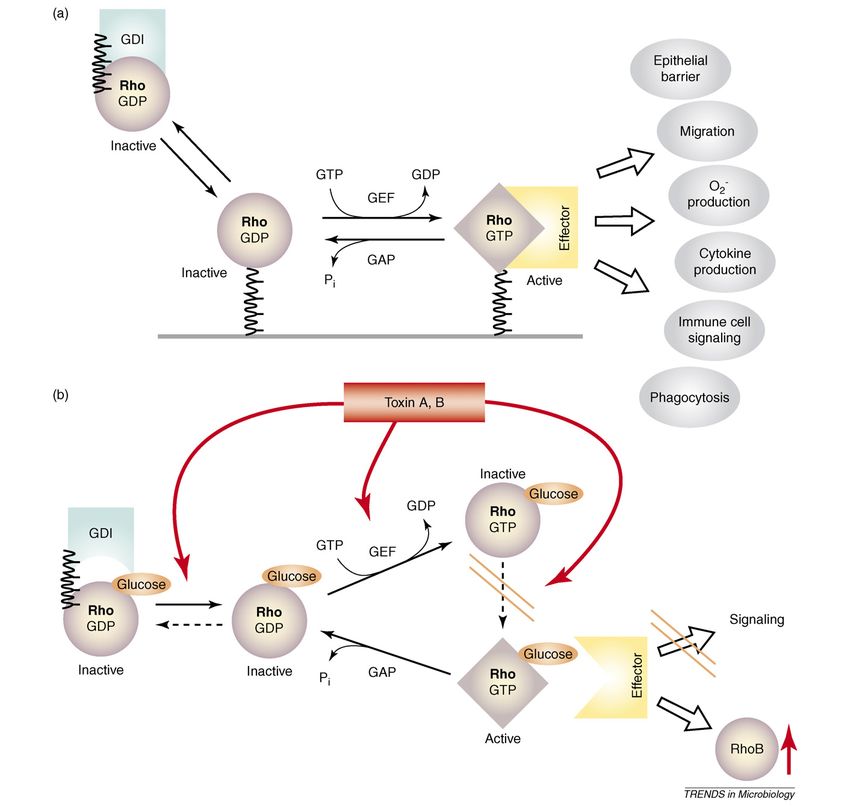
Review                                                                                                                            Trends in Microbiology        Vol.16 No.5

Figure 1. Domain structure of clostridial glucosylating toxins. The ABCD model (labeled in bold at top of figure) of clostridial glucosylating toxins is shown with Clostridium
difficile toxin B as an example. The biologically active glucosyltransferase A-domain (bold) is located at the N terminus (amino acids 1–543) and has been crystallized
recently. The DXD motif, which is involved in Mn2+ coordination, is located in this domain. The C terminus of the clostridial glucosylating toxins, which consists of
polypeptide repeats, is involved in receptor binding (B-domain, bold). A fragment of the C-terminal repeats of toxin A has been crystallized, showing a solenoid-like
structure. From this rather small fragment, the structure of the whole C-terminal part of toxin A has been deduced, which consists of 39 repetitive elements [19]. According
to this model the repeats have a b-hairpin structure. Rotation of the hairpins by 1208 relative to the neighboring hairpin forms a screw-like structure. The cysteine protease
domain (residues 544–767), which is similar to Vibrio cholerae RTX toxin, is located adjacent to the glucosyltransferase domain. This C-domain (bold) is involved in
processing and cutting of the toxin. The cysteine protease C-domain can be characterized by the catalytic triad consisting of Asp587, His653 and Cys698 (DHC). Alternatively,
the DXG (D1665) motif was suggested to be part of an aspartate protease domain, possibly involved in processing of the toxins. In the middle part of the protein is a short
hydrophobic region (residues 956–1128), which might be involved in pore formation and delivery of the catalytic domain into the cytosol (D-domain, bold). Images were
created using PyMOL (www.pymol.org).

Gala1–3Galb1–4GlcNAc-glycan [19]. The carbohydrate-                                       A. However, structural data are not available to date and
binding groove is formed between the junction of a large                                  the nature of the receptor of C. difficile toxin B is even
repeat and the hairpin turn of the following small repeat                                 further from being defined.
by several amino acids. These residues are not conserved
in the other clostridial glucosylating toxins and might                                   Toxin uptake: a question of cutting and delivery
have a crucial role in carbohydrate receptor specificity.                                 Following receptor binding, the clostridial glucosylating
However, human tissue generally does not produce a-                                       toxins are endocytosed [25] (Figure 2) – the precise endo-
anomeric galactose bonds [23], indicating that the carbo-                                 cytosis pathway is not known. After endocytosis, the toxins
hydrate structure Gala1–3Galb1–4GlcNAc cannot be part                                     translocate through the early endosomal membrane into
of intestinal receptors in humans. Therefore the disac-                                   the cytosol. This process depends on the acidification of
charide Galb1–4GlcNAc, which is present in humans, has                                    endosomes by vesicular H+-ATPase. Bafilomycin, which
been suggested to be part of a possible glycan receptor.                                  blocks the H+-ATPase, inhibits cytosolic entry of the toxin
Whether a glycoprotein or glycosphingolipid [24] (either                                  and intoxication of cells [26]. Therefore, C. difficile toxins A
with or without an additional protein receptor) comprises                                 and B belong to the short trip toxins group (which includes
the intestinal receptor remains to be clarified. The C-                                   diphtheria toxin) that enter the cytosol of host cells from an
terminal part of toxin B is probably similar to that of toxin                             early endosomal compartment. These protein toxins can be

                                                                                                                                                                           223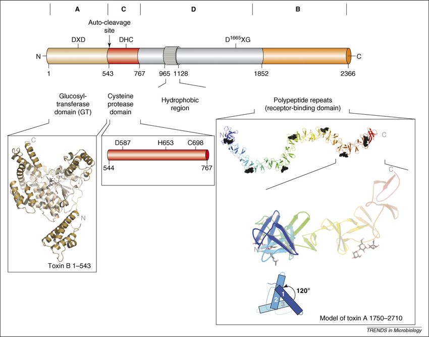
Review                                                                                                                          Trends in Microbiology Vol.16 No.5

Figure 2. Processing of clostridial glucosylating toxins. Clostridial glucosylating toxins bind to the cell membrane through the C terminus. The receptors for the toxins are
not yet defined. For C. difficile toxin A carbohydrates are proposed as receptors. After binding, the toxins are endocytosed to reach an acidic endosomal compartment. Here,
conformational changes occur enabling insertion of the toxin into the endosomal membrane and subsequent pore-formation. The toxins are processed by autocatalytic
cleavage, which in the case of toxin B depends on the presence of inositol hexakisphosphate (InsP6). Only the glucosyltransferase domain of the N terminus of the toxins is
released into the cytosol. In the cytosol, Rho GTPases are glucosylated at Thr37 (e.g. RhoA) or at Thr35 (e.g. Rac or Cdc42).

distinguished from the long trip toxins (which includes                                  pore formation is directly involved in delivery of the toxin
cholera toxin) that travel retrograde from endosomes to the                              into the cytosol remains unclear, but it is thought that the
Golgi and from there to the ER, where they eventually                                    hydrophobic region (residues 956–1128 of toxin B) is part of
enter the cytosol [27]. The low pH of early endosomes                                    the delivery domain.
induces conformational changes of the clostridial toxins,                                   The precise mechanism of the translocation process
resulting in exposure of a hydrophobic region of the protein                             remains one of the most enigmatic puzzles of the action
toxins, enabling membrane insertion [28]. It has been                                    of these toxins. Recent studies indicate that the toxins
suggested that residues 956–1128 of toxin B are part of                                  must be processed to reach the cytosol. Only the catalytic
the hydrophobic region, which is involved in membrane                                    domain of the toxins, including the N-terminal 543 amino
insertion [13]. A short-term decrease in extracellular pH of                             acids, is delivered into the cytosol [30,31]. The search for a
the medium of cell cultures mimics the low pH of endo-                                   host protease possibly involved in toxin processing resulted
somes and enables the toxins to enter host cells directly                                in the unexpected finding that the clostridial glucosylating
through the cell membrane [26,28]. Membrane insertion is                                 toxins are auto-proteolytically processed [32]. Even more
paralleled by formation of pores. This can be shown by                                   surprising, inositol hexakisphosphate (InsP6, phytic acid)
86
  Rb+ ion release from 86Rb+-loaded host cells when the pH                               was found to be an essential factor for activation of proteol-
of the cell medium is reduced to pH 5 [26]. Toxin B mutants                              ysis [32], however its functional role is not clear. Polypho-
and N-terminal truncated toxins consisting of the C termi-                               sphorylated inositol, a common inositol metabolite in
nus and the middle part of the protein (including the hydro-                             mammalian cells, is highly charged and has diverse bio-
phobic residues 956–1128) are sufficient for pore formation                              logical functions, including mRNA traffic king, binding to
[26]. It has been suggested that the C terminus, which is                                clathrin-assembly protein AP-2, inhibition of protein phos-
involved in binding, is not essential for pore formation but                             phatases and stimulation of protein kinases [33,34]. InsP6
enhances toxin–membrane interaction. The efficacy of pore                                might be involved in stabilization of toxin protein confor-
formation induced by toxin A is largely cholesterol-depend-                              mation, which is essential for protease activity and/or
ent. Cholesterol depletion of membranes with methyl-b-                                   proper cleavage.
cyclodextrin inhibits 86Rb+ efflux and cholesterol repletion                                Recently it was suggested that toxin B possesses
reconstituted pore-forming activity of toxin A [29]. Whether                             aspartate protease activity, which is responsible for toxin

224
Review Trends in Microbiology Vol.16 No.5
cleavage because the aspartate protease inhibitor EPNP The N-terminal enzyme domain
(1,2-epoxy-3-p-nitrophenoxypropane) blocked the proces- The N terminus harbors the glucosyltransferase activity of
sing of toxin B and labeled aspartate 1665 as part of a the toxins and is the biological activity domain [37]. The 543
short Asp-Xaa-Gly (DXG) motif observed in many aspar- amino acid residues, which are delivered into the cytosol of
tate proteases [32]. Another hypothesis describes a host cells, form the glucosyltransferase domain. Recently,
cysteine protease activity as being responsible for the the 3D-structure of the catalytic domain (residues 1–543) of
processing of clostridial glucosylating toxins. There are toxin B has been solved [38]. The catalytic core is formed by a
several lines of evidence supporting this hypothesis. First, mixed a/b-fold with mostly parallel b-strands (Figure 3a).
as observed frequently for cysteine proteases, dithiothrei- The overall structure of the catalytic core is similar to other
tol activates the auto-catalytic cleavage of the toxin. bacterial glycosyltransferases, such as Neisseria meningiti-
Second, the autocatalytic activity is blocked by N-ethylma- dis galactosyltransferase LgtC (lipooligosaccharide glycosyl
leimide, a typical inhibitor of cysteine proteases. Third, the transferase C) [39] and Bacillus subtilis glycosyltransferase
clostridial glucosylating toxins share sequence similarity SpsA [40], but also to eukaryotic glycosyltransferases, such
with a novel, recently identified autocatalytic cysteine as bovine galactosyltransferase a3GalT [41]. All of these
protease domain within the structure of the RTX (repeats transferases belong to the glycosyltransferase A (GT-A)
in toxins) toxin of Vibrio cholerae. Several essential cata- family [42]. Mainly based on the sequence homology
lytic residues, including the putative catalytic triad D587, described here, the family of clostridial glucosylating toxins
H653 and C698, are conserved [35,36] (Figure 1). Accord- has been designated glycosyltransferase family 44 as
ing to this model, a cysteine protease cutting domain, defined by Henrissat et al. (http://www.cazy.org/). This
which is adjacent to the glucosyltransferase domain, is family also includes genes from Escherichia coli and Chla-
responsible for processing of the toxin. mydia trachomatis coding for putative glycosyltransferases.
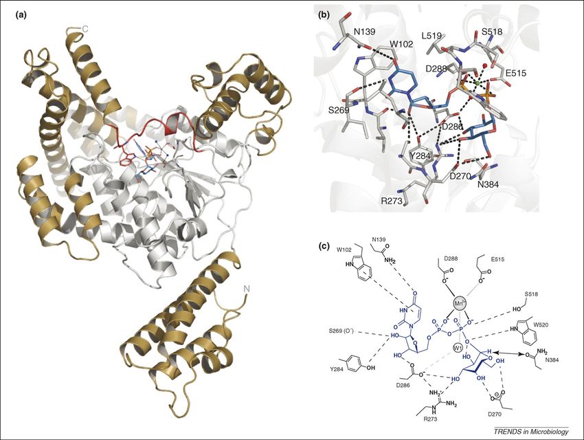
Figure 3. The glucosyltransferase domain of Clostridium difficile toxin B. (a) Structural model of the glycosyltransferase domain of C. difficile toxin B, where UDP-glucose
(blue) is attached to the catalytic cleft via Mn2+ (brown) and to the residues of the DXD motif (ball-and-stick model); water molecules are shown as small blue spheres. The
GT-A type fold catalytic core is presented in white and the additional subdomains are shown in brown. The putative ‘flexible loop’ region is shown in red with Trp520
hydrogen bonding to the b-phosphate of UDP-glucose. (b) The catalytic cleft of toxin B, as in (a) but rotated 458. Several amino acids involved in co-substrate binding are
shown. (c) Schematic representation of the catalytic cleft shown in (b). Images were created using PyMOL (www.pymol.org).
225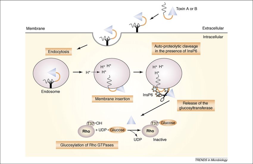
Review                                                                                        Trends in Microbiology Vol.16 No.5

    The catalytic core of toxin B consists of 234 residues       [54,55]. A SNi (internal return) mechanism has been
plus >300 additional residues. These additional residues         suggested for the transferase reaction. This mechanism
are mainly helices. The four N-terminal helices are              is characterized by an oxocarbenium-like transition state
particularly prominent and seem to be an independent             [38].
subdomain, possibly involved in membrane interaction.               The clostridial glucosylating toxins exhibit high co-sub-
Mesmin and coworker showed for lethal toxin from C.              strate specificity. Whereas toxin A and B and also the
sordellii (the closest homolog of toxin B) that the first        related lethal toxin from C. sordellii use UDP-glucose as
18 amino acids (correlating to the first helix of the N-         a donor substrate, the co-substrate of a-toxin from Clos-
terminal subdomain) are crucial for lipid bilayer attach-        tridium novyi is UDP-N-acetylglucosamine (UDP-GlcNAc)
ment [43]. Also, truncations beyond amino acid Lys65             [56]. Biochemical studies revealed that Ile383 and Glu385
[44] lead to inactivation of the glucosyltransferase and         are responsible for co-substrate specificity. These residues
glucohydrolase activity, demonstrating the importance of         limit the space of the catalytic cleft for binding of the co-
the N-terminal helical bundle for catalytic activity.            substrate in toxin B. Exchange of these residues changes
Recently, it was shown that domain C1 of Pasteurella             the co-substrate specificities of the toxins. Thus, the toxin
multocida toxin (PMT) is structurally similar (41% sim-          B double mutant Ile383Ser, Gln385Ala accepts UDP-
ilarity with toxin B) to this subdomain of toxin B. A role       GlcNAc as a co-substrate [57] and changing Ser385 and
in membrane interaction has also been proposed for PMT           Ala387 to Ile and Glu, respectively, turns a-toxin into a
[45].                                                            UDP-glucose-accepting transferase.
    The crystal structure of the catalytic domain of toxin B
revealed a set of essential amino acid residues involved in      Interaction of toxin B with Rho GTPases
glucosyltransferase reaction or in substrate binding. The        Glycosyltransferases are region-selective enzymes. In
Asp-Xaa-Asp (DXD) motif is characteristic of GT-A family         the case of the clostridial glucosylating toxins a
members (Asp286 and Asp288 in toxin B) [46,47]. This             specific threonine residue in the substrate proteins
motif is involved in Mn2+, UDP and glucose binding.              (small GTP-binding proteins) is monoglucosylated
Whereas Asp288 complexes directly with Mn2+, interaction         [54,58]. This threonine senses the integrity of the nucleo-
of Asp286 with Mn2+ occurs via a water molecule. Asp286          tide GTP, which is bound to the GTPase in the active
also interacts with the 30 hydroxyl group of the UDP-ribose      conformation and to the hydrolyzed GDP form in the
and with the 30 hydroxyl group of glucose, so it is of major     inactive conformation. Sensing occurs via binding to a
importance for proper positioning of UDP-glucose in the          Mg2+ ion, which is also complexed with the phosphates of
catalytic cleft of the enzyme. Trp102, which is also essen-      the nucleotide. Glucosylation prevents the sensor func-
tial for enzyme activity, stabilizes the uracil ring of UDP by   tion and thereby prevents the fundamental confor-
aromatic stacking. In addition to the DXD motif and              mational switch of the GTP-binding proteins necessary
Trp102, Asp270, Arg273, Tyr284, Asn384 and Trp520 were           for interaction with diverse effectors or regulator
identified by alanine scanning as being essential for            proteins [59] (Box 1).
enzyme activity [48]. Asp270 and Arg273 are conserved               Substrate specificity of the GTP-binding proteins for the
in several GT-A type glycosyltransferases and form with          toxins has not been structurally defined to date. Never-
the help of Asp286, a highly defined hydrogen-bond net-          theless, distinct amino acids or regions on Rho GTPases
work for proper adjustment of the glucose moiety of the          have been shown to have a role in defining specificity. The
donor substrate [41,49]. A common structural feature of all      substrate properties of RhoA versus RhoD are defined by
GT-A type glycosyltransferase is the ‘flexible loop’, which      the residues Ser73 and Phe85, respectively, located on the
switches from an open, disordered conformation (apo-             switch II region [60]. The differential recognition of Rac
enzyme) to a closed, ordered conformation on UDP-sugar           and RhoA by lethal toxin, toxin B1470 and toxin B8864, is
binding. Co-substrate binding creates a deep pocket that         attributed to the N-terminal region around amino acids
serves as a binding site for the acceptor substrate [50]         22–27 of the GTPases where Lys27 of Rho has a major role
(Figure 3a). The deep burial of the UDP-sugar could pre-         [61].
vent water molecules from acting as acceptors and hydro-            Earlier studies showed that the C-terminal part of the
lyzing the nucleotide sugar. It was proposed that the            catalytic domain of the toxins (residues 364–516) confers
‘flexible loop’ helped release the reaction products, leading    substrate specificity [62]. Recent data indicate that amino
to suitable turnover of the enzyme [42,51]. Trp520 is well       acids Arg455, Asp461, Lys463 and Glu472, and residues of
conserved in all of the clostridial glucosylating toxins and     helix a17 (e.g. Glu449) of toxin B are essential for enzyme–
resides on the putative ‘flexible loop’ formed by amino acids    protein substrate recognition [48]. Introduction of helix
510 to 523 and interacts with the scissile bond of the donor     a17 of toxin B into C. sordellii lethal toxin prevents the
substrate (b-phosphate oxygen of UDP). It was shown for          modification of Ras subfamily proteins but enables gluco-
other glycosyltransferases that the corresponding trypto-        sylation of RhoA. Crystallographic and biochemical results
phan residue swings out thereby opening the conformation         led to a docking model in which the GTPase consensus
while releasing UDP [52,53].                                     binding region (suggested for effector or regulator binding
    Glycosyltransferases are strictly stereospecific             [63]) interacts in a similar manner with the glucosyltrans-
enzymes. The known clostridial glucosylating toxins              ferase toxins (Figure 4). In this model, the membrane-
are retaining glucosyltransferases, because the sugar            associated regions of the GTPase and the region of the
is attached to the target protein in the same a-                 toxin, which is supposed to interact with the membrane,
anomeric configuration as in the substrate UDP-glucose           are located on the same side [48].

226
Review Trends in Microbiology Vol.16 No.5
Box 1. Functional consequences of glucosylation of Rho GTPases by clostridial toxins
The clostridial glucosylating toxins modify Rho proteins (Figure I). oxide production, cytokine secretion and immune cell signaling. The
These are low molecular mass GTPases, which control numerous clostridial glucosylating toxins modify Rho GTPases at threonine 35
signaling pathways [64,65] (Figure Ia). Approximately 20 Rho or 37, which is located in the switch-I region of the GTPases [58,66]
GTPases have been described, including RhoA, Rac1 and Cdc42, the (Figure Ib). The toxin-catalyzed glucosylation of Rho proteins results
best studied targets of the toxins. Rho GTPases are inactive in the in inhibition of effector coupling and subsequent blocking of signal
GDP-bound form and associated with guanine nucleotide dissociation transduction pathways [67]. It also blocks nucleotide exchange by
inhibitors (GDI), which keep the GTPases in the cytosol. Guanine GEFs [67] and inhibits intrinsic and GAP-stimulated GTPase activity
nucleotide exchange factors (GEFs) activate Rho GTPases. This [67]. Glucosylated Rho is no longer able to interact with GDI and is
enables interaction with different effectors to control numerous therefore found at the plasma membrane [68]. However, crystal-
signaling processes. The active state of Rho GTPases is terminated lographic [54] and NMR [55] data obtained from the Rho-related Ras
by hydrolysis of bound GTP facilitated by GTPase-activating proteins protein, which has been glucosylated by the lethal toxin of C. sordellii,
(GAP). Rho proteins regulate the actin cytoskeleton, enzyme activa- indicate that glucosylation prevents formation of the active ‘GTP
tion (e.g. protein kinases, phospholipases), cell polarity, gene conformation’ of the GTPases, whereas binding to GTP itself is
transcription and cell proliferation (for review see [65]). However, possible. Surprisingly, the glucosylating toxins cause up-regulation of
considering host–pathogen interactions and immune cell activities, it Rho B, which is an immediate-early gene product [69]. Some Rho B
is of note that Rho proteins are essential for epithelial barrier seems to be able to escape modification by toxins A or B and might
functions, immune cell migration, adhesion, phagocytosis, super- have important signaling functions [70].
Figure I. Regulation and signaling of Rho proteins (a) and functional consequences of the modification of Rho proteins by clostridial glucosylating toxins (b).
227Review Trends in Microbiology Vol.16 No.5
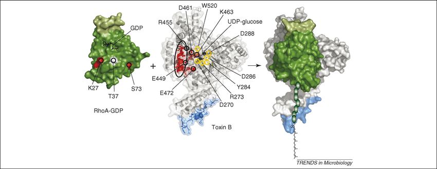
Figure 4. Model of toxin B interaction with RhoA – docking model of the glycosyltransferase domain toxin B (white) with its substrate RhoA (green) according to reference
[48]. RhoA is shown as a surface representation with the acceptor amino acid threonine 37 (T37) shown in white. Ser73 and Lys27 of RhoA defining the specificity towards
the toxins are marked and shown in red. The positions of amino acids in toxin B necessary for protein substrate recognition and helix a17 are circled in black. The different
locations of amino acids important for enzyme activity are circled in yellow. The N-terminal 18 amino acids of toxin B involved in membrane attachment are shown in blue.
UDP-glucose in toxin B and GDP in RhoA are marked and shown in stick representation (black). The C-terminal linker peptide of RhoA with its isoprenyl membrane anchor is
schematically represented. Images were created using PyMOL (www.pymol.org).
Concluding remarks 5 Voth, D.E. and Ballard, J.D. (2005) Clostridium difficile toxins:
mechanism of action and role in disease. Clin. Microbiol. Rev. 18,
Studies from recent years have yielded important progress
247–263
in our knowledge of the structure and mode of action of 6 Just, I. and Gerhard, R. (2004) Large clostridial cytotoxins. Rev.
clostridial glucosylating toxins. These results indicate the Physiol. Biochem. Pharmacol. 152, 23–47
need for a revision of the current AB-model of clostridial 7 Mehlig, M. et al. (2001) Variant toxin B and a functional toxin A
glucosylating toxins. Characterization of clostridial gluco- produced by Clostridium difficile C34. FEMS Microbiol. Lett. 198,
171–176
sylating toxins according to this model seems to be a scien-
8 Chaves-Olarte, E. et al. (1999) A novel cytotoxin from Clostridium
tific Procrustes’ bed. Thus, we suggest that an ABCD model difficile serogroup F is a functional hybrid between two other large
is more appropriate for describing the structure and func- clostridial cytotoxins. J. Biol. Chem. 274, 11046–11052
tion of these toxins. However, clostridial toxins are large and 9 Rupnik, M. et al. (1998) A novel toxinotyping scheme and correlation of
their full 3D-structure remains unknown, we also have yet toxinotypes with serogroups of Clostridium difficile isolates. J. Clin.
Microbiol. 36, 2240–2247
to understand the precise functions of all toxin domains. 10 Von Eichel-Streiber, C. et al. (1996) Large clostridial cytotoxins - a
This model will probably need to be revised again in the family of glycosyltransferases modifying small GTP-binding proteins.
future. Nevertheless, recent results on the structural Trends Microbiol. 4, 375–382
analysis of the toxins have increased our knowledge about 11 Busch, C. and Aktories, K. (2000) Microbial toxins and the
this medically important toxin family, and also offer glucosylation of Rho family GTPases. Curr. Opin. Struct. Biol. 10,
528–535
multiple options for new therapeutic strategies. For 12 Von Eichel-Streiber, C. et al. (1992) Evidence for a modular structure
example, scavenging the toxins using compounds interact- of the homologous repetitive C-terminal carbohydrate-binding
ing specifically with the receptor-binding domain is a feas- sites of Clostridium difficile toxins and Streptcoccus mutans
ible approach [19]. Another option is the development of glucosyltransferases. J. Bacteriol. 174, 6707–6710
13 Barroso, L.A. et al. (1994) Mutagenesis of the Clostridium difficile toxin
specific inhibitors of the autocatalytic cleavage or the search
B gene and effect on cytotoxic activity. Microb. Pathog. 16, 297–303
for compounds that specifically inhibit the glucosyltransfer- 14 Collier, R.J. (2001) Understanding the mode of action of diphtheria
ase activity of these toxins. To this end, solving the 3D- toxin: a perspective on progress during the 20th century. Toxicon 39,
structure of other members of the family of glucosylating 1793–1803
toxins in addition to the crystal structure of the enzyme– 15 Moncrief, J.S. and Wilkins, T.D. (2000) Genetics of Clostridium difficile
toxins. Curr. Top. Microbiol. Immunol. 250, 35–54
substrate–cosubstrate complex is highly desirable.
16 Dove, C.H. et al. (1990) Molecular characterization of the Clostridium
difficile toxin A gene. Infect. Immun. 58, 480–488
References 17 Von Eichel-Streiber, C. and Sauerborn, M. (1990) Clostridium difficile
1 Bartlett, J.G. and Perl, T.M. (2005) The new Clostridium difficile - what toxin A carries a C-terminal structure homologous to the carbohydrate-
does it mean? N. Engl. J. Med. 353, 2503–2505 binding region of streptococcal glycosyltransferase. Gene 96, 107–113
2 McDonald, L.C. et al. (2005) An epidemic, toxin gene-variant strain of 18 Ho, J.G. et al. (2005) Crystal structure of receptor-binding C-terminal
Clostridium difficile. N. Engl. J. Med. 353, 2433–2441 repeats from Clostridium difficile toxin A. Proc. Natl. Acad. Sci. U. S. A.
3 Pepin, J. et al. (2005) Mortality attributable to nosocomial Clostridium 102, 18373–18378
difficile-associated disease during an epidemic caused by a 19 Greco, A. et al. (2006) Carbohydrate recognition by Clostridium difficile
hypervirulent strain in Quebec. CMAJ 173, 1037–1042 toxin A. Nat. Struct. Mol. Biol. 13, 460–461
4 Perelle, S. et al. (1997) Production of a complete binary toxin (actin- 20 Fernandez-Tornero, C. et al. (2001) A novel solenoid fold in the cell wall
specific ADP-ribosyltransferase) by Clostridium difficile CD196. Infect. anchoring domain of the pneumococcal virulence factor LytA. Nat.
Immun. 65, 1402–1407 Struct. Biol. 8, 1020–1024
228Review Trends in Microbiology Vol.16 No.5
21 Krivan, H.C. et al. (1986) Cell surface binding site for 45 Kitadokoro, K. et al. (2007) Crystal structures reveal a thiol protease-
Clostridium difficile enterotoxin: evidence for a glycoconjugate like catalytic triad in the C-terminal region of Pasteurella multocida
containing the sequence Gala1-3Galß1-4GlcNAc. Infect. Immun. 53, toxin. Proc. Natl. Acad. Sci. U. S. A. 104, 5139–5144
573–581 46 Wiggins, C.A.R. and Munro, S. (1998) Activity of the yeast MNN1 a-1,3-
22 Tucker, K.D. and Wilkins, T.D. (1991) Toxin A of Clostridium difficile mannosyltransferase requires a motif conserved in many other
binds to the human carbohydrate antigens I, X, and Y. Infect. Immun. families of glycosyltransferases. Proc. Natl. Acad. Sci. U. S. A. 95,
59, 73–78 7945–7950
23 Larsen, R.D. et al. (1990) Frameshift and nonsense mutations in a 47 Busch, C. et al. (1998) A common motif of eukaryotic
human genomic sequence homologous to a murine UDP-Gal:b-D- glycosyltransferases is essential for the enzyme activity of large
Gal(1,4)-D-GlcNAc a(1,3)-galactosyltransferase cDNA. J. Biol. Chem. clostridial cytotoxins. J. Biol. Chem. 273, 19566–19572
265, 7055–7061 48 Jank, T. et al. (2007) Clostridium difficile glucosyltransferase toxin B -
24 Teneberg, S. et al. (1996) Molecular mimicry in the recognition of essential amino acids for substrate binding. J. Biol. Chem. 282, 35222–
glycosphingolipids by Gala3Galß4GlcNAcß-binding Clostridium 35231
difficile toxin A, human natural anti a-galactosyl IgG and the 49 Negishi, M. et al. (2003) Glucosaminylglycan biosynthesis: what we can
monoclonal antibody Gal-13: characterization of a binding-active learn from the X-ray crystal structures of glycosyltransferases GlcAT1
human glycosphingolipid, non- identical with the animal receptor. and EXTL2. Biochem. Biophys. Res. Commun. 303, 393–398
Glycobiology 6, 599–609 50 Breton, C. et al. (2006) Structures and mechanisms of
25 Florin, I. and Thelestam, M. (1983) Internalization of Clostridium glycosyltransferases. Glycobiology 16, 29R–37R
difficile cytotoxin into cultured human lung fibroblasts. Biochim. 51 Qasba, P.K. et al. (2005) Substrate-induced conformational changes in
Biophys. Acta 763, 383–392 glycosyltransferases. Trends Biochem. Sci. 30, 53–62
26 Barth, H. et al. (2001) Low pH-induced formation of ion channels by 52 Kubota, T. et al. (2006) Structural basis of carbohydrate
Clostridium difficile toxin B in target cells. J. Biol. Chem. 276, 10670– transfer activity by human UDP-GalNAc: polypeptide alpha-N-
10676 acetylgalactosaminyltransferase (pp-GalNAc-T10). J. Mol. Biol. 359,
27 Sandvig, K. et al. (2004) Pathways followed by protein toxins into cells. 708–727
Int. J. Med. Microbiol. 293, 483–490 53 Ramakrishnan, B. et al. (2002) Crystal structure of beta1,4-
28 Qa’Dan, M. et al. (2000) pH-induced conformational changes in galactosyltransferase complex with UDP-Gal reveals an
Clostridium difficile toxin B. Infect. Immun. 68, 2470–2474 oligosaccharide acceptor binding site. J. Mol. Biol. 318, 491–502
29 Giesemann, T. et al. (2006) Cholesterol-dependent pore formation of 54 Vetter, I.R. et al. (2000) Structural consequences of mono-glucosylation
Clostridium difficile toxin A. J. Biol. Chem. 281, 10808–10815 of Ha-Ras by Clostridium sordellii lethal toxin. J. Mol. Biol. 301, 1091–
30 Pfeifer, G. et al. (2003) Cellular uptake of Clostridium difficile toxin B: 1095
translocation of the N-terminal catalytic domain into the cytosol of 55 Geyer, M. et al. (2003) Glucosylation of Ras by Clostridium sordellii
eukaryotic cells. J. Biol. Chem. 278, 44535–44541 lethal toxin: consequences for the effector loop conformations observed
31 Rupnik, M. et al. (2005) Characterization of the cleavage site and by NMR spectroscopy. Biochemistry 42, 11951–11959
function of resulting cleavage fragments after limited proteolysis of 56 Selzer, J. et al. (1996) Clostridium novyi a-toxin-catalyzed
Clostridium difficile toxin B (TcdB) by host cells. Microbiology 151, incorporation of GlcNAc into Rho subfamily proteins. J. Biol. Chem.
199–208 271, 25173–25177
32 Reineke, J. et al. (2007) Autocatalytic cleavage of Clostridium difficile 57 Jank, T. et al. (2005) Change of the donor substrate specificity of
toxin B. Nature 446, 415–419 Clostridium difficile toxin B by site-directed mutagenesis. J. Biol.
33 Shears, S.B. (2001) Assessing the omnipotence of inositol Chem. 280, 37833–37838
hexakisphosphate. Cell. Signal. 13, 151–158 58 Just, I. et al. (1995) Glucosylation of Rho proteins by Clostridium
34 Voglmaier, S.M. et al. (1992) Inositol hexakisphosphate receptor difficile toxin B. Nature 375, 500–503
identified as the clathrin assembly protein AP-2. Biochem. Biophys. 59 Herrmann, C. et al. (1998) Functional consequences of
Res. Commun. 187, 158–163 monoglucosylation of H-Ras at effector domain amino acid
35 Egerer, M. et al. (2007) Auto-catalytic cleavage of Clostridium difficile threonine-35. J. Biol. Chem. 273, 16134–16139
toxins A and B depends on a cysteine protease activity. J. Biol. Chem. 60 Jank, T. et al. (2006) Exchange of a single amino acid switches the
282, 25314–25321 substrate properties of RhoA and RhoD towards glucosylating and
36 Sheahan, K-L. et al. (2007) Autoprocessing of the Vibrio cholerae RTX transglutaminating toxins. J. Biol. Chem. 281, 19527–19535
toxin by the cysteine protease domain. EMBO J. 26, 2552–2561 61 Müller, S. et al. (1999) Impact of amino acids 22-27 of Rho-subfamily
37 Hofmann, F. et al. (1997) Localization of the glucosyltransferase GTPases on glucosylation by the large clostridial cytotoxins TcsL-1522,
activity of Clostridium difficile toxin B to the N-terminal part of the TcdB-1470 and TcdB-8864. Eur. J. Biochem. 266, 1073–1080
holotoxin. J. Biol. Chem. 272, 11074–11078 62 Hofmann, F. et al. (1998) Chimeric clostridial cytotoxins: identification
38 Reinert, D.J. et al. (2005) Structural basis for the function of of the N-terminal region involved in protein substrate recognition.
Clostridium difficile Toxin B. J. Mol. Biol. 351, 973–981 Infect. Immun. 66, 1076–1081
39 Persson, K. et al. (2001) Crystal structure of the retaining 63 Dvorsky, R. and Ahmadian, M.R. (2004) Always look on the bright site
galactosyltransferase LgtC from Neisseria meningitidis in complex of Rho: structural implications for a conserved intermolecular
with donor and acceptor sugar analogs. Nat. Struct. Biol. 8, 166– interface. EMBO Rep. 5, 1130–1136
175 64 Burridge, K. and Wennerberg, K. (2004) Rho and Rac take center stage.
40 Charnock, S.J. and Davies, G.J. (1999) Structure of the nucleotide- Cell 116, 167–179
diphospho-sugar transferase, SpsA from Bacillus subtilis, in native and 65 Etienne-Manneville, S. and Hall, A. (2002) Rho GTPases in cell biology.
nucleotide-complexed forms. Biochemistry 38, 6380–6385 Nature 420, 629–635
41 Gastinel, L.N. et al. (2001) Bovine alpha1,3-galactosyltransferase 66 Just, I. et al. (1996) Inactivation of Ras by Clostridium sordellii lethal
catalytic domain structure and its relationship with ABO histo- toxin-catalyzed glucosylation. J. Biol. Chem. 271, 10149–10153
blood group and glycosphingolipid glycosyltransferases. EMBO J. 67 Sehr, P. et al. (1998) Glucosylation and ADP-ribosylation of Rho
20, 638–649 proteins - effects on nucleotide binding, GTPase activity, and
42 Unligil, U.M. and Rini, J.M. (2000) Glycosyltransferase structure and effector-coupling. Biochemistry 37, 5296–5304
mechanism. Curr. Opin. Struct. Biol. 10, 510–517 68 Genth, H. et al. (1999) Monoglucosylation of RhoA at Threonine-37
43 Mesmin, B. et al. (2004) A phosphatidylserine-binding site in the blocks cytosol-membrane cycling. J. Biol. Chem. 274, 29050–29056
cytosolic fragment of Clostridium sordellii lethal toxin facilitates 69 Fritz, G. et al. (1995) The Ras-related small GTP-binding protein RhoB
glucosylation of membrane-bound Rac and is required for is immediate- early inducible by DNA damaging treatments. J. Biol.
cytotoxicity. J. Biol. Chem. 279, 49876–49882 Chem. 270, 25172–25177
44 Spyres, L.M. et al. (2003) Mutational analysis of the enzymatic domain 70 Gerhard, R. et al. (2005) Clostridium difficile toxin A induces
of Clostridium difficile toxin B reveals novel inhibitors of the wild-type expression of the stress-induced early gene product RhoB. J. Biol.
toxin. Infect. Immun. 71, 3294–3301 Chem. 280, 1499–1505
229You can also read