Fluorescence in situ hybridisation (FISH) - rarechromo.org
←
→
Page content transcription
If your browser does not render page correctly, please read the page content below
Fluorescence in situ
hybridisation (FISH)

               rarechromo.org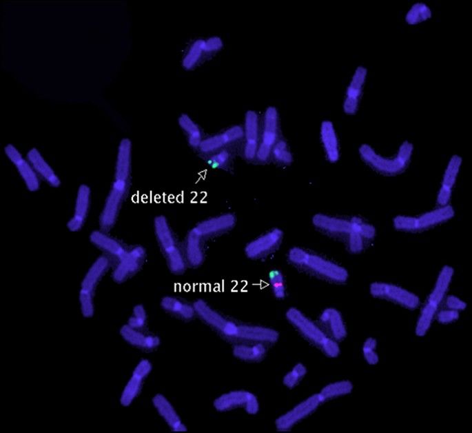
Fluorescence in situ hybridization (FISH)
Chromosomes
Chromosomes are structures that contain the genetic information (DNA) that tells the
body how to develop and function. The cells in the body have 46 chromosomes which are
arranged in pairs. Each pair contains a chromosome from each parent and they are
arranged approximately according to size and numbered from 1 to 22. The last pair
contains the sex chromosomes. Girls have two Xs (XX) and boys have an X and a Y (XY).
Cytogenetic (chromosome) testing looks for changes in the number and structure of the
chromosomes and for losses (deletions) and gains (duplications) of genetic material.
People with these types of changes in their cells may have an increased risk of birth
defects, developmental delay, behavioural problems and intellectual disability.

Looking at chromosomes (chromosome analysis)
Chromosomes can be seen if they are stained and magnified
under the microscope. Chromosomes have a short (p) arm and a
long (q) arm held together at a structure called the centromere.
Each chromosome has a distinct pattern of light and dark bands
which are numbered outwards from the centromere. The
chromosome diagram on the right shows the banding pattern of
chromosome 10. Clinical scientists use microscope analysis to                  p arm
look for changes in the shape of the chromosomes and their
banding patterns. If the change is large enough,
rearrangements and imbalances (deletions and duplications)
involving chromosomes or parts of the chromosomes can be                       centromere
seen. Clinical scientists who do the analysis are very skilled at
detecting very small and often very subtle changes. However,
chromosome rearrangements and the loss or gain of material
can often be extremely small and frequently cannot be observed
with a routine chromosome test even by the most skilled of
scientists. FISH testing allows some of these changes to be                    q arm
detected.

What is FISH?
FISH is a method that can be used to detect small deletions and
duplications that are not visible using microscope analysis. It
can also be used to detect how many chromosomes of a certain
type are present in each cell and to confirm rearrangements
that are suspected after microscope analysis. FISH looks
specifically at the one specific area of a chromosome only. It
uses a very small chemical that glows brightly when it detects the
specific region on a chromosome. A scientist uses a special
microscope to look at the chromosome and see how many bright
spots are present. When a person has a deletion, only one bright   Chromosome 10
spot can be seen instead of two (one on each chromosome). When a person has a
duplication, three bright spots can be seen instead of just two.
How does FISH work?
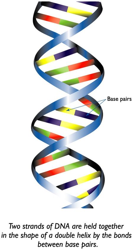
The DNA in our cells contains two strand-like molecules coiled
together into a structure known as a double helix (see right).
Each strand has a sequence containing a mixture of four bases
(A, T, G and C). The bases in each strand are able to bind to
each other and hold the DNA together, but can only do so if
they are properly matched, or complementary, to those in the
other strand (an A can only bind to a T and a C can only bind to
a G). When two complementary sequences find each other they
will bind together, or hybridise. FISH works by exploiting the
ability of one DNA strand to hybridise specifically to another
DNA strand.
FISH uses small DNA strands called probes that have a
fluorescent label attached to them. The probes are
complementary to specific parts of a chromosome. When DNA
is heated, the patient’s two DNA strands break apart, or
denature, and the probes are able to hybridise to their
complementary sequence in the patient’s DNA (see diagram
below).
If a small deletion is present in the region complementary to the probe, the probe will not
hybridise. If a duplication is present, more of the probe is able to hybridise.
patient’s DNA
heating
denaturation
probe with
fluorescent
label
cooling
hybridisation
An overview of the FISH test
FISH testing for a deletion. Two probes are usually used;The first probe
(green) is a control probe used to identify both copies of the chromosome under test. It
hybridises to a sequence that is not part of the deletion, so a signal is observed on each
chromosome. The second probe (red) hybridises to the sequence that may be deleted. A
deletion is usually found in only one of the chromosomes in a pair, therefore the probe
can bind to the intact chromosome, but is unable to bind to the deleted chromosome and
only one signal is seen.

What types of probes are used?
Many FISH probes bind to specific gene-containing parts of the DNA and are used to
detect small imbalances. These are known as locus-specific probes. Other probes bind to
the centromere or the telomeres (the ends of the chromosome arms) and are used to
count the number of certain chromosomes and to detect rearrangements. Paint probes
are also available which bind to the whole chromosome.

What samples are needed for FISH testing?
FISH is most commonly performed on blood samples from adults and children. FISH can
also be used as a prenatal test for aneuploidy (extra copies of whole chromosomes) using
amniotic fluid from an amniocentesis or placental samples from chorionic villus
sampling (CVS). Less commonly it is used as a prenatal test for deletions, also using
amniotic fluid or CVS samples.
Why has FISH been offered for your child?
FISH is often performed alongside standard microscope analysis if your child has
characteristics that are strongly suggestive of a particular deletion syndrome or another
syndrome for which a FISH test is available. Your geneticist may request microscope
analysis and FISH testing together or may request FISH testing if microscope analysis
gives a normal result.

How will we be given the results?
The results are most likely to be given to you by your geneticist who will talk you through
your child’s results and you will almost certainly receive a follow up letter.
FISH results are often included with the results of microscope analysis in a combined
report. The first line of a report shows the results as a karyotype. If your child’s
chromosomes are not deleted, the karyotype may look something like this:
46,XX.ish 22q11.2(TUPLE1x2)(ARSAx2)
46             The total number of chromosomes present
XX             The sex chromosomes present (XX for girls, XY for boys)
ish            The analysis was by fluorescent in situ hybridisation (FISH)
22             The FISH test was performed on chromosome 22
q11.2          The chromosome has two breakpoints, both in band 22q11.2 and
               material between these two breakpoints is missing
(TUPLE1x2)     The DNA segment (probe) called TUPLE is present in two copies (as
               expected)
(ARSAx2)       The DNA segment (probe) called ARSA2 is present in two copies (as
               expected)

If a deletion is present, the karyotype is written slightly differently, as shown below

46,XX.ish del(22)(q11.2q11.2)(TUPLE1-)
46             The total number of chromosomes present
XX             The sex chromosomes present (XX for girls, XY for boys)
ish            The analysis was by fluorescent in situ hybridisation (FISH)
del            A deletion, or material is missing
(22)           The deletion is from chromosome 22
(q11.2q11.2)   The chromosome has two breakpoints, both in band 22q11.2 and
               material between these two breakpoints is missing
(TUPLE1-)      One copy of the DNA segment (marker) called TUPLE is missing

How long do the results take?
With blood testing, results are usually available in 4 weeks and can be available in under
two weeks for priority cases such as newborn babies. Results may be available much
sooner if a blood sample has been provided previously for microscope analysis, as the
same sample can be used for the FISH test.
What are the advantages of FISH? FISH is able to detect many small deletions, duplications and rearrangements that are not visible with standard microscope analysis A diagnosis from FISH may avoid your child having to undergo many other tests What are the benefits of FISH? It may help you and your doctor watch for common health problems associated with your child’s chromosome imbalance It may help to predict what to expect as your child gets older It may show which specific genes are included in your child’s deletion or duplication. If the gene(s) has been associated with a particular feature or health problem it may help to guide management or treatment for your child It can help you to obtain specialist services for your child You can choose to join a support group to meet other parents facing similar challenges Parents and other family members can be tested to see if they are carriers of changes in their DNA that put them at risk of having more children with a chromosome change What are the limitations of FISH? FISH testing does not usually screen all chromosomes for changes. Most FISH probes are specific for one particular deletion or duplication within one band of one chromosome so it will only find what it is looking for FISH probes are only available for the most well characterised deletion and duplication syndromes. Your child may have a small imbalance that cannot be detected with the standard FISH tests currently available FISH has limited ability to precisely define which genes and breakpoints are involved in an imbalance It can be difficult for clinical scientists to see a duplication using FISH, as the attachment of extra probes is not always easy to see What other tests are available if FISH does not reveal any rearrangements? If microscope analysis and FISH testing do not reveal a rearrangement, your child may be referred for microarray-based comparative genomic hybridisation (array CGH). This test examines all chromosomes for losses and gains of DNA and can detect smaller imbalances than those detected with FISH or microscope analysis.
Notes
Support and Information

                             Rare Chromosome Disorder Support Group,
                             PO Box 2189, Caterham, Surrey CR3 5GN, UK
                             Tel/Fax: +44(0)1883 330766
                             info@rarechromo.org I www.rarechromo.org

Unique is a charity without government funding, existing entirely on donations
and grants. If you are able to support our work in any way, however small,
please make a donation via our website at
www.rarechromo.org/html/MakingADonation.asp
Please help us to help you!

                                     Produced with the support of
                                     EuroGentest

This leaflet is not a substitute for personal medical advice. Families should consult a
medically qualified clinician in all matters relating to genetic diagnosis, management and
health. The information is believed to be the best available at the time of publication. It
was compiled by Unique and reviewed by Dr Shehla Mohammed and Dr Caroline Ogilvie,
Guy’s Hospital, London. UK and Professor Maj Hultén, Professor of Reproductive
Genetics, University of Warwick, UK. 2011, 2013 Version 1.1 (SW)
Copyright © Unique 2013

Rare Chromosome Disorder Support Group Charity Number 1110661
Registered in England and Wales Company Number 5460413
You can also read