Microscopy investigation of the surface behaviour of different materials after H- irradiation in different conditions - A. T. Pérez Fontenla ...
←
→
Page content transcription
If your browser does not render page correctly, please read the page content below
Microscopy investigation
of the surface behaviour
of different materials
after H- irradiation in
different conditions
A. T. Pérez Fontenla (EN-MME-MM)
on behalf of the spare RFQ Project team
Outline

1. Linac4 RFQ in-situ inspection

2. Additional study on collimator

3. Proposed studies

4. Results

5. Summary

             08/03/2021             2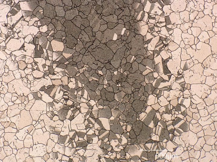
Linac4 RFQ in-situ inspection
  L4 tunnel         Optical microscopy at vane front face            Endoscopy at front face and vacuum port

                                                        Worm-like features

              08/03/2021                                                                               3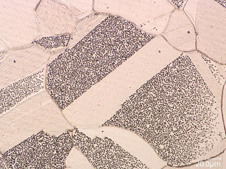
Linac4 RFQ in-situ inspection
 L4 tunnel         Optical microscopy at vane front face              Endoscopy at front face and vacuum port

                                                                                 CLIC TD24_R05 (tested in XBox_1)

                                                         Worm-like features

                                            Linac2 vane front face               CLIC Crab Cavity (tested in XBox_2)

                                                                                    Image presented in HG2016
                                                                                   courtesy of Enrique Rodríguez

             08/03/2021                                                                                        4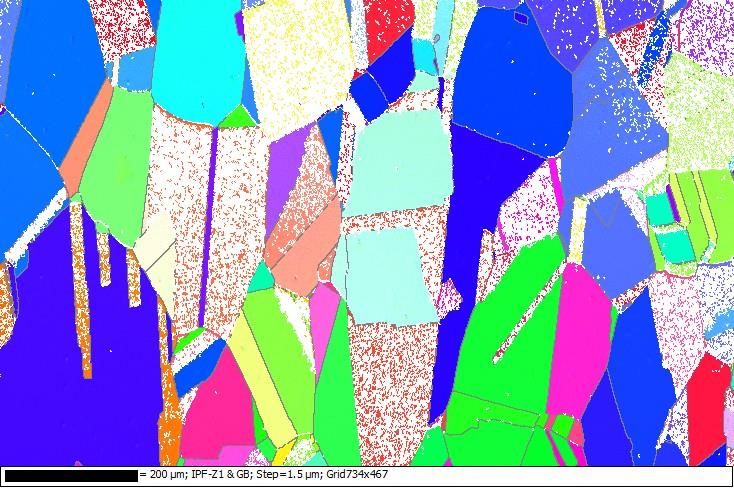
Additional study on Cu-OFE collimator
                                               Collimator/mark fabricated from cold rolled
                                               Cu-OFE plate (3 mm thickness) was installed
                                               and tested at 1.7 x 10¹⁹ H- ions at 45 keV
                                               (number of total impacting ions calculated
                                               from the electrical signal)

                                               Microscope inspection of the Cu surface
                                               around the square was performed before and
                                               after testing

   Halo perceptible during visual inspection
       Approximate diameter ≈ 20 mm
          Estimated area ≈ 2.5 cm2

                 08/03/2021                                                        5
Additional study on Cu-OFE collimator
                       Before testing
Comparison before
and after testing
pointed out the
presence of blisters

                       After testing

                                        Micrometric protrusions/blisters?
                                        Only appreciable at SEM with > 1kx

                          08/03/2021                                         6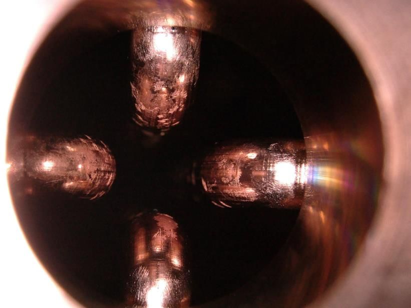
Additional study on Cu-OFE collimator
                       SEM top view

Removed volume:
10 x 8 x 5 µm3

Superficial layer of
Cu (at ≈ 0.5 µm
                                      initiation
depth from the
surface) heavily                                                                         Pt layer
affected by pores
                                                                                            Cu
Similar features are
reported in the
bibliography due to
penetration of                             nano-voids coalescence   micrometric cavity
relatively insoluble
gases (hydrogen)                          FIB-SEM cross section

                         08/03/2021                                                         7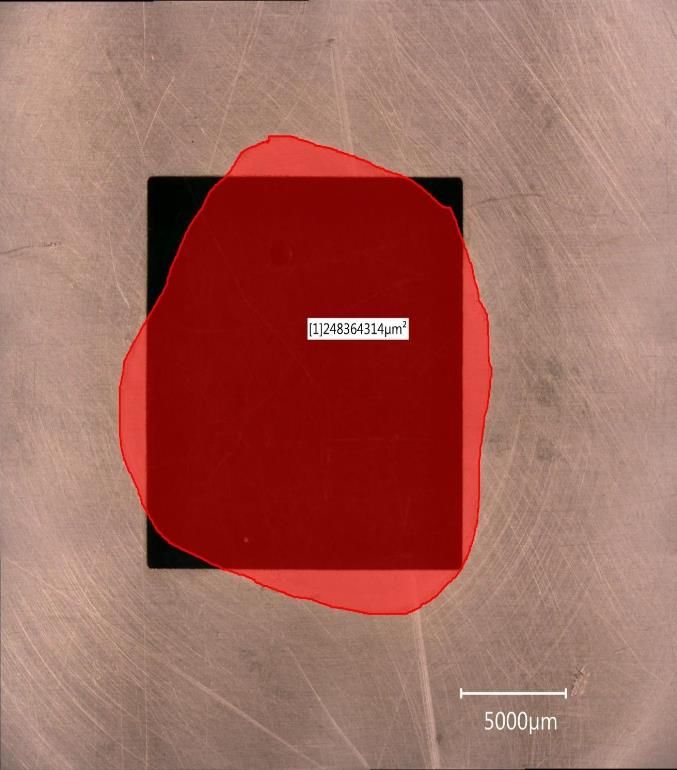
Proposed studies
 • A list of candidate materials was carefully defined as alternative to
   Cu-OFE for manufacturing of a future RFQ with better performance
   Cu-OFE         CuCr1Zr              Cu98Be2             Nb               Ta              Ti6Al4V
 Forged and       Forged,           Hard temper         RF purity         Melted           Premium
  annealed        solution          (precipitation      (RRR300)          quality            grade
(current base   annealed and         hardening)                          Annealed          β-forged
  material)         aged

 • The materials were selected based on:
    • Their usability for meter-long high gradient RF cavities and,
    • Their potential resistance to blistering and breakdown phenomena;
                                                                     •   Purity
                •   Radiation dose
                                                                     •   Grain size
 Relevant       •   Ion energy                        Relevant
  testing                                            metallurgical   •   Pre-existing defects
parameters      •   Angle of incidence                aspects
                                                                     •   Mechanical properties
                •   Temperature of the “target”
                                                                     •   Crystal orientation at surface

                       08/03/2021                                                                   8
Proposed studies
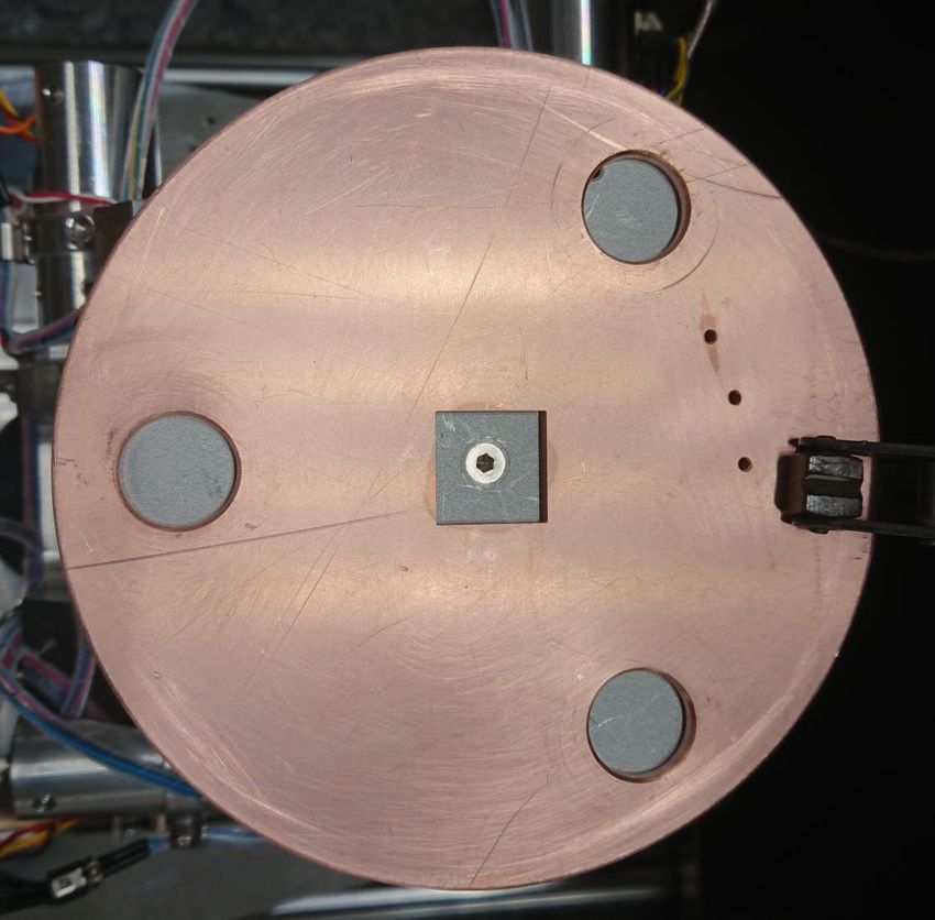
• Combined H- source test stand + pulsed DC:
• Particle type H- ions at 45 keV
• Different incidence angles are possible
• Electrodes of Ø80 mm and Ø60 mm
• Helsinki’s system:
• Particle type H2+ at 90 keV
• Different incidence angles are possible
• Sample holder ~ Ø100 mm simultaneously 4 samples (30 mm x 30 mm)
Image courtesy of
Mizohata Kenichiro
Combined H- source + DC testing at CERN Helsinki’s system
Cu-OFE CuCr1Zr Cu98Be2 Nb Ta Ti6Al4V Cu-OFE CuCr1Zr Cu98Be2 Nb Ta Ti6Al4V
1
~ Dose
1.0 x 10¹⁹
p/cm2 4 n. a.
Tested Manufacturing completed Material purchased
08/03/2021 9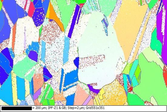
Results: Cu-OFE electrode
After thermal treatment in vacuum at 650 ºC for 2 hours
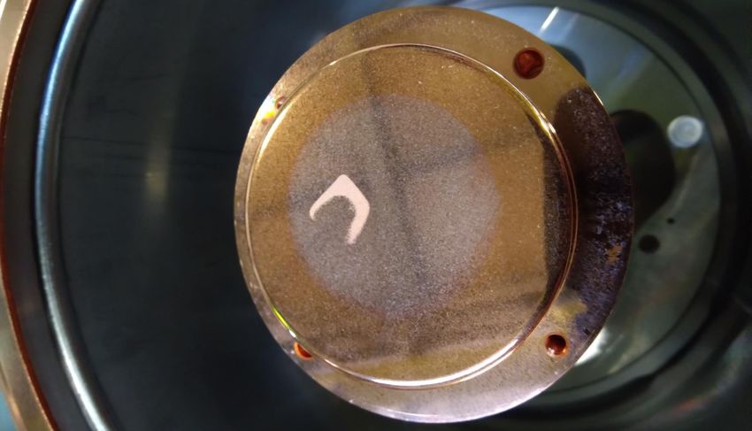
Cathode: After irradiation test with an estimated total dose of 1.2E19 p delivered during 40 hours (data courtesy of Giulia Bellodi).

                                                                     Outer ring

                                                 “C-shaped”

Previous affected surface estimation on collimators
   test was 2.5 cm2 and in the electrode 0.5 cm2

                                                          Central region

                                                                                                               Image courtesy of Sebastien Bertolo

                                      08/03/2021                                                                                        10
Results: Cu-OFE electrode
In the transition from
irradiated spot to non-
irradiated zones,
some grains are more
affected than others
All the C-shaped area
presents blisters
Why C-shaped?
Investigations are on-
going
C-shaped area Non/affected zone
Transition area
08/03/2021 11Results: Cu-OFE electrode
When observed at Transition area
high magnification
faceting of the Cu is
noticeable
The blisters present
small variations in
size diameter and
morphology
depending on the
grain orientation
C-shaped area
08/03/2021 12Results: Cu-OFE electrode
Blisters in different
phases of growing
are perceptible and Opening most probably throw the cap thickness
in many sites
coalescence of two
or three blisters is
observable
Coalescence
Some of the blisters
present openings
(holes)
Initial phase
08/03/2021 13Results: Cu-OFE electrode
The relation between the blisters location and crystallographic orientation was evaluated experimentally
using the non-destructive technique Electron Back-Scattered Diffraction (EBSD) into the SEM.
Using the common color-codes for cubic materials, it
can be observed that in general the blue and green
grains (corresponding to crystal directions [111] and
[101] respectively) were less affected by blisters than
the red grains (corresponding to crystal direction [001])
Inverse Pole Figures coloring maps in Z direction (IPF-Z)
08/03/2021 14Results: Cu-OFE specimen (Helsinki)
Presence of blisters randomly distributed on the Cu-OFE Cu-OFE Collimator CERN (1.7E19p/cm^2)
surface was confirmed with both used doses in Helsinki (is
not possible to correlate with grain orientations at the
transition areas due to the surface roughness)
Size, density and shape of the blisters on the collimator are
similar to the ones observed at the lower dose test region
In the case of higher dose, different features appeared (larger
blisters?) in addition to the smaller blisters
Some blisters present holes/openings at the cap
Cu-OFE Helsinki (4E18p/cm^2) Cu-OFE Helsinki (1E18p/cm^2)
Holes
15Results: Cu-OFE specimen (Helsinki)
Cu-OFE Collimator CERN (1.7E19p/cm^2)
Nanopores observed in the collimator are only visible
punctually in the affected layer on the Cu-OFE
irradiated samples in Helsinki
The blister cap thickness on the collimator is similar to
the lower dose test in Helsinki and similar to some
features in the higher dose (initiation/nucleation sites?);
Presence of larger blisters containing smaller events is
confirmed and a carbon layer is visible;
Cu-OFE Helsinki (4E18p/cm^2) Cu-OFE Helsinki (1E18p/cm^2)
Pt deposit
Pt deposit
Larger events when
enough dose/time? Initiation sites?
16Cu-OFE First test in Helsinki (4E18p/cm^2)
Results: Cu-OFE specimen (Helsinki)
Cu-OFE Helsinki (4E18p/cm^2)
Thinning due to plastic deformation
The cap is thicker at the central part
17CuCr1Zr First test in Helsinki (4E18p/cm^2)
Results: CuCr1Zr specimen (Helsinki)
CuCr1Zr Helsinki (4E18p/cm^2) CuCr1Zr Helsinki (1E18p/cm^2)
Cr precipitates
Blisters
18Results: CuCr1Zr specimen (Helsinki)
CuCr1Zr Helsinki (4E18p/cm^2)
Thin layer
Aspect of fragile fracture at the blister base
~ 35 wt. % C
~ 10 wt. % C
19Results: CuCr1Zr specimen (Helsinki)
CuCr1Zr Helsinki (4E18p/cm^2)
Pt deposit
The cap is thicker at the central part
Thinning due to plastic deformation
Microstructure is visible
20Results: Nb and Ti6Al4V (Helsinki)
Nb Helsinki (4E18p/cm^2) Pt deposit
C layer
Ti6Al4V Helsinki (4E18p/cm^2)
Pt deposit
C layer
Microstructure is visible
21Summary
• The in-situ test in Linac4 RFQ by optical means pointed out the presence of local damage on the Cu surface
(BKD craters and “worm-like features”);
• Further investigations with collimators/masks confirmed the presence of blisters induced by the interaction
with the beam;
• The size of those events differs but they could be connected a deeper look to better understand the blister-
BKD location relation is needed;
• Combined testing (irradiation + pulsed DC system) was performed on RFQ base material (Cu-OFE). The
presence of blister was confirmed and their aspect and location were characterized by SEM and EBSD;
• In addition, other candidate materials for RFQ production were identified and are being studied in similar way
in order to compare their performance and resistance to blistering and BKD phenomena;
• The cost and leading time to fabricate and test at CERN large electrodes oriented us to look for testing
alternatives and the preliminary results obtained with Helsinki’s system compared well with CERN studies in
collimators/masks;
• This flexible and reproducible system allowed us to test simultaneously Cu-OFE, CuCr1Zr, Nb and Ti6Al4V
specimens with two different doses. Nevertheless, some questions raised up (C layer, bigger blisters at higher
doses/time, surface preparation…) more tests needs to be performed;
• So far, both cuprous materials tested presented blisters (in the case of the CuCr1Zr only at higher dose or on
deformed areas) and Nb and Ti6Al4V were free of features even at higher doses;
• It’s being a continuous learning process and it is important to mention the implication of a wide range of
experts at CERN and Helsinki to evaluate the findings until now and the project support to hire a PhD student.
08/03/2021 22Thank you for your attention!
Extra slides

         08/03/2021   24
Extra slides
EDS comparison at the central region (SOI-5) and external region (SOI-7)
08/03/2021 25Extra slides
08/03/2021 26Extra slides
High dose Low dose
08/03/2021 27Faceting on the Cu surface is visible independently of the region.
That is a consequence of the thermal treatment performed on the electrode before testing.
Faceting is related with selective evaporation of atoms in non–energetically favourable crystal position thus the
appearance varies depending on the grain orientation
Mo rich inclusions are visible all over the Cu
surfaceVery superficial features and are visible all over the Cu surface
When observed at higher magnification:
When observed at higher magnification:

SOI-5: Central region                                SOI-7: Outer ring region

                            SOI-2: C-shaped region
Comparison of Nb sample same location before and after irradiation (dose 4E18 H/cm^2)
Top view
Before irradiation
Nb surface present pitting after etching
Top view
After irradiationTiAl6V4_Dose 4E18 H/cm^2 Top view After irradiation Cross section After irradiation
Extra slides

         08/03/2021   34
You can also read


