FOURTH ANNUAL EUROPEAN 3D ADVANCED FIBER DISSECTION COURSE: ACQUIRING THE MENTAL IMAGERY NECESSARY TO OPERATE THE BRAIN - Santander. 21, 22 and 23 ...
←
→
Page content transcription
If your browser does not render page correctly, please read the page content below
FOURTH ANNUAL EUROPEAN
3D ADVANCED FIBER DISSECTION COURSE:
ACQUIRING THE MENTAL IMAGERY
NECESSARY TO OPERATE THE BRAIN
Santander. 21, 22 and 23 October 2021

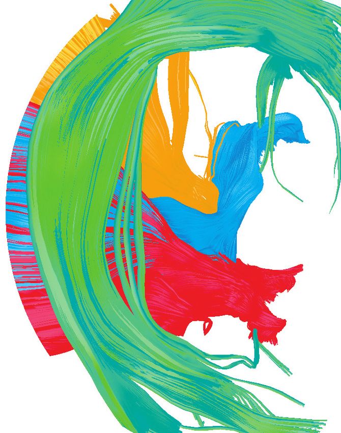
The white matter of the cerebrum underlies the outer cortex of gray
matter, and is composed of densely packed axons that are organized in
fascicles or fiber tracts. These tracts have a complex three-dimensional (3D)
configuration within the hemispheres, the brainstem and the spinal cord.
A detailed knowledge of the architectural anatomy of the white matter
tracts is paramount, for strategically planning for surgical management of
parenchymal brain lesions, such as gliomas. Neuroanatomical laboratory
training is very valuable to study and understand the anatomy of white matter
fibers. In particular, cortex-sparing fiber dissection facilitates knowledge of
this complex anatomy. None of the recently developed surgical guides
such as neuronavigation, intraoperative magnetic resonance imaging or
ultrasonography can provide a similar comprehensive understanding of
the 3D fiber pathways organization.

In the present course, the participants will learn the technique of cortex-
sparing fiber dissection in order to acquire the mental imagery of the
main white matter tracts. We wanted to give a practical perspective to the
course; therefore, in the second and third days, the participants will directly
apply the knowledge acquired to practice surgical approaches in the
laboratory. We choose two challenging approaches to eloquent areas: an
insular approach to a fronto-temporo-insular glioma, and a posterior basal
temporal approach to the parahippocampus and cingulum.

The congress will be held in the prestigious School of Medicine at the
University of Cantabria.

We look forward to welcoming you in Santander.

Juan Martino
Course director
COURSE DIRECTOR                           HONORED GUEST
Professor Juan Martino                    Professor Hugues Duffau
Neurosurgery Department.                  Neurosurgery Department.
Hospital Universitario Marqués de         Centre Hospitalier Universitaire de
Valdecilla. Santander. Spain              Montpellier. Montpellier. France
Mail: juan.martino@hotmail.com

CO-DIRECTORS
Professor Juan A. Montero Simon
Department of Anatomy and
Cellular Biology. Cantabria University.
Santander. Spain.

Professor Rubén Martin-Láez
Neurosurgery Department.
                                          SCIENTIFIC COMMITTEE
Hospital Universitario Marqués de         Dr. Emmanuel Mandonnet
Valdecilla. Santander. Spain              Neurosurgery Department.
                                          Hospital Lariboisiere. Paris. France

                                          Dr. Alejandro Fernández-Coello
                                          Neurosurgery Department.
                                          Hospital Universitari de Bellvitge.
                                          Barcelona. Spain

                                          Dr. Pablo González-López
                                          Neurosurgery Department.
                                          Hospital General de Alicante. Alicante.
                                          Spain
Dr. David Mato                            Dr. Jesús Esteban
Neurosurgery Department.                  Neurosurgery Department.
Hospital Universitario Marqués de         Hospital Universitario Marqués de
Valdecilla. Santander. Spain              Valdecilla. Santander. Spain

Dr. Cristian de Quintana Schmidt          Dr. Carlos Santos
Neurosurgery Department.                  Neurosurgery Department.
Hospital de la Santa Creu i Sant Pau.     Hospital Universitario Marqués de
Barcelona. Spain                          Valdecilla. Santander. Spain

Dr. Carlos Velasquez                      Dra. Patricia López
Neurosurgery Department.                  Neurosurgery Department.
Hospital Universitario Marqués de         Hospital Universitario Marqués de
Valdecilla. Santander. Spain              Valdecilla. Santander. Spain

Dr. Enrique Marco de Lucas                Dra. Carla Mora
Neuroradiology Department. Hospital       Neurosurgery Department.
Universitario Marqués de Valdecilla.      Hospital Universitario Marqués de
Santander. Spain                          Valdecilla. Santander. Spain.

Monserrat Fernández-Calderón              Dra. Aranzazu Sánchez
Department of Anatomy and                 Neurosurgery Department. Hospital
Cellular Biology. Cantabria University.   Universitario Marqués de Valdecilla.
Santander. Spain                          Santander. Spain.
Maximal number of participants per course: 12 Target Audience and Objectives:
This activity was designed for Neurosurgeons, Neurologists,
Neuroradiologists, Residents/Fellows in these specialties,
Course dates: 21, 22 and 23 October 2021
and Neuro-nurses.
After the conclusion of this activity, participants will be able to:
Course equipment and facilities: • Identify the anatomy of the white matter fiber tracts.
• Course equipment and facilities: • Comprehensive understanding of the 3D anatomical
• Anatomy laboratory at the University of Cantabria. relationships between the white matter connections.
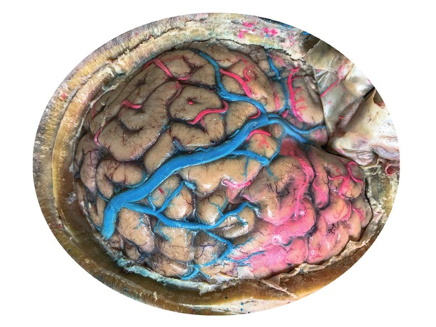
• Two cerebral hemispheres for each participant. The • Evaluate surgical approaches to challenging areas: the
specimens were previously selected to ensure the quality dominant insular lobe and the posterior parahippocampus.
for dissection. The specimen’s vessels were injected with • Discuss surgical cases and analyze different treatment
red and blue colorants for greater similarity with a real options of tumors located within eloquent areas.
brain.
• 3D Microscope Kinevo 900 Carl Zeiss: one for the course.
• Microscopes (techno-scopes) Carl Zeiss: one for each Course venue:
participant. Anatomy Laboratory.
• Ultrasonic aspirators CUSA Clarity (Integra): one for each Department of Anatomy and Cellular Biology.
participant. School of Medicine. Cantabria University.
Av. Herrera Oria, s/n. 39011. Santander (Cantabria). Spain.
• 3D television (75 inches, Full HD): one for the course.
• Medtronic StealthViz Neuronavigation system: one for the
course. Registration fees:
• Medtronic Echography system: one for the course. • Full hands-on registration: 2.100 euros + VAT. Includes
lectures attendance, dissection of cerebral hemispheres
• Video camera: one for the course.
and simulation of surgical approaches, lunch and
• 3D Glasses: one for each participant. refreshments breaks, and course dinner on Friday.
• Instruments for dissection for each participant.
Technical Secretariat:
Accreditation: AFORO CONGRESOS
• This teaching activity is accredited by the Commission Pasaje de Peña 2, 3º C. Edificio Simeón
for Continuing Education of the Health Professions of the 39008 Santander. Spain
Community of Cantabria. 6,5 credits in 2019. Phone: + 34 942 23 06 27
• The scientific and educational content of this event has Email: zulema@aforocongresos.com
been endorsed by the Sociedad Española de Neurocirugía www.aforocongresos.com
(SENEC).
PROGRAM. Thursday, 21st of October 2021
09:00 Registration.
09:10 Opening.
Dr. Juan A. Montero Simon, Dr. Juan Martino
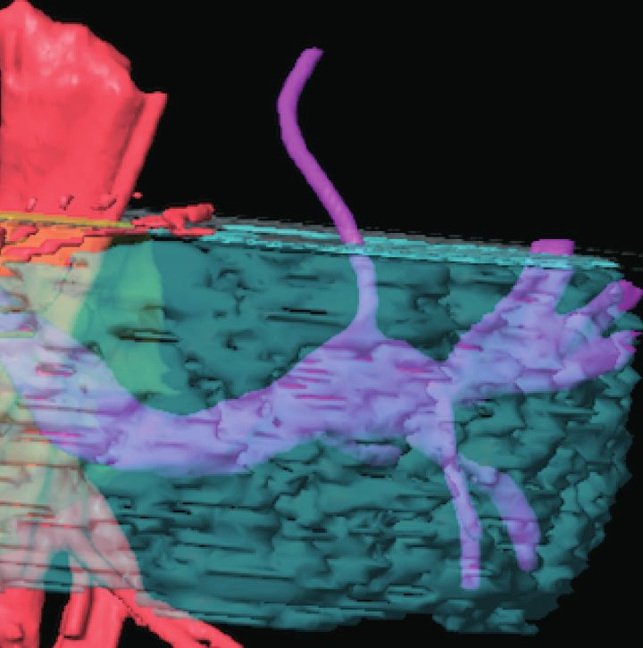
09:15 3D LECTURE: Introduction to brain Diffusion tensor imaging (DTI) tractography.
Dr. Christian de Quintana Schmidt
09:45 Hands-on. Introduction to StealthViz software.
Dr. Christian de Quintana Schmidt
10:15 3D LECTURE: DTI tractography. How I do it.
Dr. Christian de Quintana Schmidt
11:15 Hands-on. DTI tractography reconstruction of the main white matter connections.
Dr. Christian de Quintana Schmidt
12:15 DTI tractography as an important tool to study the subcortical anatomy and presurgical planning of glioma
surgery.
Dr. Enrique Marco de Lucas
13:00 Lunch
14:00 3D LECTURE: How to prepare the brains for fiber dissection.
Dr. David Mato
14:15 3D LECTURE: sulco-gyral anatomy. The cerebral lobes.
Dr. Pablo González-Lopez
15:00 3D LECTURE: anatomy of the dorsal associative tracts of the brain: superior longitudinal fasciculus, arcuate
fasciculus, middle longitudinal fasciculus.
Dr. Juan Martino
15:30 Functional roles of the dorsal associative tracts of the brain.
Dr. Alejandro Fernandez-Coello
15:45 Hands-on: Fiber dissection of the dorsal associative tracts. Each participant will have one hemisphere to dissect.
The participants will learn how to remove the arachnoid membranes and the cortex without damaging the
underling white matter. The participants will dissect the dorsal associative tracts: the subcomponents of the
superior longitudinal fasciculus, arcuate fasciculus and middle longitudinal fasciculus.
Professor Hugues Duffau, Dr. Juan Martino, Dr. Alejandro Fernandez-Coello, Dr. Emmanuel Mandonnet, Dr. Pablo
González-Lopez, Dr. David Mato, Dr. Carlos Velasquez, Dr. Carlos Santos, Dra. Carla Mora, Dr. Jesus Esteban, Dra. Patricia
Lopez, Dra. Aranzazu Sanchez, and Monserrat Fernández-Calderón.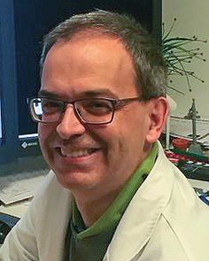
PROGRAM. Friday, 22nd of October 2021

09:00 3D LECTURE: anatomy of the ventral associative tracts and the fascicles related to the insula: inferior longitudinal
      fasciculus, inferior fronto-occipital fasciculus and uncinate fasciculus.
      Dr. Juan Martino
09:30 Functional roles of the ventral associative tracts and the fascicles related to the insula.
      Dr. Alejandro Fernandez-Coello
09:45 3D LECTURE: anatomy of the brain isthmus and the temporal stem: a crucial anatomical concept that is often
      forgotten.
      Dr. Carlos Velasquez and Dr. Pablo Gonzalez
10:15 Hands-on: Fiber dissection of the ventral associative tracts and the fascicles related to the insula. Each
      participant will have one hemisphere to dissect. The participants will dissect the ventral associative tracts and
      the tracts related to the insula region: inferior longitudinal fasciculus, inferior fronto-occipital fasciculus and
      uncinate fasciculus.
      Professor Hugues Duffau, Dr. Juan Martino, Dr. Alejandro Fernandez-Coello, Dr. Emmanuel Mandonnet, Dr. Pablo
      González-Lopez, Dr. David Mato, Dr. Carlos Velasquez, Dr. Carlos Santos, Dra. Carla Mora, Dr. Jesus Esteban, Dra. Patricia
      Lopez, Dra. Aranzazu Sanchez, and Monserrat Fernández-Calderón.
13:00 Lunch.

14:00 Intraoperative electrical stimulation mapping of associative fiber pathways.
      Professor Hugues Duffau
14:30 3D LECTURE: limbic and paralimbic tumors. Anatomy and related surgical approaches.
      Dr. Pablo González-Lopez
15:00 3D LECTURE: presentation of the parahippocampal surgical case.
      Dr. Juan Martino
15:15 Hands-on: Approach to the posterior parahippocampus and cingulum. Professor Hugues Duffau will perform
      a step by step posterior basal and temporal approach to the posterior parahippocampus and cingulum.
      Simultaneously, each participant will perform the approach in the specimens. The participant will use a real
      MRI of a glioma infiltrating the basal temporal lobe to guide the resection. In this approach, the deep functional
      connections are the arcuate fasciculus, inferior longitudinal fasciculus, inferior fronto-occipital fasciculus and
      optic radiations.
      Professor Hugues Duffau, Dr. Emmanuel Mandonnet, Dr. Juan Martino, Dr. Alejandro Fernandez-Coello, Dr. Pablo
      González-Lopez, Dr. David Mato, Dr. Carlos Velasquez, Dr. Carlos Santos, Dra. Carla Mora, Dr. Jesús Esteban, Dra. Patricia
      Lopez, Dra. Aranzazu Sanchez, and Monserrat Fernández-Calderón.
21:00 Course dinner.
PROGRAM. Saturday, 23rd of October 2021

09:00 Surgical anatomy of the insula.
      Dr. Emmanuel Mandonnet
09:30 Insula glioma surgery: the transopercular approach.
      Professor Hugues Duffau
10:00 3D LECTURE: presentation of the insular surgical case.
      Dr. Juan Martino
10:15 Hands-on: Transopercular approach to the insula. Professor Hugues Duffau will perform a step by step
      transopercular approach to the insula in the specimen. Simultaneously, each participant will perform the
      approach in the specimens. The participants will use a real magnetic resonance image (MRI) of a fronto-
      temporo-insular glioma to guide the resection. We will have a unique opportunity to ask Professor Duffau
      many questions about the challenges of this approach: how to preserve the deep functional connections
      (inferior fronto-occipital fasciculus, uncinate fasciculus, pyramidal pathway, etc.), and vascular structures
      (lenticulostriate arteries).
      Professor Hugues Duffau, Dr. Emmanuel Mandonnet, Dr. Juan Martino, Dr. Alejandro Fernandez-Coello, Dr. Pablo
      González-Lopez, Dr. David Mato, Dr. Carlos Velasquez, Dr. Carlos Santos, Dra. Carla Mora, Dr. Jesús Esteban, Dra. Patricia
      Lopez, Dra. Aranzazu Sanchez, and Monserrat Fernández-Calderón.

13:00 Lunch.

14:00 Hands-on: Transopercular approach to the insula. Professor Hugues Duffau and participants will complete the
      transopercular approach to the insula.

19:00 Closing remarks.
5Th Mapping Course Bellvitge 2021

The present course is intimately linked with the “Bellvitge Mapping Course” that
takes place at the Bellvitge University Hospital in Barcelona, and that is organized
by Andreu Gabarros. The Santander’s course delves in subcortical anatomical study,
and simulation of surgical approaches in the cadaver. On the other hand, Bellvitge´s
course is more focused in intraoperative mapping techniques, neurophysiologic
intraoperative monitoring, and live broadcast surgeries. Therefore, in order to have
a broader perspective of mapping techniques for resection of brain tumors within
eloquent areas, we strongly recommend the participants to assist to both courses in
a consecutive order.

Bellow you can see a resume of the Bellvitge Mapping Course program.

Juan Martino and Andreu Gabarros

First day
Laboratory brain fiber dissection

Second day
DTI tractography

Third day
Lectures: intraoperative mapping techniques and neurophysiologic intraoperative monitoring

Fourth day
Live surgeries of tumors within eloquent areas
FOURTH ANNUAL EUROPEAN
                        3D ADVANCED FIBER DISSECTION COURSE:
                        ACQUIRING THE MENTAL IMAGERY
                        NECESSARY TO OPERATE THE BRAIN
                        Santander. 21, 22 and 23 October 2021

                                                                  © MBA SURGICAL EMPOWERMENT
                                                                  V1-202102
                                                                  21mk008
                        Technical Secretariat:
                        AFORO CONGRESOS
                        Pasaje de Peña 2, 3º C. Edificio Simeón
                        39008 Santander. Spain
                        Phone: + 34 942 23 06 27
                        Email: zulema@aforocongresos.com
                        www.aforocongresos.com

Sponsoring companies:
You can also read