From Child Abuse to Developing Borderline Personality Disorder Into Adulthood: Exploring the Neuromorphological and Epigenetic Pathway - Cureus
←
→
Page content transcription
If your browser does not render page correctly, please read the page content below
Open Access Review
                                         Article                                                  DOI: 10.7759/cureus.9474

                                         From Child Abuse to Developing Borderline
                                         Personality Disorder Into Adulthood:
                                         Exploring the Neuromorphological and
                                         Epigenetic Pathway
                                         Pranita Mainali 1, 2 , Tehrima Rai 3 , Ian H. Rutkofsky 2

                                         1. Psychiatry, Washington DC Veterans Affairs Medical Center, Washington, DC, USA 2. Psychiatry,
                                         California Institute of Behavioral Neurosciences & Psychology, Fairfield, USA 3. Pediatrics, California
                                         Institute of Behavioral Neurosciences & Psychology, Fairfield, USA

                                         Corresponding author: Pranita Mainali, pranita_mainali@icloud.com

                                         Abstract
                                         Borderline personality disorder (BPD) is one of the most common personality disorders seen in
                                         the general population. Among multiple identified risk factors, one of the most influential
                                         elements is exposure to an adverse childhood experience in terms of emotional, physical, or
                                         sexual abuse. A cascade of neuromorphological and epigenetic changes occurs in response to
                                         these childhood stressors, which may have a strong link to the development of BPD. PubMed
                                         and Google Scholar were searched for articles relevant to child abuse and the development of
                                         BPD. The search was not restricted to any time frame or geographic location. Significant
                                         epigenetic and neuromorphological changes are seen with child abuse, contributing to the
                                         development of BPD. Chronic stressors lead to hypothalamic-pituitary axis (HPA) activation,
                                         releasing cortisol that acts on the prefrontal cortex, amygdala, and hippocampus, producing the
                                         behavioral patterns seen in BPD. Overstimulation of gray matter leads to permanent
                                         neuromorphological changes, which can be visualized in functional MRI/brain scans.
                                         Hypermethylation of messenger ribonucleic acid in various sites suggests the impact of child
                                         abuse on the genetic level. Interestingly, the prevalence of BPD is seen equally in both genders
                                         but is diagnosed more frequently in females because they tend to be more likely to seek help.
                                         Understanding the impact of early age life stressors into adulthood calls for serious focus on
                                         early diagnosis and intervention. This implies the need for more studies in patients with BPD
                                         with or without any childhood traumatic experience and a better understanding of the changes
                                         that occur biopsychologically and genetically in response to trauma.

                                         Categories: Neurology, Pediatrics, Psychiatry
                                         Keywords: child abuse, early life stressor, borderline personality disorder, adverse childhood
Received 05/26/2020                      experience
Review began 06/28/2020
Review ended 07/12/2020
Published 07/30/2020                     Introduction And Background
© Copyright 2020                         Nearly 7.5 million children are victims of abuse in the United States (US) annually, where 74.9%
Mainali et al. This is an open access
                                         are victims of neglect, 18.3% are physically abused, 8.6% are sexually abused, and 7.1% are
article distributed under the terms of
                                         psychologically maltreated [1]. Children younger than one year, especially males, have the
the Creative Commons Attribution
License CC-BY 4.0., which permits        highest rate of child abuse. Nearly 50% of childhood fatalities occur in the first year of life [2].
unrestricted use, distribution, and      However, abuse remains a concern in all age groups, with 27% of victims being younger than
reproduction in any medium, provided     three years of age, and 20% are aged three to four years [3]. Child abuse is a public concern, as it
the original author and source are
                                         is estimated that five to six children die every day because of domestic violence, physical
credited.
                                         abuse, sexual trauma, or neglect [4]. About 80% of victims develop a psychiatric illness,

                                         How to cite this article
                                         Mainali P, Rai T, Rutkofsky I H (July 30, 2020) From Child Abuse to Developing Borderline Personality
                                         Disorder Into Adulthood: Exploring the Neuromorphological and Epigenetic Pathway. Cureus 12(7): e9474.
                                         DOI 10.7759/cureus.9474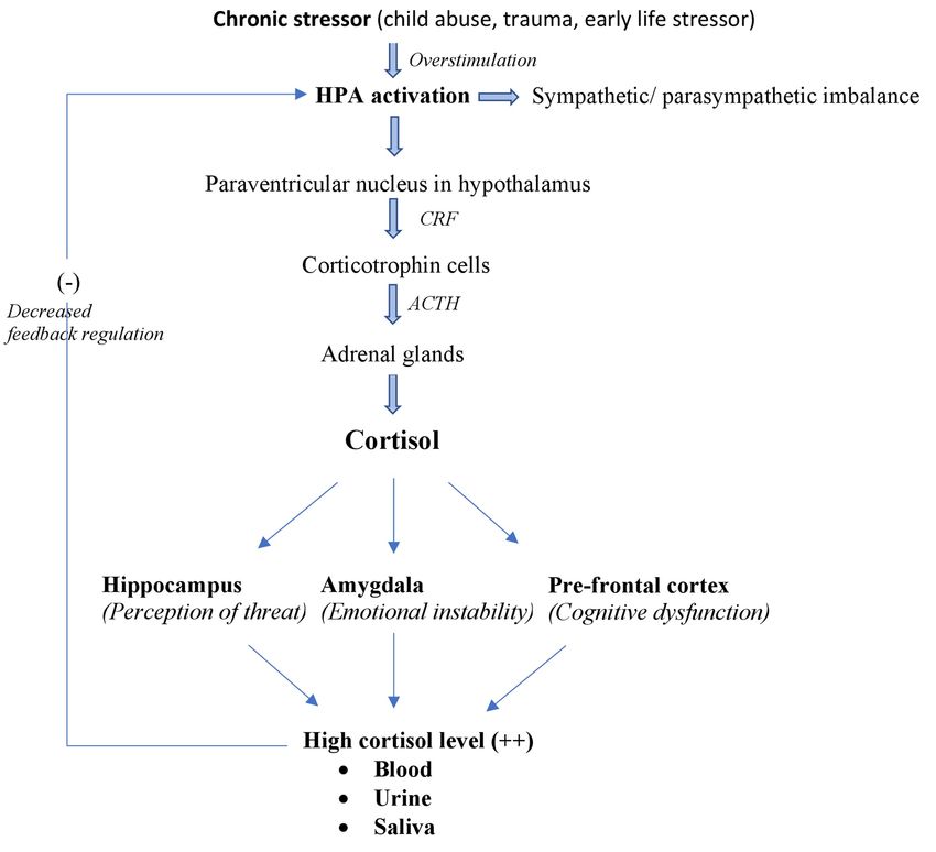
behavioral, and emotional issues before the age of 21 years [5]. Recent literature has suggested
                                    a significant relationship between child abuse and its persistence with the development and
                                    severity of maladaptive personality traits into adulthood [6,7]. Among all personality disorders,
                                    30% to 90% of patients who meet the criteria for borderline personality disorder (BPD) have a
                                    history of child abuse or trauma [8].

                                    BPD is a common personality disorder seen in 1% to 3% of the general population, 10% in
                                    outpatient settings, 20% in inpatient settings, and 9% to 27% in the emergency department
                                    [9,10]. It is characterized by a marked pervasive pattern of emotional dysregulation, impulsive
                                    behavior, identity disturbances, and interpersonal conflicts [11]. Predominantly, it is seen three
                                    to four times higher in females in various clinical settings, even though the gender prevalence
                                    remains nearly equal in the community [12]. A study has shown that early biological and
                                    neurological stressors lead to DNA methylation, which impairs usual brain functioning,
                                    interfering with emotional regulation and stability, impulse control, coping skills,
                                    interpersonal skills, cognition, and other core skills, as seen in BPD [13]. However, limited
                                    studies have been conducted to establish a clear correlation between child abuse or trauma and
                                    the development of BPD later in life. The fact that men are less likely to seek help also creates a
                                    challenge to understanding the prevalence and severity of BPD with child abuse [14]. The
                                    purpose of this study is to explore the possible epigenetic modifications and neurobiological
                                    changes due to early life stressors and related to the development of BPD into adulthood. This
                                    study also seeks to assess these neuromorphological changes and understand the sex
                                    distribution of BPD.

                                    Review
                                    Method
                                    PubMed and Google Scholar were searched for articles relevant to child abuse and the
                                    development of BPD later in life. A comprehensive search was done to understand the
                                    association of epigenetic and neurobiological effects of childhood trauma with the
                                    development of BPD, along with the impact of duration and severity on both sexes. Medical
                                    Subject Heading (MeSH) terms used were “child abuse”, “adult survivor of child abuse”, and
                                    “child abuse, sexual” which returned 399 peer-reviewed articles; “early life stressor” returned
                                    533 articles, “childhood trauma” returned 16,657 publications, and “borderline personality
                                    disorder” returned 6,439 publications. Keeping the search relevant to the topic, MeSH terms
                                    were combined with other phrase choices consisting of “childhood trauma and borderline
                                    personality disorder” which returned 204 articles; “trauma exposure in children and borderline
                                    personality disorder” returned two systematic reviews, “borderline personality disorder and
                                    gender” returned 525 articles, and combining all the above-mentioned MeSH terms yielded
                                    eight articles. All articles were reviewed including the abstracts for the restricted one. The
                                    search was not restricted to any timeframe or geographic location.

                                    Result
                                    This review shows that any form of child abuse can lead to long-term neurobiological and
                                    permanent morphological changes in the brain of the victim. Overactivation of the
                                    hypothalamic-pituitary axis (HPA) leads to excess cortisol production. This mechanism
                                    consistently prepares the body for a flight or fight response and misinterprets standard
                                    environmental signals as a threat. Overstimulation of gray matter leads to a reduction in the
                                    volume of the hippocampus, activation of the amygdala, and impairment of the prefrontal,
                                    frontal limbic, and parietal areas. All these changes lead to the personality changes seen with
                                    individuals with BPD. BPD may be recorded more frequently in women who tend to seek
                                    psychopharmacological treatment, but in reality, both sexes are equally affected [15]. Due to
                                    differences in the nature and presentation of the condition among men and women, males with
                                    BPD are more frequently incarcerated due to aggressive or drug-seeking behavior, and females

2020 Mainali et al. Cureus 12(7): e9474. DOI 10.7759/cureus.9474                                                                  2 of 11
are more often evaluated in healthcare settings [16]. It is important to evaluate every abused
child regardless of sex, as the impact is not only seen clinically but also on the genetic level,
triggering hypermethylation of messenger ribonucleic acid in various sites, as reflected in
individuals with BPD [17,18].
Neurobiological variations
Child abuse leads to long-term repercussions as stressors trigger chronic “hyperarousal” of the
HPA, which mediates cascades of neurohormonal changes in response [19]. The paraventricular
nucleus in the hypothalamus releases corticotropin-releasing factor, which stimulates
corticotrophin cells to release adrenocorticotropic hormone (ACTH) [20]. ACTH then acts on the
adrenal glands to stimulate the secretion of cortisol, the primary stress hormone. Cortisol
functions as the major hormone for the fight or flight response; it activates the autonomic
nervous system (ANS), leading to an arousal state through the sympathetic nervous system [21].
Under chronic stress, unregulated cortisol secretion could lead to disruption of the ANS, as
supported by a meta-analysis conducted by Koenig et al. [22]. The study draws attention to the
disinhibition symptoms of BPD like impulsivity, self-harming behavior, emotional
dysregulation, and its link to the lower resting-state vagal tone measured by vagally mediated
heart rate variability.
The hyperarousal of the hippocampus by cortisol leads to misinterpretation of signals perceived
from the natural environment, individuals, and situations, as constant threats and sends the
wrong message to the amygdala. On the other hand, activation of the amygdala, which
regulates fear and aggression, results in unstable and unpredictable intense emotional turmoil
in response to minor stress with a more extended latency for returning to baseline; this is
common in BPD. Overfunctioning of the prefrontal cortex explains the loss of rationality,
reasoning, and decision-making capacity in BDP (Doctoral Dissertation: Human C.
Neuropsychological Deficits in Borderline Personality Disorder, University of Johannesburg;
1998) [23,24].
Many studies have shown an increase in urinary, salivary, or blood cortisol levels in individuals
with BPD with or without comorbid posttraumatic stress disorder (PTSD), which is a hallmark
of HPA activation. In 2002, Rinne et al. conducted a study in 39 patients with or without
childhood trauma and with a BPD diagnosis [25]. The study showed a higher level of ACTH (and
hence cortisol level) in the blood of patients with BPD, who had also sustained early life
stressors. In 2007, Wingenfeld et al. conducted a study on 21 women with BPD and 24 controls
without BPD with the conclusion that overnight urinary cortisol level was much higher in
women with BPD in comparison to those in the control group [26]. Similarly, another study
conducted by Lieb et al. linked BPD to overactivation of the adrenal glands and decreases in the
HPA regulatory feedback mechanism, as evidenced by a significant elevation in salivary cortisol
level in 23 unmedicated women with BPD in comparison to 24 control women (Figure 1) [27].
2020 Mainali et al. Cureus 12(7): e9474. DOI 10.7759/cureus.9474 3 of 11FIGURE 1: Impact of the chronic childhood stress on HPA
activation
HPA, hypothalamic-pituitary axis; CRF, corticotrophin-releasing factor; ACTH, adrenocorticotrophic
hormone.
Neuromorphological variation
Many studies have shown visual evidence of morphological changes, mainly in the
hippocampal area, amygdala, hypothalamus, and prefrontal cortex in patients with BPD [28-
30]. In 2001, Herpertz et al. conducted a study to scan brain activity using functional MRI in six
women with BPD and without other psychiatric comorbidities with six matched control women
[31]. The study showed increased blood oxygenation levels on both sides of the amygdala along
with activation of the medial and inferolateral prefrontal cortex. This association explains the
emotional instability in BPD patients even with low stressors and increased latency for the
emotion to return to baseline. Similarly, in 2013, an analysis by Kuhlmann et al. on 30 BPD
patients and 33 matched controls showed decreased hippocampal volumes in those with BPD
along with increased volume in the hypothalamus. Remarkably, the size of the hypothalamus
has a direct relationship with exposure to childhood trauma in patients with BPD [32].
Soloff et al. in 2012 conducted a more extensive study, including 68 participants with BPD (16
men and 52 women) and 52 healthy control group (28 men and 24 women), with suicidal
attempts or suicidal behaviors [33]. A structural MRI scan showed a significant decrease in gray
matter in the left insula, right mid-superior temporal gyrus, right mid-inferior orbitofrontal
2020 Mainali et al. Cureus 12(7): e9474. DOI 10.7759/cureus.9474 4 of 11gyrus, right insular cortex, left fusiform gyrus, left lingual gyrus, and right parahippocampal
gyrus in patients with BPD and lethal suicidal attempts. Using three-dimensional (3D) MRI, Irle
et al. found that 30 women with BPD and severe childhood trauma had a drastic reduction of
hippocampal volume by 17% and a reduction in parietal cortex volume by 11% in comparison to
control group [34]. This study showed a direct proportional correlation between the severity of
trauma exposure in childhood and the amount of volume reduction in the mentioned areas of
the brain. Therefore, in line with the findings of a study by Dannlowski et al., there is a
possibility that the symptoms of BPD may be due to overstimulation of the hippocampal area
leading to the gradual reduction in gray matter volume, as is commonly seen in patients who
are victims of early traumatization and PTSD (Table 1) [31-35].
Study
Author Implications Sample size Result
year
Overstimulation of gray matter leads to a decrease in 148 healthy subjects Functional MRI analysis showed reduced gray matter volume in the hippocampus,
Dannlowski
2012 hippocampal volume = loss of memory, flexible cognition, screened for childhood insula, orbitofrontal cortex, anterior cingulate gyrus, and caudate in subject with high
et al. [35]
and social behavior maltreatment using CTQ CTQ score
Six BPD females without
Herpertz et Amygdala activation = intense emotion to low stressor; other psychiatric Functional MRI showed increased blood oxygenation level on both sides of the
2001
al. [31] increased attention to emotionally activating external stimuli comorbidities and six amygdala; activation of medical and inferolateral prefrontal cortex in BPD
matched control females
Decreased hippocampal volume = increased stress and 30 patients with BPD
Kuhlmann Decrease in hippocampal volume with BPD; increase in the hypothalamus compared
2013 glucocorticoid activation; increased hypothalamus volume = (unmedicated), 33 healthy
et al. [32] to control
overstimulation of central stress regulation controls
68 with BPD (16 males, 52
Structural abnormality: in prefrontal and frontolimbic region Diminished gray matter concentration in the left insula, right mid-superior temporal
Soloff et al. females) and 52 healthy
2012 = emotional instability, cognitive functioning impairment, gyrus, right mid-inferior orbitofrontal gyrus, right insular cortex, left fusiform gyrus, left
[33] controls (28 males, 24
and impulsive behavior lingual gyrus, and right parahippocampal gyrus in BPD with lethal suicidal attempts
females)
Parietal atrophy = misinterpretation of sensory/visual 30 BPD females with a
Irle et al. information and constant perception of threat to normal history of childhood trauma 3D structural MRI showed decreased in hippocampal area by 17% and in the parietal
2005
[34] stimuli; abnormality in temporoparietal cortex = psychotic and 25 matched healthy region by 11% in females with BPD and trauma history
symptoms controls
TABLE 1: Summary of neuromorphological changes seen in patients with borderline
personality disorder.
BPD, borderline personality disorder; CTQ, childhood trauma questionnaire; 3D, three dimensional.
Epigenetic modifications with childhood maltreatment
Along with the overstimulation of the HPA axis, there is increased methylation of
glucocorticoid receptor gene NR3C1, as observed by Martin-Blanco et al. in an analysis
conducted in 2014 [36]. Among 281 subjects with BPD, higher NR3C1 methylation was seen in
patients with BPD who had exposure to childhood trauma [32]. Similarly, Dammann et al.
analyzed DNA methylation in a patient with BPD and compared the results with the control
group [17]. Of 14 neurogenes, they found increased methylation of HTR2A (by 0.8%), NR3C1
2020 Mainali et al. Cureus 12(7): e9474. DOI 10.7759/cureus.9474 5 of 11(by 1.8%), MAOA (by 1.5%), MAOB (by 1.4%), and soluble COMT (S-COMT). Overall, the
methylation of these genes was 1.7% higher in patients with BPD and childhood trauma (p <
0.0001) in comparison to patients in the control group. A similar finding was reported by
Teschler et al., who described 1.26-fold higher methylation in an analyzed gene associated CpG
sites in the blood sample of 24 female BPD patients in comparison to 11 female healthy
controls [18].
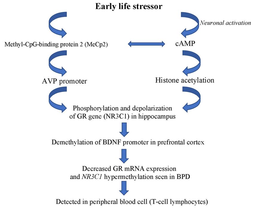
The influence of environmental stressors leading to epigenetic modification was summarized
by Gescher et al. in a meta-analysis published in 2018 (Figure 2) [37].
FIGURE 2: Environmental stressor leading to epigenetic
modification.
AVP, arginine vasopressin gene; cAMP, adenosine 3’,5’-cyclic monophosphatase; BDNF, brain-
derived neurotrophic factor gene; GR gene, glucocorticoid receptor gene; mRNA, messenger
ribonucleic acid; NR3C1, nuclear receptor subfamily 3, group C, member 1; BPD, borderline
personality disorder.
Gender prevalence
An analysis by Sansone and Sansone proposed that men with BPD are more likely to be found
incarcerated due to violent temperament or substance use disorders, while women with BPD are
more likely to be seen in healthcare settings for seeking treatment for eating disorders, mood
disorders, anxiety, or PTSD [16]. Tadić et al. investigated the gender difference between axis I
and axis II patients with BPD diagnosis. Among 110 women and 49 men with BDP, in axis I,
men had more alcohol dependence (65% vs. 43%), and more women suffered from anorexia
2020 Mainali et al. Cureus 12(7): e9474. DOI 10.7759/cureus.9474 6 of 11nervosa (21% vs. 4%). Men presented more often with “anger issues” (74% vs. 49%), and women
presented more often with “emotional instability” (94% vs. 82%) [38].
Zanarini et al. discussed a strong correlation between gender and expression of axis II
comorbidities. The analysis concluded that more males with BPD diagnosis meet the criteria for
paranoia, passive-aggressive, sadistic, narcissistic, or antisocial personality disorders [39].
Interestingly, Marchetto, in 2006, found no significant gender difference with regards to self-
harming behavior such as repetitive skin cutting [40]. Although there are differences in the
presentation of BPD in men and women, the difference in the level of impairment remains
insignificant [15]. However, as women with BPD tend to seek more pharmacotherapy and
psychotherapy in their lifetime as compared to men with BPD, this might account for the higher
prevalence of BPD seen in females (3:1) as opposed to males [41,42].
This literature review sheds light on the impact of child abuse and early life stressors on the
neurogenetic level, raising concerns for cautiousness in screening children with any form of
abuse. The consequences of early life stress are multidirectional; however, with a focus on
developing BPD into adulthood, initial screening and diagnosis can lead to timely intervention
and prevention in the long run. This paper encapsulates the neurogenetic and morphological
changes seen in both sexes, as detected in serum studies, through genetic testing and brain
scans. This can also be applied to guiding BPD diagnosis in the various developmental stages of
childhood. Additionally, this knowledge can be used to understand the impact of therapy or
trauma-focused intervention.
Understanding the link between child abuse and the development of any type of personality
disorder can change the approach to such situations. Long-term follow-up from the day of
screening and diagnosis is equally crucial, along with developing case-specific individual
treatment modalities. These interventions can lead to preventing or mitigating some of the
molecular-level changes that lead to the development of BPD. Child abuse in itself is a public
concern, and understanding the short-term and long-term mental and physical consequences
can be valuable in improving the quality of life of victims immediately and in the long run.
This study is not without limitations. This review focuses mainly on understanding the
correlation between the early life stressor of child abuse and the development of BPD later into
adulthood. This study does not specify the various types of trauma experiences (emotional,
physical, or sexual abuse) and does not examine exposure to each type of abuse and their effect
on the genetic and neurological levels. Moreover, it does not account for various demographic
variables such as the age of the child when exposed to trauma, duration of exposure, stressor
severity, family history, and socioeconomic history, factors that play a vital role in the
development of personality disorders.
Conclusions
Child abuse is a risk factor for the development of BPD and is a public concern. Understanding
the impact of early age negative life stressors on adulthood calls for serious focus on early
diagnosis and intervention. This implies the need for more research focusing on patients with
BPD with or without childhood traumatic experience and understanding the changes that occur
in response to trauma. A detailed study of the impact of the nature and severity of trauma on
children of various age groups may lead to a better understanding of how to modulate
treatment based on individual needs. We need to understand and explore the risk of offspring
developing BPD with epigenetic changes in parents. Future studies addressing whether
intervention at an early age can halt or reverse any unwanted changes in the victim will play an
essential role among patients of both sexes who may be at risk for BPD.
2020 Mainali et al. Cureus 12(7): e9474. DOI 10.7759/cureus.9474 7 of 11Additional Information
Disclosures
Conflicts of interest: In compliance with the ICMJE uniform disclosure form, all authors
declare the following: Payment/services info: All authors have declared that no financial
support was received from any organization for the submitted work. Financial relationships:
All authors have declared that they have no financial relationships at present or within the
previous three years with any organizations that might have an interest in the submitted work.
Other relationships: All authors have declared that there are no other relationships or
activities that could appear to have influenced the submitted work.
References
1. Child maltreatment statistics in the U.S. (2018). Accessed: April 10, 2020:
http://americanspcc.org/child-abuse-statistics/.
2. Barth RP, Blackwell DL: Death rates among California's foster care and former foster care
populations. Child Youth Serv Rev. 1998, 20:577-604. 10.1016/S0190-7409(98)00027-9
3. Gray JD, Cutler CA, Dean JG, Kempe CH: Prediction and prevention of child abuse and neglect .
J Soc Issues. 1979, 35:127-139. 10.1111/j.1540-4560.1979.tb00805.x
4. Oswald SH, Heil K, Goldbeck L: History of maltreatment and mental health problems in foster
children: a review of the literature. J Pediatr Psychol. 2010, 35:462-472. 10.1093/jpepsy/jsp114
5. U.S. Department of Health and Human Services, Administration for Children and Families,
Administration on Children, Youth and Families, Children’s Bureau. Child maltreatment.
(2012). Accessed: July 24, 2020: https://www.acf.hhs.gov/cb/resource/child-maltreatment-
2012.
6. Kaplow JB, Widom CS: Age of onset of child maltreatment predicts long-term mental health
outcomes. J Abnorm Psychol. 2007, 116:176-187. 10.1037/0021-843X.116.1.176
7. Martin-Blanco A, Soler J, Villalta L, et al.: Exploring the interaction between childhood
maltreatment and temperamental traits on the severity of borderline personality disorder.
Compr Psychiatry. 2014, 55:311-318. 10.1016/j.comppsych.2013.08.026
8. Yen S, Shea MT, Battle CL, et al.: Traumatic exposure and posttraumatic stress disorder in
borderline, schizotypal, avoidant, and obsessive-compulsive personality disorders: findings
from the collaborative longitudinal personality disorders study. J Nerv Ment Dis. 2002,
190:510-518.
9. Biskin RS: The lifetime course of borderline personality disorder . Can J Psychiatry. 2015,
60:303-308. 10.1177/070674371506000702
10. Shaikh U, Qamar I, Jafry F, Hassan M, Shagufta S, Odhejo YI, Ahmed S: Patients with
borderline personality disorder in emergency departments. Front Psychiatry. 2017, 8:136.
10.3389/fpsyt.2017.00136
11. American Psychiatric Association: Diagnostic and Statistical Manual of Mental Disorders .
American Psychiatric Publishing, Washington, DC; 2013.
10.1093/acrefore/9780199975839.013.104
12. Paris J: Estimating the prevalence of personality disorders in the community . J Pers Disord.
2010, 24:405-411. 10.1521/pedi.2010.24.4.405
13. Elliott JC, Stohl M, Wall MM, et al.: Childhood maltreatment, personality disorders and 3‐year
persistence of adult alcohol and nicotine dependence in a national sample. Addiction. 2016,
111:913-923. 10.1111/add.13292
14. Oliver MI, Pearson N, Coe N, Gunnell D: Help-seeking behaviour in men and women with
common mental health problems: cross-sectional study. Br J Psychiatry. 2005, 186:297-301.
10.1192/bjp.186.4.297
15. Zlotnick C, Rothschild L, Zimmerman M: The role of gender in the clinical presentation of
patients with borderline personality disorder. J Pers Disord. 2002, 16:277-282.
10.1521/pedi.16.3.277.22540
16. Sansone RA, Sansone LA: Gender patterns in borderline personality disorder . Innov Clin
Neurosci. 2011, 8:16-20.
17. Dammann G, Teschler S, Haag T, Altmüller F, Tuczek F, Dammann RH: Increased DNA
methylation of neuropsychiatric genes occurs in borderline personality disorder. Epigenetics.
2020 Mainali et al. Cureus 12(7): e9474. DOI 10.7759/cureus.9474 8 of 112011, 6:1454-1462. 10.4161/epi.6.12.18363
18. Teschler S, Bartkuhn M, Künzel N, Schmidt C, Kiehl S, Dammann G, Dammann R: Aberrant
methylation of gene associated CpG sites occurs in borderline personality disorder. PLoS One.
2013, 8:e84180. 10.1371/journal.pone.0084180
19. Balbernie R: Circuits and circumstances: the neurobiological consequences of early
relationship experiences and how they shape later behaviour. J Child Psychother. 2001,
27:237-255. 10.1080/00754170110087531
20. Pompili M, Serafini G, Innamorati M , et al.: The hypothalamic-pituitary-adrenal axis and
serotonin abnormalities: a selective overview for the implications of suicide prevention. Eur
Arch Psychiatry Clin Neurosci. 2010, 260:583-600.
21. Harris BN, Carr JA: The role of the hypothalamus-pituitary-adrenal/interrenal axis in
mediating predator-avoidance trade-offs. Gen Comp Endocrinol. 2016, 230-231:110-142.
10.1016/j.ygcen.2016.04.006
22. Koenig J, Kemp AH, Feeling NR, Thayer JF, Kaess M: Resting state vagal tone in borderline
personality disorder: a meta-analysis. Prog Neuropsychopharmacol Biol Psychiatry. 2016,
64:18-26. 10.1016/j.pnpbp.2015.07.002
23. Lupien SJ, Maheu F, Tu M, Fiocco A, Schramek TE: The effects of stress and stress hormones
on human cognition: implications for the field of brain and cognition. Brain Cogn. 2007,
65:209-237. 10.1016/j.bandc.2007.02.007
24. Linehan MM: Cognitive-Behavioral Treatment of Borderline Personality Disorder . Guilford
Publications, New York; 2018.
25. Rinne T, de Kloet ER, Wouters L, Goekoop JG, DeRijk RH, van den Brink W:
Hyperresponsiveness of hypothalamic-pituitary-adrenal axis to combined
dexamethasone/corticotropin-releasing hormone challenge in female borderline personality
disorder subjects with a history of sustained childhood abuse. Biol Psychiatry. 2002, 52:1102-
1112. 10.1016/S0006-3223(02)01395-1
26. Wingenfeld K, Driessen M, Adam B, Hill A: Overnight urinary cortisol release in women with
borderline personality disorder depends on comorbid PTSD and depressive psychopathology.
Eur Psychiatry. 2007, 22:309-312. 10.1016/j.eurpsy.2006.09.002
27. Lieb K, Rexhausen JE, Kahl KG, Schweiger U, Philipsen A, Hellhammer DH, Bohus M:
Increased diurnal salivary cortisol in women with borderline personality disorder . J Psychiatric
Res. 2004, 38:559-565. 10.1016/j.jpsychires.2004.04.002
28. Ruocco AC, Amirthavasagam S, Zakzanis KK: Amygdala and hippocampal volume reductions
as candidate endophenotypes for borderline personality disorder: a meta-analysis of magnetic
resonance imaging studies. Psychiatry Res. 2012, 201:245-252.
10.1016/j.pscychresns.2012.02.012
29. Kreisel SH, Labudda K, Kurlandchikov O, et al.: Volume of hippocampal substructures in
borderline personality disorder. Psychiatry Res. 2015, 231:218-226.
10.1016/j.pscychresns.2014.11.010
30. Driessen M, Herrmann J, Stahl K, et al.: Magnetic resonance imaging volumes of the
hippocampus and the amygdala in women with borderline personality disorder and early
traumatization. Arch Gen Psychiatry. 2000, 57:1115-1122. 10.1001/archpsyc.57.12.1115
31. Herpertz SC, Dietrich TM, Wenning B, et al.: Evidence of abnormal amygdala functioning in
borderline personality disorder: a functional MRI study. Biol Psychiatry. 2001, 50:292-298.
10.1016/S0006-3223(01)01075-7
32. Kuhlmann A, Bertsch K, Schmidinger I, Thomann PA, Herpertz SC: Morphometric differences
in central stress-regulating structures between women with and without borderline
personality disorder. J Psychiatry Neurosci. 2013, 38:129-137. 10.1503/jpn.120039
33. Soloff PH, Pruitt P, Sharma M, Radwan J, White R, Diwadkar VA: Structural brain
abnormalities and suicidal behavior in borderline personality disorder. J Psychiatr Res. 2012,
46:516-525. 10.1016/j.jpsychires.2012.01.003
34. Irle E, Lange C, Sachsse U: Reduced size and abnormal asymmetry of parietal cortex in women
with borderline personality disorder. Biol Psychiatry. 2005, 57:173-182.
10.1016/j.biopsych.2004.10.004
35. Dannlowski U, Stuhrmann A, Beutelmann V, et al.: Limbic scars: long-term consequences of
childhood maltreatment revealed by functional and structural magnetic resonance imaging.
Biol Psychiatry. 2012, 71:286-293. 10.1016/j.biopsych.2011.10.021
36. Martín-Blanco A, Ferrer M, Soler J, et al.: Association between methylation of the
2020 Mainali et al. Cureus 12(7): e9474. DOI 10.7759/cureus.9474 9 of 11glucocorticoid receptor gene, childhood maltreatment, and clinical severity in borderline
personality disorder. J Psychiatric Res. 2014, 57:34-40. 10.1016/j.jpsychires.2014.06.011
37. Gescher DM, Kahl KG, Hillemacher T, Frieling H, Kuhn J, Frodl T : Epigenetics in personality
disorders: today's insights. Front Psychiatry. 2018, 9:579. 10.3389/fpsyt.2018.00579
38. Tadić A, Wagner S, Hoch J, et al.: Gender differences in axis I and axis II comorbidity in
patients with borderline personality disorder. Psychopathology. 2009, 42:257-263.
10.1159/000224149
39. Zanarini MC, Frankenburg FR, Dubo ED, Sickel AE, Trikha A, Levin A, Reynolds V: Axis II
comorbidity of borderline personality disorder. Compr Psychiatry. 1998, 39:296-302.
10.1016/S0010-440X(98)90038-4
40. Marchetto MJ: Repetitive skin-cutting: parental bonding, personality and gender . Psychol
Psychother. 2006, 79:445-459. 10.1348/147608305X69795
41. Goodman M, Patil U, Steffel L, Avedon J, Sasso S, Triebwasser J, Stanley B: Treatment
utilization by gender in patients with borderline personality disorder. J Psychiatr Pract. 2010,
16:155-163. 10.1097/01.pra.0000375711.47337.27
42. Skodol AE, Bender DS: Why are women diagnosed borderline more than men? . Psychiatr Q.
2003, 74:349-360.
2020 Mainali et al. Cureus 12(7): e9474. DOI 10.7759/cureus.9474 10 of 11You can also read