From neurology to neuropathology and back
←
→
Page content transcription
If your browser does not render page correctly, please read the page content below
Free Neuropathology 2:10 (2021)                                                                                                                                                Arnulf H. Koeppen
doi: https://doi.org/10.17879/freeneuropathology-2021-3333                                                                                                                            page 1 of 6

                                                                                                                                                                             Reflections

From neurology to neuropathology and back
Arnulf H. Koeppen

Research Service (151), VA Medical Center, Albany, NY, USA

Corresponding author:
Arnulf H. Koeppen · Research Service (151) · VA Medical Center · 113 Holland Avenue · Albany, NY 12208 · USA
arnulf.koeppen@va.gov

Submitted: 13 April 2021                  ·           Accepted: 17 April 2021                    ·        Copyedited by: Deanna C. Fang                       ·       Published: 21 April 2021

Keywords: Neuropathology, Neurology, Reflections, Autobiography

Chemistry or medicine 1958-1963?                                                                            ternship of 18 months, I joined the neurology train-
                                                                                                            ing program at Montefiore Hospital and Medical
     In the fifties, high school students in Germany                                                        Center in New York City in 1965. Neurology educa-
had to select their career goals without much guid-                                                         tion was disappointing, but I met the late Drs. Harry
ance or college education. I was a student at a gym-                                                        Zimmerman, Robert Terry, and Asao Hirano who
nasium of mathematics and natural sciences in Os-                                                           stimulated my interest in neuropathology and,
terode, a provincial town near the Harz Mountains                                                           above all, clinicoanatomic correlation in neurology
in Lower Saxony of Germany. My father, a general                                                            and neurosurgery. After one year at Montefiore, I
practitioner in a small town in the Harz, had differ-                                                       was recruited to the Department of Neurology and
ent ideas. He wanted me to be a medical student                                                             Psychiatry of Northwestern University School of
and ultimately take over his practice. The university                                                       Medicine in Chicago. My first assignment was to the
town, Göttingen, was located 50 km from our home.                                                           Veterans Affairs (VA) Medical Center in Hines, Illi-
Göttingen was then a small town, but medical edu-                                                           nois, where the late Dr. Kevin D. Barron was the
cation was excellent and diverse. I stayed in a room                                                        chief neurologist. Dr. Barron had been trained by
near the Max Planck Institute where Otto Hahn, the                                                          Drs. Zimmerman and Terry and continued to prac-
Nobel laureate in nuclear fission, was still active. My                                                     tice neuropathology at Northwestern and the VA
inaugural dissertation involved measurements of                                                             Medical Center. Dr. Barron was a master of clinico-
nuclear sizes in rat liver, adrenals, and endometrium                                                       anatomic correlation and stressed that neurologists
after the animals were treated with drugs affecting                                                         should have training in neuropathology. His training
the autonomic nervous system (Koeppen, 1963). My                                                            had a major impact on my further career as a neu-
interest in the neurosciences began during my last                                                          rologist and neuropathologist. While opposition to
year of medical school. The inaugural dissertation                                                          neurologists and psychiatrists practicing neuropa-
received the annual faculty award in 1963; and the                                                          thology was evolving, we were reminded of non-
sum of 800 Deutschmarks was sufficient to defray                                                            pathologists who had made major contributions to
my cost of a trip by ocean liner to the United States                                                       the practice of neuropathology. We thought of Alois
where a non-profit organization, the Ventnor Foun-                                                          Alzheimer and his 1906 illustrations of neurofibril-
dation, had offered me a position as a medical assis-                                                       lary tangles, and of King Engel who gave us modified
tant (intern) at a New Jersey hospital. After an in-                                                        trichrome in the diagnosis of mitochondrial

Copyright: © 2021 The author(s). This is an open access article distributed under the terms of the Creative Commons Attribution 4.0 International License (https://creativecommons.org/licenses/by/4.0/),
which permits unrestricted use, distribution, and reproduction in any medium, provided the original author and source are credited, a link to the Creative Commons license is provided, and any changes are
indicated. The Creative Commons Public Domain Dedication waiver (https://creativecommons.org/publicdomain/zero/1.0/) applies to the data made available in this article, unless otherwise stated.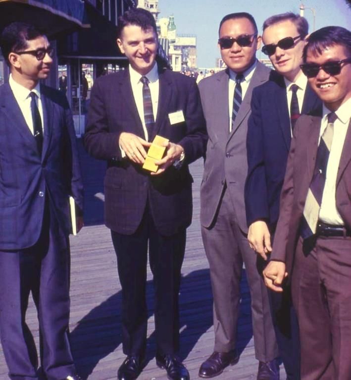
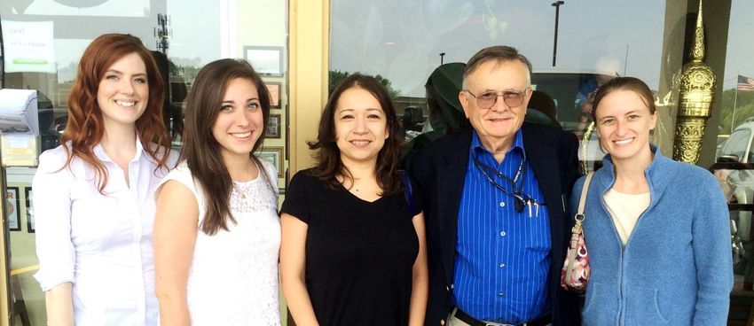
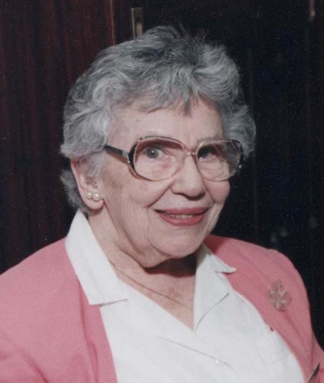
Free Neuropathology 2:10 (2021)                                                                                             Arnulf H. Koeppen
doi: https://doi.org/10.17879/freeneuropathology-2021-3333                                                                         page 2 of 6

Figure 1. Laboratory staff in Albany, NY. From left to right: Alyssa Sossei, research assistant; Sarah Bjork, medical student; R. Liane Ramirez,
research assistant; the author; Pamela Kruger, post-doctoral scientist.

myopathy. My training in neuropathology from                                 PLP protein is unusual because it is soluble in a mix-
1969 to 1971 was enough to gain acceptance to the                            ture of chloroform and methanol (Folch and Lees,
neuropathology board examination in 1980, and,                               1951). Through the work on PMD, I met the co-dis-
since that time, I was convinced that I was a better                         coverers of PLP, the late Dr. Marjorie B. Lees (figure
neurologist because I was also a neuropathologist.                           2), and the late Prof. Franz Seitelberger who had ad-
During my time in Chicago, Dr. Barron insisted that I                        vanced the existence of a “connatal” form of PMD.
should learn neurochemistry as well, and I was as-                           Prof. Seitelberger invited me to the Neurological In-
signed to the research laboratory of the late Dr. Jo-                        stitute in Vienna where I gave a lecture on PLP and
seph Bernsohn. Over a one-year period, I learnt                              PMD. Profs. Kurt Jellinger and Herbert Budka were
standard and advanced laboratory techniques, and                             in audience. I also discovered that the late neuropa-
my initial interest in chemistry came to belated frui-                       thologist, Dr. Wolfgang Zeman (figure 3), had writ-
tion. Since joining the Department of Neurology at                           ten to Dr. Lees in 1963, proposing that PLP was a
Albany Medical College in 1969, I have maintained a                          likely candidate in the genetic mutation underlying
neurochemical and neuropathological laboratory at                            the disease. Pelizaeus-Merzbacher disease is an X-
the Albany VA Medical Center (figure 1) while also                           linked disorder, and Pelizaeus, a German balneolo-
running the clinical neurology service. Among my                             gist, understood the mechanism of X-linked trans-
valued neurochemical discoveries was a sensitive as-                         mission before the existence of an X-chromosome
say of brain malonyl-coenzyme A, a critical molecule                         was known. Pelizaeus (1885) stated succinctly that
in the biosynthesis of fatty acids (Mitzen and Koep-                         the “disease goes through the mother but does not
pen, 1984). The autopsy practice generated access                            do anything to her”. It is peculiar that at the time of
to specimens from unusual cases, such as Pelizaeus-                          my work on PMD, a spontaneous X-linked central
Merzbacher disease (PMD). The neurochemical la-                              nervous system myelin deficiency in rats was discov-
boratory helped me identify the cause of the dis-                            ered in Albany, NY. The animal model was called
ease, namely, a deficiency of proteolipid protein                            "myelin-deficient (md) rat", and the neuropatho-
(PLP) (Koeppen, et al., 1987). The mutation in this                          logic and genetic similarity to Pelizaeus-Merzbacher
patient with PMD was a point mutation (Hudson, et                            disease was remarkable (Koeppen et al, 1988). PMD
al., 1989) though later, the mutation in most cases                          patients also show a lack of other myelin proteins,
was identified as a duplication of the PLP gene. The                         and the injection of a white matter homogenate into
Free Neuropathology 2:10 (2021)                                                                     Arnulf H. Koeppen
doi: https://doi.org/10.17879/freeneuropathology-2021-3333                                                 page 3 of 6

guinea pigs (type 13) did not cause experimental al-
lergic encephalomyelitis (EAE). Antibodies to PLP
were not commercially available, and I isolated the
protein from frozen normal human white matter. In-
jection into rabbits caused encephalitis that closely
resembled EAE. One rabbit with advanced encepha-
litis generated a high-titer anti-PLP antibody that
worked very well on paraffin sections of human cen-
tral nervous tissue. The antibody became very pop-
ular, and I forwarded samples to several foreign and
domestic myelin investigators.

                                                             Figure 3. Wolfgang Zeman

                                                             CAG trinucleotide repeat expansions. The expan-
                                                             sions are expressed by changing the length of poly-
Figure 2. Marjorie B. Lees                                   glutamine in the cognate proteins, hence, became
                                                             known as polyglutamine (or polyQ) diseases. Anti-
                                                             bodies raised against the polyQ stretch of the nor-
                                                             mal TATA-binding protein, designated “1C2”, be-
A new goal: the hereditary ataxias                           came useful in the visualization of intranuclear inclu-
                                                             sion bodies in the ataxic polyQ diseases and Hun-
      In 1972, a patient with autosomal dominant
                                                             tington's chorea. Stimulated by the experience with
ataxia presented with an unusual disturbance of oc-
                                                             a single patient with supranuclear pseudoophthal-
ular motility that became known as supranuclear
                                                             moplegia, I traveled to Scotland, his ancestral home,
pseudoophthalmoplegia. Many years later, the mu-
                                                             to search for other members of this family. Over a
tation was identified as a cytosine-adenine-guanine
                                                             period of 40 days, I could not find any but discovered
(CAG) trinucleotide repeat expansion on chromo-
                                                             unrelated patients with an identical disturbance of
some 12, and the disease was named spinocerebel-
                                                             eye movements. At that time, commercial genetic
lar ataxia type 2 (SCA-2). The mutation was like the
                                                             testing for SCA was not yet fully available, and the
CAG trinucleotide expansions in SCA-1 and SCA-3
                                                             lack of saccades in some patients with ataxia be-
(Machado-Joseph disease). SCA-6, SCA-7, and SCA-
                                                             came a "genetic marker" of the disease. The
17 were later also identified as the consequence of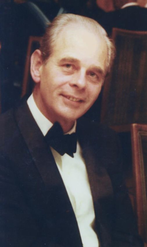
Free Neuropathology 2:10 (2021)                                                                     Arnulf H. Koeppen
doi: https://doi.org/10.17879/freeneuropathology-2021-3333                                                 page 4 of 6

discovery of supranuclear pseudoophthalmoplegia              cells of the pancreas. Many clinicians and FA re-
remains important but due to its variability can no          searchers consider FA a "neurodegenerative" dis-
longer be called a genetic marker. The Scottish ex-          ease, implying the existence of onset, progression,
perience (Koeppen, et al, 1977) prompted me to ap-           and death. The term neurodegenerative also means
ply to the Alexander-von-Humboldt Foundation for             atrophy, and clinical onset is thought to be related
a research fellowship. The Foundation agreed, and I          to a degree of atrophy that translates to nervous
moved to the University of Hamburg, Germany,                 system dysfunction. Neuropathologists, however,
where I found a warm welcome in the Department               will agree that FA is also a hypoplastic disease as far
of Human Genetics and the neuropathological insti-           as the spinal cord and dorsal root ganglia are con-
tute of the Eppendorf University Hospital. The plan          cerned. Sectioning of the spinal cord longitudinally
was to backtrack from autopsy records to surviving           to represent dorsal root entry zones as Obersteiner
family members. The director of Eppendorf neuro-             and Redlich did in 1894 in Vienna invariably showed
pathology was Prof. Hans-Joachim Colmant, a psy-             loss of demarcation between the peripheral nervous
chiatrist. He made available a huge archive of neu-          tissue of dorsal roots and the central nervous tissue
ropathological specimens in the basement of the              of the spinal cord (Koeppen, et al, 2017). A surprising
hospital that had survived the bombings of World             observation in FA was the protrusion of glial tissue
War II. The chief psychiatrist during the war, Prof.         from the spinal cord into dorsal roots, in support of
Hans Bürger-Prinz, decided that the collection of            dysfunction of a local barrier called the boundary
slides, reaching back to 1891, should be protected           cap. This work received the 2018 Stout award from
against the Allied bombings and moved the collec-            the United States and Canadian Academy of Pathol-
tion to a secure location. Beyond specimens of               ogy for “resolving a scientific medical problem by
ataxia, the collection also contained invaluable             studying its anatomic features”. This award is my
other cases, including Jakob's original 1920 speci-          most prized recognition.
mens of prion disease. It was an exciting experience
to study these slides and be allowed to make photo-          The mystery of brain iron
graphs. German neuropathologists favored Cresyl
violet as their routine stain, and it was apparent that           In 1967, I participated in the examination of a
this stain does not properly show the sponginess             brain that was incrusted by hemosiderin. The diag-
that is readily apparent on hematoxylin-and-eosin.           nosis, superficial siderosis, was not difficult, but the
My stay in Hamburg was intermittent, but I benefit-          disorder was thought to be rare, and magnetic reso-
ted from the collection at the Eppendorf Hospital            nance imaging with its extraordinary ability to de-
over an aggregate period of 2 years. On return to Al-        tect brain iron, was still to come. Dr. Kevin D. Barron
bany, NY, I began to collect autopsy specimens of            and I studied the specimen in detail, and I entered a
hereditary ataxia in a tissue repository and, to this        10-year period of research in normal and pathologi-
day, make available fixed and frozen samples to              cal brain iron (Koeppen et al, 1992-2002). This iron
other ataxia investigators.                                  work correlated well with my later studies in FA be-
                                                             cause other researchers thought that this disease
Another change in direction: From                            was due to iron accumulation in mitochondria of all
dominant to recessive ataxia, 1996-                          affected organs.
2021
                                                             America, 1964-2021
     After Dr. Massimo Pandolfo and his group dis-
covered the mutation in Friedreich ataxia (FA) in                  My fifty-seven years in the United States con-
1996, my interest changed, and my research now fo-           stitute two thirds of my life. My early youth was
cuses on this autosomal recessive disease. Though            troubled by the experience of World War II, America
FA is a monogenic disease, the pathology offers a            has been good to me. I waved goodbye to Europe
fascinating array of lesions in multiple organs: brain,      when the Maasdam, a passenger liner of the Hol-
spinal cord, dorsal root ganglia, sensory peripheral         land-America Line put out to sea in late January
nerves, eyes, heart, and the insulin-producing beta          1964, and I greeted the Statue of Liberty 10 days
Free Neuropathology 2:10 (2021)                                                                             Arnulf H. Koeppen
doi: https://doi.org/10.17879/freeneuropathology-2021-3333                                                         page 5 of 6

later. I was never homesick, but occasionally think of               continues to fascinate me, and, during my Chicago
my time as a refugee at the end of World War II. In                  years, the sky of the Midwest seemed higher and
January 1945, my mother packed some essentials                       bluer. I did not forget my European origin or my
and took me, two brothers, and a sister by bus to the                years of medical education in Germany. I am still tri-
west, leaving the then German province of Silesia. At                lingual (English, German, and Spanish) and enjoy the
that time, our father was a prisoner of war in an                    occasional opportunity to practice three languages.
American camp near Naples, Italy, and did not re-                    I also became an amateur radio operator (a “ham”
turn to Germany until 1947. Things did not stabilize                 in American lingo), and my last conversation with my
until 1949 when the family walked across the border                  late father was by ham radio. While on sabbatical in
between the newly established Communist German                       Hamburg, I received the call sign of DJ0RF wherein
Democratic Republic and the American zone of West                    the “DJ0” was reserved for foreign nationals who
Germany. Food, drink, and clothing were scarce, and                  had an amateur radio license in their home coun-
we spent one year in a displaced-persons camp. The                   tries. Ham radio will be my main pursuit after I finally
privations ended in 1950 when the German econ-                       retire from research or the practice of neurology and
omy improved with the outstanding help of the Mar-                   neuropathology.
shall Plan. After I became more established in the
United States, I raised a family and met many good                   A final word: Do we teach enough clini-
people. I want to thank my teachers of neurology
and neuropathology, among whom my friend and
                                                                     coanatomic correlation?
colleague for 47 years, the late Dr. Kevin D. Barron,
                                                                           We hear a lot from proponents of “evidence-
was the most important (figure 4). The United States
                                                                     based” medicine but rarely about the art of medi-
                                                                     cine. Clinicoanatomic correlation benefits from
                                                                     both, and thinking of brain structure while consider-
                                                                     ing a differential diagnosis is a rewarding exercise.
                                                                     Senior physicians will agree that anatomy and pa-
                                                                     thology are the basis of medicine. Telehealth has ar-
                                                                     rived and is perhaps not in line with the art of medi-
                                                                     cine. Its supporters may argue that it is more artful
                                                                     than face-to-face and hands-on medicine. Whatever
                                                                     we prefer as clinicians, we should strive to empha-
                                                                     size clinicoanatomic correlation in our teaching and
                                                                     convey the joy of a successful diagnosis based on
                                                                     traditional knowledge of anatomy. Such thought
                                                                     should, of course, precede the ordering of advanced
                                                                     imaging. In a recent survey of neurology training
                                                                     programs in the United States, 86% of program di-
                                                                     rectors considered a rotation through neuropathol-
                                                                     ogy essential (Rayi, et al, 2020). Let me add my voice
                                                                     to this goal.

Figure 4. Kevin D. Barron and former trainees. From left to right;
Suhandsu Chokroverty, MBBS; the late Kevin D. Barron, MD, the
late Cesar Mayo, MD; AHK; and the late Artemio Ordinario, MD.
The group photo was taken during the combined 1967 meeting
of the American Association of Neuropathologists and the
American Neurological Association in Atlantic City, New Jersey.
Free Neuropathology 2:10 (2021) Arnulf H. Koeppen
doi: https://doi.org/10.17879/freeneuropathology-2021-3333 page 6 of 6
References
Folch J, Lees M. Proteolipides, a new type of tissue lipoproteins. Their Koeppen A.H., Dickson A.C., Chu R.C., Thach R.E.: The pathogenesis of
isolation from brain. J Biol Chem 1951; 191 807-17 superficial siderosis of the central nervous system. Ann Neurol1993; 34:
646-53
Hudson L.D., Puckett C., Berndt .J, Chan J., Gencic S. Mutation of the
proteolipid protein (PLP) gene in human X-linked myelin disorder. Proc Koeppen A.H.: The history of iron in the brain. J Neurol Sci 1995; 134
Natl Acad Sci 1989; 86: 8128-31 (Suppl.): 1-9
Koeppen A.H.: (transl.) The influence of pharmacological agents of the Koeppen A.H.: A brief history of brain iron research. J Neurol Sci 2002;
autonomic nervous system on the cellular nuclei of the liver, uterus, and 207: 95-7
adrenal cortex measured by karyometric methods). Inaugural
dissertation of University of Göttingen Medical School, 1963 Koeppen A.H., Becker A.B., Qian J., Gelman B.B., Mazurkiewicz J.E.:
Friedreich ataxia: Developmental failure of the dorsal root entry zone. J
Koeppen A.H., Hans M.B., Shepherd D.I., Best P.V.: Adult-onset Neuropathol Exp Neurol 2017; 76 (11): 969-77
hereditary ataxia in Scotland. Arch Neurol 1977; 34: 611-18
Mitzen E.J., Koeppen A.H.: Malonate, malonyl-coenzyme A and acetyl-
Koeppen A.H., Ronca N.A., Greenfield E.A., Hans M.B.: Defective coenzyme A in developing rat brain. J Neurochem 1984; 43: 499-506
biosynthesis of proteolipid protein in Pelizaeus-Merzbacher disease.
Ann Neurol 1987; 21: 159-70 Obersteiner H., Redlich E. (transl): On the nature and pathogenesis of
the tabetic dorsal column degeneration. Arbeit Neurol Inst Wien Univ
Koeppen A.H., Barron K.D., Csiza C.K., Greenfield E.A.: Comparative 1894; 1-3: 158-72
immunocytochemistry of Pelizaeus-Merzbacher disease, the jimpy
mouse, and the myelin-deficient rat. J Neurol Sci 1988; 84: 315-27 Pelizaeus F.: (transl) On a peculiar form of spastic paralysis with brain
symptoms on hereditary basis (multiple sclerosis). Arch Psychiat
Koeppen A.H., Borke R.C.: Experimental superficial siderosis of the Nervenkrankh 1885; 16: 698-710
central nervous system. I. Morphological observations, J Neuropathol
Exp Neurol.1991; 50: 579-9 Rayi A. et al.: How are residents trained in neuropathology? A survey of
neurology program directors in the United States. J Neuropathol Exp
Koeppen A.H., Hurwitz C.G., Dearborn R.E., Dickson A.C., Borke R.C., Chu Neureol 2020; 79:1218-22
R.C.: Experimental superficial siderosis of the central nervous system:
biochemical correlates. J Neurol Sci 1992; 112: 38-45You can also read