Transfer RNA derived fragment tRF 28 QSZ34KRQ590K in plasma exosomes may be a potential biomarker for atopic dermatitis in pediatric patients
←
→
Page content transcription
If your browser does not render page correctly, please read the page content below
EXPERIMENTAL AND THERAPEUTIC MEDICINE 21: 489, 2021

           Transfer RNA‑derived fragment tRF‑28‑QSZ34KRQ590K
               in plasma exosomes may be a potential biomarker
                    for atopic dermatitis in pediatric patients
                          LI MENG1*, LONG JIANG2*, JIAN CHEN1, HONGJIN REN1, ZHIQIN GAO1,
                                     FEI WU3, YIYANG WEN1 and LIANJUAN YANG1

                Departments of 1Dermatological Mycology, 2Dermatological Surgery and 3Dermatological Pathology,
                Shanghai Skin Disease Hospital, Tongji University School of Medicine, Shanghai 200443, P.R. China

                                        Received July 6, 2020; Accepted January 28, 2021

                                                  DOI: 10.3892/etm.2021.9920

Abstract. Atopic dermatitis (AD) is a common chronic               may be a potential biomarker for pediatric patients with AD.
relapsing inflammatory disease. There is substantial evidence      The present study enhanced the understanding of the patho‑
suggesting that noncoding RNAs have indispensable roles            genesis of AD and provided a novel marker for the diagnosis
in the pathogenesis of AD. Exosomal transfer RNA‑derived           and targeted treatment of AD.
fragments (tRFs) have been identified as potential biomarkers
for various disorders. However, the role of tRFs in AD has         Introduction
remained to be elucidated, which was thus the aim of the present
study. Plasma samples from 23 pediatric patients with AD and       Atopic dermatitis (AD) is a common dermatological disease
23 healthy controls were collected. Exosomes were success‑         with prevalence rates of 15‑30% in children and 2‑10% in
fully isolated from plasma according to the manufacturer's         adults (1). The combination of genetic, immune and environ‑
protocol. Small RNA sequencing was performed in 3 patients         mental stimuli appears to contribute to the manifestation of
with AD and 3 controls, and 135 significantly differentially       AD. Hereditary susceptibility is considered a primary factor.
expressed plasma exosomal tRFs were identified, including          To elucidate the genetic factors of AD, multiple genetic studies
36 upregulated and 99 downregulated tRFs. Using the                have been performed and a series of susceptibility genes/loci
miRanda and RNAhybrid databases, 58,227 target genes of            have been identified, which indicate two major biological
these 135 differentially expressed tRFs were predicted. Gene       pathways responsible for the etiology of AD, including skin
ontology and Kyoto Encyclopedia of Genes and Genomes               epithelial dysfunction and innate/adaptive immune response
pathway analyses suggested that these target genes of tRFs         dysregulation, which affect each other (2). However, its exact
are involved in multiple functions and pathways associated         pathogenesis has remained largely elusive.
with AD. Downregulation of tRF‑28‑QSZ34KRQ590K and                     Exosomes are thought to have important roles in intercel‑
tRF‑33‑Q99P9P9NH57SD3 were validated in 20 patients                lular communication and have been proven to be potential
with AD and 20 controls by reverse transcription‑quantitative      markers for numerous diseases, such as tumours (3) and renal
PCR and tRF‑28‑QSZ34KRQ590K exhibited significance                 disease (4). Along with the identification of RNA in exosomes
in the receiver operating characteristic curve analysis.           in 2007 and the development of high‑throughput techniques
The present study was the first to provide a tRF profile in AD     for nucleic acid analyses, there have been a growing number
and implied that plasma exosomal tRF‑28‑QSZ34KRQ590K               of studies on RNA sequences in exosomes (5). One class of
                                                                   small noncoding (snc)RNAs, transfer RNA‑derived fragments
                                                                   (tRFs), which are generated from transfer RNA (tRNA), have
                                                                   been suggested to have roles in the regulation of gene expres‑
                                                                   sion, cell proliferation, RNA processing, modulation of the
Correspondence to: Professor Lianjuan Yang, Department             DNA damage response, priming of viral reverse transcriptase,
of Dermatological Mycology, Shanghai Skin Disease Hospital,        neurodegeneration and tumour suppression (6). tRFs have
Tongji University School of Medicine, 1278 Baode Road, Jingan,
                                                                   been implicated in various infections, such as human T‑cell
Shanghai 200443, P.R. China
E‑mail: lianjuanyang@163.com
                                                                   leukaemia virus type 1 (7) and HIV‑1‑infected cells (8).
                                                                   Different tRFs have been identified in cellular proliferation
*
    Contributed equally                                            and tRF signatures have been described in myelodysplastic
                                                                   syndromes (9) and various cancer types, such as prostate
Key words: atopic dermatitis, biomarker, plasma, exosome,          cancer (10) and squamous cell carcinoma (11). These studies
sequence, transfer RNA‑derived fragments                           highlighted the potential role of tRFs in disease progres‑
                                                                   sion. A database of tRFs has been developed where each
                                                                   unique tRF has been given a name following the progress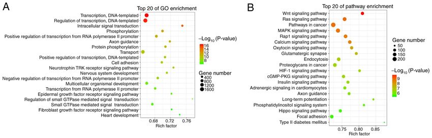
2                                        MENG et al: ROLES OF tRFs IN ATOPIC DERMATITIS

of high‑throughput sequencing and analyses of small tRNA            PBS at room temperature; ix) 10 µl phosphotungstic acid was
fragments (12). According to their mapped positions on the          dropped on a copper net, precipitated for 1 min, before the
primary or mature tRNA transcript, tRFs may be classified           floating liquid was absorbed with filter paper at room tempera‑
into various types, such as tRNA halves (tiRNA/tiR) (13),           ture; and x) this was fried at room temperature for 2 min,
tRF‑1, tRF‑3, tRF‑5 (14), and endogenous tRFs (i‑tRFs) (15).        before TEM was performed (JEM‑1200EX, Japan Electronics
    However, to the best of our knowledge, no previous study        Corporation).
has characterized tRFs in AD. In the present study, the possible
roles of tRFs in AD were explored, which may provide novel          RNA extraction, small RNA library preparation and
information regarding the pathogenesis of AD.                       sequencing. Total RNA from plasma exosomes was extracted
                                                                    with TRIzol reagent (Invitrogen; Thermo Fisher Scientific,
Materials and methods                                               Inc.). Subsequently, purified RNAs were sent to Yingbio for
                                                                    constructing small RNA libraries and performing small RNA
Ethics statement and human specimens. This study was                sequencing analysis. In brief, the small RNA was bound with
approved by the ethics committee of Shanghai Skin Disease           3'‑ and 5'‑adapters and complementary (c)DNA constructs
Hospital (Shanghai, China) and was conducted according              were created by reverse transcription (RT) followed by PCR.
to the principles of the Declaration of Helsinki. A total of        The small RNA fragments (15‑40 nt) were excised and purified
23 pediatric patients with AD meeting the Hanifin‑Rajka             and the purified libraries were quantified and validated. Small
diagnostic criteria (16) and 23 healthy controls were enrolled      RNA sequencing was performed on an Illumina HiSeq 2500
from Shanghai Skin Disease Hospital (Shanghai, China)               (Illumina, Inc.) (18).
between December 1, 2017 and October 1, 2018 in the present
study. All controls were evaluated by experienced doctors and       Data analysis. Low‑quality reads and short reads (1 or
EXPERIMENTAL AND THERAPEUTIC MEDICINE 21: 489, 2021 3
Table I. Clinical features of subjects in the AD and control a complete membrane structure (Fig. S1), which indicated the
groups. successful isolation of exosomes from human plasma.
Item AD group (n=23) Control group (n=23) Sequencing of tRFs from plasma exosomes. A total of
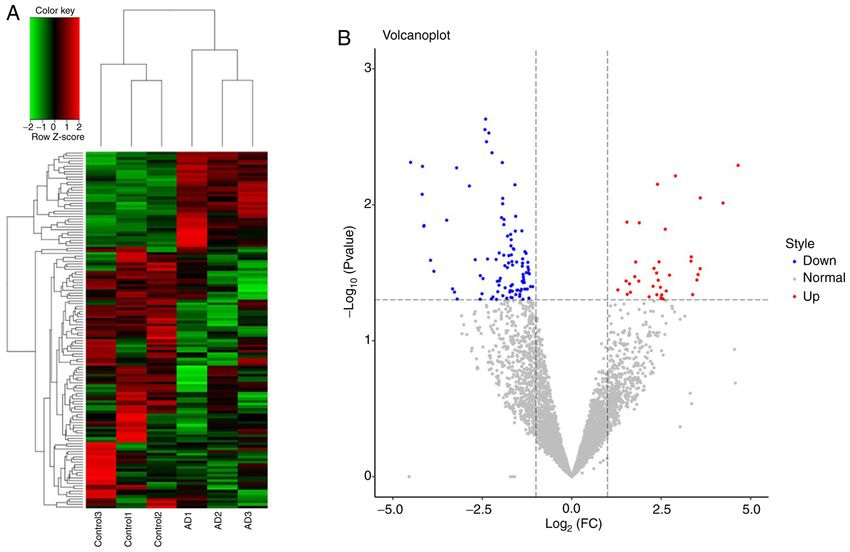
4,637 tRFs were identified between 3 patients with AD and
Age (years) 9.5 (3‑13) 9.0 (6‑12) 3 controls. Of these tRFs, 135 were significantly differentially
Sex (female/male) 12/11 10/13 expressed, including 36 upregulated and 99 downregulated
SCORAD ‑ tRFs (Fig. 1); the details are provided in Table SI. A total of
Mild 7 ‑ 6 types of tRFs were identified, namely tRF‑1, tRF‑3, tRF‑5,
Moderate 10 ‑ 3'‑halves, 5'‑halves and i‑tRF (Table SII).
Severe 6 ‑
Target gene prediction and functional analysis of differentially
Values are expressed as mean (range) or n. As the control group, expressed tRFs. Analyses with the miRanda and RNAhybrid
subjects with no atopic disease history in individuals and three gener‑ databases identified 58,227 target genes associated with 135
ations of immediate family members were used. As an indicator of differentially expressed tRFs. GO analysis revealed that these
AD severity, the SCORAD system was used as follows: 0~24, mild; target genes were functionally enriched in transcription, DNA
25‑50, moderate; 51‑103, severe. SCORAD, SCORing atopic derma‑ templating, regulation of transcription, intracellular signal
titis; AD, atopic dermatitis.
transduction, transport and epidermal growth factor receptor
signalling pathway (Fig. 2A). KEGG pathway analysis
revealed that these target genes were enriched in the Wnt
signalling pathway, Ras signalling pathway, MARK signalling
on an ABI Q6 detection system (Applied Biosystems; Thermo pathway, Rap1 signalling pathway, calcium signalling pathway,
Fisher Scientific, Inc.). The thermocycling conditions were as endocytosis and hypoxia‑inducible factor (HIF)‑1 signalling
follows: 95˚C for 10 min; followed by 95˚C for 15 sec, then to pathway (Fig. 2B).
60˚C for 60 sec, for 45 cycles. U6 was selected as the internal
control. The PCR specific primer sequences were as follows: Validation of differentially expressed tRFs. To further
tRF‑33‑Q99P9P9NH57SD3 forward 5'‑GCAG GCT TCTGT confirm the tRFs, 10 differentially expressed tRFs, including
AGTGTAGTGGTTA‑3' and reverse, 5'‑AGTG CGTGTCGT 6 downregulated tRFs (tRF‑34‑YKWIR959MIL0HV,
GGAGTCG ‑3'; tRF‑28‑QSZ34KRQ590K forward, 5'‑AGG tRF‑34‑LZNL7L73V6M9I3, tRF‑28‑QSZ34KRQ590K,
CTCGTTG GTC TAG GGGTA‑3' and reverse, 5'‑AGTG CG tRF‑33‑Q99P9P9NH57SD3, tRF‑36‑Q99P9P9NH57S36D
TGTCGTGGAGTCG‑3'; and U6 forward, 5'‑CGATACAGA and tRF‑37‑Q99P9P9NH57S362) and 4 upregulated tRFs
GAAGATTAGCATG GC‑3' and reverse, 5'‑AACG CTTCA (tR F‑28‑D89NJ4S2I7DM, tR F‑30‑PNR8YO9LON4V,
CGAATTTGCGT‑3'. The 2‑ΔΔCq method was used to quantify tRF‑22‑79MP9PMNI and tsRNA‑1042), were subjected
the tRFs (19). to RT‑qPCR analysis in plasma exosomes from 3 patients
with AD and 3 controls. A total of four tRFs, namely
Statistical analysis. Spearman's nonparametric correla‑ tRF‑28‑QSZ34KRQ590K, tRF‑33‑Q99P9P9NH57SD3,
tion analysis was used to analyse the correlation between tRF‑37‑Q99P9P9NH57S362 and tsRNA‑1042, exhibited
the expression of tRFs and disease severity using SPSS 23 differential expression (P= 0.037, 0.030, 0.047 and 0.041
(IBM Corp.). The predictive value of biomarkers regarding respectively; Fig. 3A). Of these, tRF‑28‑QSZ34KRQ590K
the probability of AD was determined by receiver operating and tRF‑33‑Q99P9P9NH57SD3 were further selected to be
characteristic (ROC) curve analysis, which was performed validated by RT‑qPCR in 20 patients with AD and 20 controls.
using MedCalc software (version 10.4.7.0; MedCalc). The The results indicated that the expression of both of the tRFs
area under the ROC curve (AUC) was calculated to evaluate exhibited significant differences between the AD and control
the diagnostic potential of tRFs for AD. The optimal diag‑ groups (P4 MENG et al: ROLES OF tRFs IN ATOPIC DERMATITIS
Figure 1. (A) Hierarchical clustering of differentially expressed tRFs between the AD (n=3) and control groups (n=3). The colour scale represents high (red)
to medium (black) to low (green) relative expression. (B) Volcano plot displaying the differentially expressed tRFs displaying significantly upregulated tRFs
(red), significantly downregulated tRFs (blue) and nonsignificant tRFs (grey). AD, atopic dermatitis; tRFs, transfer RNA‑derived fragments; FC, fold change.
Figure 2. Bubble graphs of (A) GO and (B) KEGG pathway enrichment analyses each displaying 20 identified top pathways for target genes of differentially
expressed tRFs (larger circles represent a larger number of genes). The colour scale represents low (green) to high (red) P‑values. tRFs, transfer RNA‑derived
fragments; GO, Gene Ontology; KEGG, Kyoto Encyclopedia of Genes and Genomes.
Discussion in plasma exosomes from patients with AD was performed.
Exosomes were isolated successfully from plasma with
Exosomes are thought to have important roles in intercellular combined centrifugation and were then further identified by
communication and have been proven to be potential markers TEM. A total of 135 differentially expressed plasma exosomal
for numerous diseases. The discovery of tRFs in exosomes tRFs were identified in 3 subjects with AD and 3 controls.
and their importance in gene regulation have substantially GO and KEGG pathway enrichment analyses revealed a
promoted an increase in their study in recent years. They series of the possible functions and pathways of the target
are closely related to the occurrence of numerous human genes of these 135 differentially expressed tRFs. The down‑
diseases and have the potential to become novel biomarkers regulation of plasma exosomal tRF‑28‑QSZ34KRQ590K and
for diseases (20). In the present study, the first analysis of tRFs tRF‑33‑Q99P9P9NH57SD3 was further verified by RT‑qPCREXPERIMENTAL AND THERAPEUTIC MEDICINE 21: 489, 2021 5 Figure 3. Verification of tRFs and ROC curve analysis. (A) Verification of 10 tRFs in 3 subjects with AD and 3 controls by RT‑qPCR. (B) Verification of 2 tRFs in 20 patients with AD and 20 controls by RT‑qPCR. *P
6 MENG et al: ROLES OF tRFs IN ATOPIC DERMATITIS
be associated with inflammation in previous studies and the treatment of AD in the future. However, further studies are
inflammatory response has also been suggested to have roles required to better understand the roles of tRFs in AD.
in the pathogenesis of AD (25). Therefore, the present study
implied that plasma exosome tRFs may participate in the Acknowledgements
pathogenesis of AD by being involved in the inflammatory
response and the epidermal growth factor receptor signalling Not applicable.
pathway. However, further functional studies are required to
better understand the pathogenesis of AD. Funding
tRF‑28‑QSZ34KRQ590K demonstrated a significant
diagnostic value for AD in the ROC curve analysis. There This work was supported by the National Natural Science
were 28 target genes of tRF‑28‑QSZ34KRQ590K, including Foundation of China (grant nos. 81602744 and 81573063).
tachykinin receptor 1, RANBP2‑like and GRIP domain‑
containing 1, non‑SMC condensin I complex subunit H Availability of data and materials
(NCAPH), ectodysplasin‑A receptor, contactin‑associated
protein family member 5, cytochrome P450 family (CYP)27 The raw data of small RNA sequencing of plasma exosomes has
subfamily C member 1, WD repeat domain 33, POTE ankyrin been uploaded to the sequence read archive (http://www.ncbi.
domain family member F, insulin receptor substrate 1, thyroid nlm.nih.gov/bioproject/609458; submission ID: SUB7071247;
hormone receptor interactor 12, S‑antigen visual arrestin, BioProject accession no. PRJNA609458). The raw data will be
ATPase plasma membrane Ca2+ transporting 2, neurexophilin publicly available on March 1, 2021.
and PC‑esterase domain family member 3, Rho GTPase acti‑
vating protein 31, p21 (RAC1)‑activated kinase 2, midnolin, Authors' contributions
unc‑13 homolog A, potassium channel tetramerization domain
containing 15, endogenous retrovirus group V member 1 LM and LJ involved in drafting the manuscript and revising
envelope, zinc finger and SCAN domain containing 18, it critically for important intellectual content, and have made
KIAA0930 (also known as C22orf9, LSC3), SET binding substantial contributions in the conception and design of the
factor 1, forkhead box K1, AVL9 cell migration‑associated, current study. JC, HR, ZG, YW and FW were involved in the
FKBP prolyl isomerase family member 6, X‑ray repair cross acquisition of data, analysis and interpretation of data. LY was
complementing 2, phosphatase and actin regulator 1 and responsible for the conception and design of the current study
mitochondrial calcium uniporter regulator 1. A previous study and manuscript revision. LM and LY confirm the authenticity
reported on a pediatric patient with AD, mental retardation, of all the raw data. All authors read and approved the final
autistic features, epilepsy, developmental delay and abnormal manuscript.
immunological results, who carried a 7.9 Mb de novo
deletion of chromosome 22q13.2/qter, a region containing the Ethics approval and consent to participate
NCAPH2, SH3 and multiple ankyrin repeat domains 3 and
CYP2D6 genes (26). These genes are associated with the T‑cell This study was approved by the ethics committee of Shanghai
immune response and the inflammatory response has also Skin Disease Hospital (Shanghai, China) and all subjects and
been suggested to have roles in the pathogenesis of AD. Thus, their guardians provided written informed consent.
tRF‑28‑QSZ34KRQ590K may be involved in the pathogenesis
of AD by affecting the inflammatory response. Further studies Patient consent for publication
are required to uncover the role of tRF‑28‑QSZ34KRQ590K
in the pathogenesis of AD. At present, it remains undeter‑ Not applicable.
mined whether tRF‑28‑QSZ34KRQ590K is specifically
expressed in AD, although it has not been reported to be Competing interests
expressed in any other diseases, to the best of our knowledge.
However, tRF‑28‑QSZ34KRQ590K exhibited a significant The authors declare that they have no competing interests.
diagnostic value in the ROC curve analysis, which suggested
that tRF‑28‑QSZ34KRQ590K may be a potential biomarker References
for pediatric patients with AD, although further studies are
required to confirm the role of this tRF in AD. 1. Abrams EM and Sicherer S: Cutaneous sensitization to peanut
In conclusion, in the present study, 135 differentially in children with atopic dermatitis: A window to prevention of
peanut allergy. JAMA Dermatol 155: 13‑14, 2018.
expressed tRFs in the plasma exosomes of pediatric patients 2. Bin L and Leung DY: Genetic and epigenetic studies of atopic
with AD were identified, and the significantly downregulated dermatitis. Allergy Asthma Clin Immunol 12: 52, 2016.
tRF‑28‑QSZ34KRQ590K and tRF‑33‑Q99P9P9NH57SD3 3. Guescini M, Genedani S, Stocchi V and Agnati LF: Astrocytes
and Glioblastoma cells release exosomes carrying mtDNA.
were further verified. tRF‑28‑QSZ34KRQ590K exhibited J Neural Transm (Vienna) 117: 1‑4, 2010.
significance in the ROC curve analysis. Thus, tRF‑28‑QSZ‑ 4. Miranda KC, Bond DT, McKee M, Skog J, Paunescu TG,
Da Silva N, Brown D and Russo LM: Nucleic acids within
34KRQ590K may be a potential biomarker for AD in pediatric urinary exosomes/microvesicles are potential biomarkers for
patients. To the best of our knowledge, the present study was renal disease. Kidney Int 78: 191‑199, 2010.
the first to report on the roles of tRFs in AD. It provided novel 5. Valadi H, Ekstrom K, Bossios A, Sjostrand M, Lee JJ and
Lotvall JO: Exosome‑mediated transfer of mRNAs and
biological information for uncovering the pathogenesis of AD microRNAs is a novel mechanism of genetic exchange between
and a novel potential source for the early diagnosis and targeted cells. Nat Cell Biol 9: 654‑659, 2007.EXPERIMENTAL AND THERAPEUTIC MEDICINE 21: 489, 2021 7
6. Kumar P, Kuscu C and Dutta A: Biogenesis and function of 17. Severity scoring of atopic dermatitis: The SCORAD index.
transfer RNA‑related fragments (tRFs). Trends Biochem Sci 41: Consensus report of the European task force on atopic dermatitis.
679‑689, 2016. Dermatology 186: 23‑31, 1993.
7. Ruggero K, Guffanti A, Corradin A, Sharma VK, De Bellis G, 18. Zhang L, Liu S, Wang JH, Zou J, Zeng H, Zhao H, Zhang B,
Corti G, Grassi A, Zanovello P, Bronte V, Ciminale V and He Y, Shi J, Yoshida S and Zhou Y: Differential expressions of
D'Agostino DM: Small noncoding RNAs in cells transformed by microRNAs and transfer RNA‑derived Small RNAs: Potential
human T‑cell leukemia virus type 1: A role for a tRNA fragment targets of choroidal neovascularization. Curr Eye Res 44:
as a primer for reverse transcriptase. J Virol 88: 3612‑3622, 2014. 1226‑1235, 2019.
8. Yeung ML, Bennasser Y, Watashi K, Le SY, Houzet L and 19. Mazzei M, Vascellari M, Zanardello C, Melchiotti E, Vannini S,
Jeang KT: Pyrosequencing of small non‑coding RNAs in HIV‑1 Forzan M, Marchetti V, Albanese F and Abramo F: Quantitative
infected cells: Evidence for the processing of a viral‑cellular real time polymerase chain reaction (qRT‑PCR) and RNAscope
double‑stranded RNA hybrid. Nucleic Acids Res 37: 6575‑6586, in situ hybridization (RNA‑ISH) as effective tools to diagnose
2009. feline herpesvirus‑1‑associated dermatitis. Vet Dermatol 30:
9. Guo Y, Bosompem A, Mohan S, Erdogan B, Ye F, Vickers KC, e491‑e147, 2019.
Sheng Q, Zhao S, Li CI, Su PF, et al: Transfer RNA detection 20. Shen Y, Yu X, Zhu L, Li T, Yan Z and Guo J: Transfer RNA‑derived
by small RNA deep sequencing and disease association with fragments and tRNA halves: Biogenesis, biological functions and
myelodysplastic syndromes. BMC Genomics 16: 727, 2015. their roles in diseases. J Mol Med 96: 1167‑1176, 2018.
10. Martens‑Uzunova ES, Jalava SE, Dits NF, van Leenders GJ, 21. Soares AR and Santos M: Discovery and function of transfer
Moller S, Trapman J, Bangma CH, Litman T, Visakorpi T and RNA‑derived fragments and their role in disease. Wiley
Jenster G: Diagnostic and prognostic signatures from the small Interdiscip Rev RNA: 8, 2017 doi: 10.1002/wrna.1423.
non‑coding RNA transcriptome in prostate cancer. Oncogene 31: 22. Zhang Z, Xiao C, Gibson AM, Bass SA and Khurana Hershey GK:
978‑991, 2012. EGFR signaling blunts allergen‑induced IL‑6 production and
11. Victoria Martinez B, Dhahbi JM, Nunez Lopez YO, Lamperska K, Th17 responses in the skin and attenuates development and
Golusinski P, Luczewski L, Kolenda T, Atamna H, Spindler SR, relapse of atopic dermatitis. J Immunol 192: 859‑866, 2014.
Golusinski W and Masternak MM: Circulating small non‑coding 23. Wu A, Chen H, Xu C, Zhou J, Chen S, Shi Y, Xu J, Gan J and
RNA signature in head and neck squamous cell carcinoma. Zhang J: miR‑203a is involved in HBx‑induced inflammation by
Oncotarget 6: 19246‑19263, 2015. targeting Rap1a. Exp Cell Res 349: 191‑197, 2016.
12. Kumar P, Mudunuri SB, Anaya J and Dutta A: tRFdb: A database 24. Kyriakis JM and Avruch J: Mammalian MAPK signal transduc‑
for transfer RNA fragments. Nucleic Acids Res 43: D141‑D145, tion pathways activated by stress and inflammation: A 10‑year
2015. update. Physiol Rev 92: 689‑737, 2012.
13. Thompson DM and Parker R: Stressing out over tRNA cleavage. 25. Hirota T, Takahashi A, Kubo M, Tsunoda T, Tomita K,
Cell 138: 215‑219, 2009. Sakashita M, Yamada T, Fujieda S, Tanaka S, Doi S, et al:
14. Lee YS, Shibata Y, Malhotra A and Dutta A: A novel class of Genome‑wide association study identifies eight new suscepti‑
small RNAs: tRNA‑derived RNA fragments (tRFs). Genes bility loci for atopic dermatitis in the Japanese population. Nat
Dev 23: 2639‑2649, 2009. Genet 44: 1222‑1226, 2012.
15. Goodarzi H, Liu X, Nguyen HC, Zhang S, Fish L and Tavazoie SF: 26. Chen CP, Lin SP, Chern SR, Tsai FJ, Wu PC, Lee CC,
Endogenous tRNA‑derived fragments suppress breast cancer Chen YT, Chen WL and Wang W: A de novo 7.9 Mb deletion
progression via YBX1 displacement. Cell 161: 790‑802, 2015. in 22q13.2→qter in a boy with autistic features, epilepsy, devel‑
16. Rajka G and Hanifin JM: Diagnostic features of atopic opmental delay, atopic dermatitis and abnormal immunological
dermatitis. Acta Dermato‑venereologica 60: 44‑47, 1980. findings. Eur J Med Genet 53: 329‑332, 2010.You can also read