Glycogen Storage Disease Ib and Severe Periodontal Destruction: A Case Report - MDPI
←
→
Page content transcription
If your browser does not render page correctly, please read the page content below
dentistry journal
Case Report
Glycogen Storage Disease Ib and Severe Periodontal
Destruction: A Case Report
Rui Ma 1 , Fardad Moein Vaziri 2 , Gregory J. Sabino 3 , Nima D. Sarmast 4, * , Steven M. Zove 5 ,
Vincent J. Iacono 5 and Julio A. Carrion 5
 1    Private Practice, 1047 Old Post Road, Fairfield, CT 06824, USA; rui.ma.dmd@gmail.com
 2    210-11808 Saint Albert Trail, Edmonton, AB T5L 4G4, Canada; fardadm@gmail.com
 3    Stony Brook University School of Dental Medicine, South Drive, Stony Brook, NY 11794, USA;
      greg.sabino@gmail.com
 4    Department of Periodontics and Dental Hygiene, The University of Texas School of Dentistry at Houston,
      7500 Cambridge Street, Suite 6427, Houston, TX 77054, USA
 5    Department of Periodontology, Stony Brook University School of Dental Medicine, South Drive,
      Stony Brook, NY 11794, USA; steven.zove@stonybrookmedicine.edu (S.M.Z.);
      vincent.iacono@stonybrookmedicine.edu (V.J.I.); julio.carrion@stonybrookmedicine.edu (J.A.C.)
 *    Correspondence: nima.d.sarmast@uth.tmc.edu; Tel.: +1-713-486-4387; Fax: +1-713-486-4393

 Received: 1 August 2018; Accepted: 28 September 2018; Published: 3 October 2018

 Abstract: Background: Glycogen storage diseases (GSDs) are genetic disorders that result from
 defects in the processing of glycogen synthesis or breakdown within muscles, liver, and other
 cell types. It also manifests with impaired neutrophil chemotaxis and neutropenic episodes
 which results in severe destruction of the supporting dental tissues, namely the periodontium.
 Although GSD Type Ib cannot be cured, associated symptoms and debilitating oral manifestations
 of the disease can be managed through collaborative medical and dental care where early detection
 and intervention is of key importance. This objective of the case report was to describe a child
 with GSD Ib and its associated oral manifestations with microbial, immunological and histological
 appearances. Case Presentation: An eight-year-old Hispanic male with a history of GSD type Ib
 presented with extensive intraoral generalized inflammation of the gingiva, ulcerations and bleeding,
 and intraoral radiographic evidence of bone loss. Tannerella forsythia was readily identifiable from
 the biofilm samples. Peripheral blood neutrophils were isolated and a deficient host response
 was observed by impaired neutrophil migration. Histological evaluation of the soft and hard
 tissues of the periodontally affected primary teeth showed unaffected dentin and cementum.
 Conclusions: This case illustrates the association between GSD Ib and oral manifestations of the
 disease. A multi-disciplinary treatment approach was developed in order to establish healthy intraoral
 conditions for the patient. Review of the literature identified several cases describing GSD and its
 clinical and radiographic oral manifestations; however, none was identified where also microbial,
 immunological, and histological appearances were described.

 Keywords: glycogen storage disease; neutrophils; chemotaxis; periodontitis; oral manifestations

1. Introduction
     Glycogen storage diseases (GSDs) are genetic disorders that result from defects in the processing
of glycogen synthesis or breakdown within muscles, liver, and other cell types [1]. It is estimated
to occur in 1 per 20,000 to 25,000 births in the United States. There are at least 10 different types
of GSDs known today. GSD I is a rare autosomal recessive disorder that leads to deficiencies of
glucose-6-phosphatase catalytic activity (Type Ia) and glucose-6-phosphate translocase (Type Ib) [2].

Dent. J. 2018, 6, 53; doi:10.3390/dj6040053                                   www.mdpi.com/journal/dentistry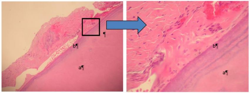
Dent. J. 2018, 6, 53 2 of 6
Clinical manifestations, such as growth retardation [3,4], short stature, doll-like face with fat cheeks,
protuberant abdomen and hepatomegaly (due to abnormal glycogen accumulation) [5], inflammatory
bowel disease [6], thyroid autoimmunity, and renal disease [7], have been observed and reported in the
literature. In addition, patients with GSD Type Ib can also develop neutropenia, as well as impaired
neutrophil function, which leads to an increased frequency and severity of bacterial infections [8].
Evidence also suggests that the neutropenia in those with GSD Ib may be caused by increased apoptosis
and migration of the neutrophils to inflamed tissues rather than by impairment in maturation [9].
In the oral cavity the neutrophil appears to perform an important role in protecting the periodontal
tissues from invasion by pathogenic bacteria resident in the dental biofilm [10].
2. Case Presentation
An eight-year-old Hispanic male presented to the Stony Brook Dental Care Center with a history
of GSD type Ib. Oral manifestations of the GSD Ib disease were observed and recorded upon the dental
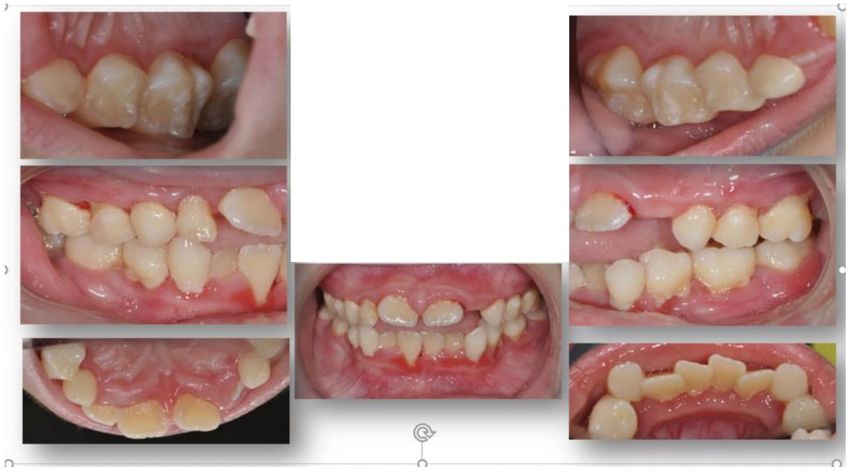
and radiographic examination. Overall, the patient presented with extensive generalized inflammation
of the gingiva, erythema, ulceration, and generalized deep periodontal pocketing with bleeding on
probing (Figure 1). Generalized severe horizontal bone loss was noted radiographically (Figure 2).
Informed consent for treatment was obtained.
Microbial samples were taken with sterile paper points at various primary and permanent teeth to
demonstrate the periodontal pathogen distribution [11]. A blood sample was drawn in order to study
systemic neutrophil migration. Peripheral blood neutrophils were isolated according to a standard
protocol [12] and suspended in HBSS + 10 mM HEPES (pH 7.4) and 1% BSA. A 48-well Boyden
chamber apparatus (Neuro Probe, Inc., Gaithersburg, MD, USA) was arranged so that 20 nM of CXCL1
(R&D Systems, Minneapolis, MN, USA), 20 nM of CXCL8 (R&D Systems), or HBSS + 10 mM HEPES
(pH 7.4) and 1% BSA was added as the chemoattractant or control in the bottom portion of the chamber.
A 5-µm 35 cellulose nitrate filter (Neuro Probe, Gaithersburg, MD, USA) was placed between the two
halves of the Boyden chamber. Neutrophils in a volume of 50 µL, at no more than 4 × 10⁶ cells/mL,
were loaded into the top chamber and allowed to migrate for 15 min at 37 ◦ C. The filter was fixed
in 100% 2-propanol, stained with Harris-type hematoxylin, clarified with xylene, and mounted for
analysis. The distance that neutrophils traveled into the filter was measured using the leading-front
method via bright-field microscopy. The microbial composition of the oral biofilm was characterized by
multiplex PCR. 16S rRNA gene was used as the primers in PCR. Sterilized deionized water was used
as negative control. Of the common putative periodontal pathogens, Tannerella forsythia was readily
identifiable from the biofilm samples (Table 1, Figure 3). In addition, a deficient host response was
observed by impaired neutrophil migration in response to the chemokines CXCL1 and CXCL8 (Figure 4).
Histological evaluation [13] of the soft and hard tissues of the periodontally affected primary teeth showed
a normal attachment apparatus, including bone, cementum, and periodontal ligament (Figure 5).
Based on the clinical findings and the understanding of the disease, a treatment plan
was developed collaboratively with the Departments of Orthodontics, Pediatric Dentistry,
and Periodontology. All remaining primary teeth had a hopeless prognosis and it was elected to
proceed with extractions after obtaining informed consent. No postoperative infections or bleeding
were reported or observed. In order to preserve the space for the remaining succedaneous teeth,
a nance appliance and lower lingual holding arch were fabricated for the maxillary and mandibular
dentitions, respectively. A two to three month recall interval for dental examinations and preventative
care has been recommended for this patient [14]. Patient was not followed up in this case report after
immediate post-operative treatment course.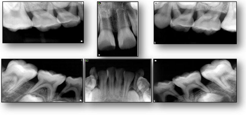
Dent. J. 2018, 6, 53                                                                                          3 of 6

      Figure 1. Clinical oral presentation of an eight-year-old male patient with a history of GSD type Ib.
      Extensive oral inflammation of the supporting periodontal tissues.

      Figure 2. Radiographic presentation of an eight-year-old with a history of GSD type Ib. Radiographic
      examination reveals severe horizontal bone loss involving the remaining primary dentition.

                             Figure 3. Tannerella forsythia distribution by tooth site.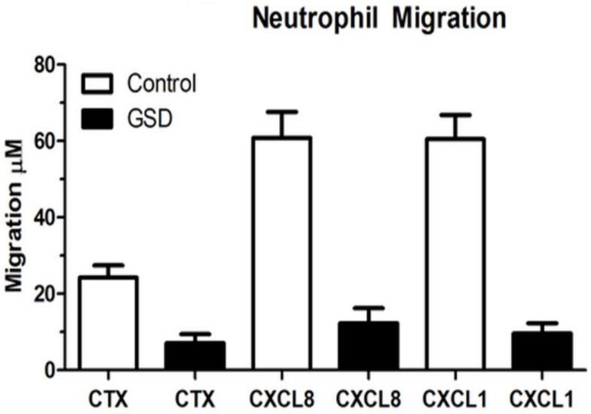
Dent. J. 2018, 6, 53                                                                                                          4 of 6

      Figure 4. Impaired neutrophil chemotaxis in GSD Ib. Decreased peripheral neutrophil response to the
      chemokines CXCL8 (IL-8) and CXCL1.

      Figure 5. Both pictures show the normal dentin (a) surrounded by normal cementum (b). The picture
      on the right magnifies the border between the two structures, which appear to be normal.

                            Table 1. Periodontal pathogen distribution table by tooth site.

                                          Sample Site         AA       PG      TD        TF
                                   1        Control#1          -        -        -        -
                                   2        Control#2          -        -        -        -
                                   3          #3(D)            -        -        -        +
                                   4         #14(D)            -        -        -        +
                                   5         #19(D)            -        -        -        +
                                   6         #30(D)            -        -        -        -
                                   7         #A(M)             -        -        -        +
                                   8          #J(M)            -        -        -        +
                                   9         #K(M)             -        -        -        +
                                   10        #T(M)             -        -        -        +
                                   11        #8(M)             -        -        -        +
                                   12        #24(M)            -        -        -        +
      AA = Aggregatibacter actinomycetemcomitans; PG = Porphyromonas gingivalis; TD = Treponema denticola;
      TF = Tannerella forsythia; “+” represents corresponding periodontal pathogen is present; “-” represents corresponding
      periodontal pathogen is absent.

3. Discussion
     Current available evidence indicates that the neutrophil serves a protective role in the
periodontium [15–18]. Thus, individuals with aberrant neutrophil production or behavior often
have early-onset, severe forms of gingivitis and/or periodontitis [19,20]. This is particularly evident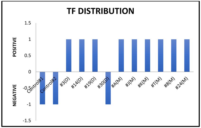
Dent. J. 2018, 6, 53                                                                                                5 of 6

in patients whose neutrophils are chemotactically defective. In this case report, two chemokines
were used to measure neutrophil migration. CXCL1 is a small cytokine, which is secreted by
human melanoma cells and expressed by macrophages, neutrophils, and epithelial cells [21,22].
Study has shown that it is critical for neutrophil-dependent bacterial elimination via induction
of reactive oxygen species [23]. CXCL8, also called interleukin 8, which is also a neutrophil
chemotactic factor and is produced by macrophages as well as other cells types. Both chemokines
are responsible to induce chemotaxis and attract neutrophils to migrate toward the site of infection.
The patient with GSD type Ib in this report had defective neutrophil chemotaxis in response to the
chemokines CXCL1 and CXCL8 in comparison to normal neutrophils. In addition, PCR analysis
indicated the presence of the “Red Complex” microorganism (which includes Porphyromonas gingivalis,
Tannerella forsythia, and Treponema denticola [24]), Tannerella forsythia, which was a major periodontal
pathogen in conjunction with a compromised host immune response that was responsible for severe
periodontal attachment destruction in this eight-year-old patient. Conversely, no histological cemental,
and dentinal abnormalities were detected. Although GSD Type Ib cannot be cured, the disease and
associated symptoms can be managed through comprehensive medical and dental care. In this case
report, the decision of removing all the remaining primary teeth was based on the severe localized
horizontal bone loss. These areas have the most plaque accumulation, clinically, as well. Due the
mobility of the primary teeth, patient was not comfortable to eat in the area. Nance appliance
and lingual holding were placed in order to minimally maintain the edentulous space and prevent
posterior teeth from shifting mesially. In patients with GSD Type Ib, dental care should be focused
on primary prevention and early recognition of dental and periodontal diseases. Understanding the
pathophysiology of GSD Ib will enhance the ability for its clinical management and, hopefully, for the
future development of a cure.

Author Contributions: All the authors have accepted responsibility for the entire content of this submitted
manuscript and approved submission.
Funding: This research received no external funding.
Acknowledgments: The authors would like to thank Stephen G. Walker, Department of Oral Biology and
Pathology, Stony Brook University School of Dental Medicine and his lab for contributions to this case report.
Conflicts of Interest: The authors declare no conflict of interest. The funding organization(s) played no role in the
study design; in the collection, analysis, and interpretation of data; in the writing of the report; or in the decision
to submit the report for publication.

References
1.    Dorland, W.A. Newman. Dorland’s Illustrated Medical Dictionary, 32nd ed.; Elsevier/Saunders: Philadelphia,
      PA, USA, 2012.
2.    Chen, Y.T. The Metabolic and Molecular Bases of Inherited Disease; McGraw Hill: New York, NY, USA, 2001;
      Volume 1, pp. 1521–1551.
3.    Weinstein, D.A.; Wolfsdorf, J.I. Effect of continuous glucose therapy with uncooked cornstarch on the
      long-term clinical course of type 1a glycogen storage disease. Eur. J. Pediatr. 2002, 161, S35–S39. [CrossRef]
      [PubMed]
4.    Mundy, H.R.; Hindmarsh, P.C.; Matthews, D.R.; Leonard, J.V.; Lee, P.J. The Regulation of Growth in Glycogen
      Storage Disease Type 1. Clin. Endocrinol. 2003, 58, 332–339. [CrossRef]
5.    Von Cudzinowski, L. Gierke’s disease: Report of case. ASDC J. Dent. Child. 1979, 45, 413–415.
6.    Visser, G.; Rake, J.P.; Labrune, P.; Leonard, J.V.; Moses, S.; Ullrich, K.; Wendel, U.; Groenier, K.H.; Smit, G.P.A.
      Granulocyte colony-stimulating factor in glycogen storage disease type 1b. Results of the European Study
      on Glycogen Storage Disease Type 1. Eur. J. Pediatr. 2002, 161, S83–S87. [CrossRef] [PubMed]
7.    Simoes, A.; Domingos, F.; Fortes, A.; Prata, M.M. Type 1 glycogen storage disease and recurrent calcium
      nephrolithiasis. Nephrol. Dial. Transplant. 2001, 16, 1277–1279. [CrossRef] [PubMed]
Dent. J. 2018, 6, 53 6 of 6
8. Weston, B.W.; Lin, J.L.; Muenzer, J.; Cameron, H.S.; Arnold, R.R.; Seydewitz, H.H.; Mayatepek, E.;
Van Schaftingen, E.; Veiga-Da-Cunha, M.; Matern, D. Glucose-6-phosphatase mutation G188R confers
an atypical glycogen storage disease type Ib phenotype. Pediatr. Res. 2000, 48, 329–334. [CrossRef] [PubMed]
9. Visser, G.; de Jager, W.; Verhagen, L.P.; Smit, G.P.; Wijburg, F.A.; Prakken, B.J.; Coffer, P.J.; Buitenhuis, M.
Survival, but not maturation, is affected in neutrophil progenitors from GSD-1b patients. J. Inherit. Metab. Dis.
2012, 35, 287–300. [CrossRef] [PubMed]
10. Miller, D.R.; Lamster, I.B.; Chasens, A.I. Role of the polymorphonuclear leukocyte in periodontal health and
disease. J. Clin. Periodontol. 1984, 11, 1. [CrossRef] [PubMed]
11. Santigli, E.; Koller, M.; Klug, B. Oral Biofilm sampling for Microbiome Analysis in healthy Children. J. Vis. Exp.
2017, 130, 56320. [CrossRef] [PubMed]
12. Denholm, E.M.; Wolber, F.M. A simple method for the purification of human peripheral blood monocytes.
A substitute for Sepracell-MN. J. Immunol. Methods 1991, 144, 247–251. [CrossRef]
13. Fischer, A.H.; Jacobson, K.A.; Rose, J.; Zeller, R. Hematoxylin and eosin staining of tissue and cell sections.
CSH Protoc. 2008. [CrossRef] [PubMed]
14. Schallhorn, R.G.; Snider, L.E. Periodontal maintenance therapy. J. Am. Dent. Assoc. 1981, 103, 227–231.
[CrossRef] [PubMed]
15. Anderson, D.C.; Mace, M.L.; Brinkley, B.R.; Martin, R.R.; Smith, C.W. Recurrent infection in glycogenosis
type Ib: Abnormal neutrophil motility related to impaired redistribution of adhesion sites. J. Infect. Dis. 1981,
143, 447–459. [CrossRef] [PubMed]
16. Burns, M.J.; Furie, M.B. Borrelia burgdorferi and interleukin-1 promote the transendothelial migration of
monocytes in vitro by different mechanisms. Infect. Immunity 1998, 66, 4875–4883.
17. Tran, S.D.; Rudney, J.D. Improved multiplex PCR using conserved and species-specific 16S rRNA gene
primers for simultaneous detection of Actinobacillus actinomycetemcomitans, Bacteroides forsythus,
and Porphyromonas gingivalis. J. Clin. Microbiol. 1999, 37, 3504–3508. [PubMed]
18. Eskan, M.A.; Jotwani, R.; Abe, T.; Chmelar, J.; Lim, J.H.; Liang, S.; Ciero, P.A.; Krauss, J.L.; Li, F.; Rauner, M.; et al.
The leukocyte integrin antagonist Del-1 inhibits IL-17-mediated inflammatory bone loss. Nat. Immunol. 2012,
13, 465–473. [CrossRef] [PubMed]
19. Roberts, H.M.; Ling, M.R.; Insall, R.; Kalna, G.; Spengler, J.; Grant, M.M.; Chapple, I.L. Impaired neutrophil
directional chemotactic accuracy in chronic periodontitis patients. J. Clin. Periodontol. 2015, 42, 1–11.
[CrossRef] [PubMed]
20. Hajishengallis, E.; Hajishengallis, G. Neutrophil homeostasis and periodontal health in children and adults.
J. Dent. Res. 2014, 93, 231–237. [CrossRef] [PubMed]
21. Ley, K.; Laudanna, C.; Cybulsky, M.I.; Nourshargh, S. Getting to the site of inflammation: The leukocyte
adhesion cascade updated. Nat. Rev. Immunol. 2007, 7, 678–689. [CrossRef] [PubMed]
22. Phillipson, M.; Heit, B.; Colarusso, P.; Liu, L.; Ballantyne, C.M.; Kubes, P. Intraluminal crawling of neutrophils
to emigration sites: A molecularly distinct process from adhesion in the recruitment cascade. J. Exp. Med.
2006, 203, 2569–2575. [CrossRef] [PubMed]
23. Stearns-Kurosawa, D.J.; Osuchowski, M.F.; Valentine, C.; Kurosawa, S.; Remick, D.G. The pathogenesis of
sepsis. Ann. Rev. Pathol. 2011, 6, 19–48. [CrossRef] [PubMed]
24. Socransky, S.S.; Haffajee, A.D.; Cugini, M.A.; Smith, C.; Kent, R.L., Jr. Microbial complexes in subgingival
plaque. J. Clin. Periodontol. 1998, 25, 134–144. [CrossRef] [PubMed]
© 2018 by the authors. Licensee MDPI, Basel, Switzerland. This article is an open access
article distributed under the terms and conditions of the Creative Commons Attribution
(CC BY) license (http://creativecommons.org/licenses/by/4.0/).You can also read


