Serum protein profiles and C-reactive protein in natural canine filariasis
←
→
Page content transcription
If your browser does not render page correctly, please read the page content below
Veterinary World, EISSN: 2231-0916                                                                     RESEARCH ARTICLE
Available at www.veterinaryworld.org/Vol.14/April-2021/7.pdf                                                 Open Access

Serum protein profiles and C-reactive protein in natural canine filariasis
                Sariya Asawakarn1     , Sujin Sirisawadi1 , Nanthida Kunnasut1    , Patchana Kamkong2      and
                                                 Piyanan Taweethavonsawat2

   1. Biochemistry Unit, Department of Veterinary Physiology, Faculty of Veterinary Science, Chulalongkorn University,
   Bangkok, 10330, Thailand; 2. Parasitology Unit, Department of Veterinary Pathology, Faculty of Veterinary Science,
                                 Chulalongkorn University, Bangkok, 10330, Thailand.
                  Corresponding author: Piyanan Taweethavonsawat, e-mail: piyanan.t@chula.ac.th,
            Co-authors: SA: sariya.a@chula.ac.th, SS: ssirisawadi@yahoo.com, NK: pui_unn@hotmail.com,
                                               PK: miint.pat@gmail.com
                   Received: 07-12-2020, Accepted: 24-02-2021, Published online: 10-04-2021

doi: www.doi.org/10.14202/vetworld.2021.860-864 How to cite this article: Asawakarn S, Sirisawadi S, Kunnasut N,
Kamkong P, Taweethavonsawat P (2021) Serum protein profiles and C-reactive protein in natural canine filariasis, Veterinary
World, 14(4): 860-864.

                                                            Abstract
Background and Aim: Canine filariasis is caused by several species of filarial worms. The pathophysiological response
to infection is mainly due to the filaria lifecycle. Laboratory detection methods to assess the pathological alterations
characteristic of filariasis are needed urgently. Serum protein profiles and C-reactive protein (CRP) levels are used widely to
diagnose several animal diseases. This study aimed to determine the serum protein profiles and CRP levels in dogs infected
with Dirofilaria immitis or Brugia pahangi or both parasites.
Materials and Methods: Blood samples were collected from 980 dogs presenting at animal hospitals and veterinary clinics
in Bangkok and its vicinity. The presence of microfilaria in samples was determined using a buffy coat smear and staining
with Wright–Giemsa. The sheathed and unsheathed microfilaria species were identified by acid phosphatase staining.
Forty positive samples were tested. The serum protein profiles were identified by agarose gel electrophoresis. The CRP
concentration was measured using a fluorescent immunoassay.
Results: Albumin levels and albumin-to-globulin ratios were significantly lower, and total protein, β2 globulin, and γ
globulin levels were significantly elevated in dogs infected with D. immitis and B. pahangi compared with reference values
in normal dogs. The average CRP concentrations in dogs infected with D. immitis or B. pahangi were 69.9 and 12.9 mg/L,
respectively.
Conclusion: The total protein and γ globulin levels increased in canine filariasis compared with the normal reference range.
The CRP concentration in dogs infected with D. immitis was extremely high, whereas that in dog infected with B. pahangi
was normal.
Keywords: Brugia pahangi, C-reactive protein, Dirofilaria immitis, dogs, serum protein.
Introduction                                                       can release an endosymbiotic bacterium, named
      Canine filariasis is a prominent mosquito-borne              Wolbachia pipientis. This bacterium is crucial to the
disease that occurs worldwide, including in Thailand.              pathophysiological response to canine heartworm
Filariasis is a considerable public health concern in              disease. The severity of infection is characterized by
tropical and subtropical areas. Several species of filar-          endocarditis, with intimal proliferation and thickened
ial worms, including Dirofilaria immitis, Dirofilaria              vessel walls [2,3]. Brugia spp. cause lymphatic fila-
repens, Brugia pahangi, and Acanthocheilonema                      riasis; in humans, these pathogens, especially Brugia
reconditum, can cause filariasis. These species have               malayi, cause elephantiasis. In pets, B. pahangi is
been reported in Thailand [1]. D. immitis infection,               transmitted by mosquitoes and its lifecycle is similar
which causes canine heartworm disease, is the most                 to that of other filarial worms. Infective larvae enter
pathogenic filarial parasite. The clinical signs of fil-           peripheral lymphatics, migrate to the nearest lymph
ariasis are exercise intolerance, coughing, ascites,               node, and develop for 2 weeks before migrating to
and heart failure. The pathophysiological response to              other lymphatics, where the larvae mature and pro-
heartworm infection is mainly due to the living adult              duce lymphadenitis, granulomatous lymphangitis, and
worms in the pulmonary arteries and right ventricle. In            lymphangiectasia. Notably, in dogs, B. pahangi does
addition, dead microfilaria and released adult worms               not cause elephantiasis as it does in humans. Infected
                                                                   dogs are generally asymptomatic, although lymphade-
Copyright: Asawakarn, et al. Open Access. This article is          nopathy and lymphedema have been reported in some
distributed under the terms of the Creative Commons Attribution
4.0 International License (http://creativecommons.org/licenses/    cases [4,5].
by/4.0/), which permits unrestricted use, distribution, and              The development of laboratory methods to
reproduction in any medium, provided you give appropriate credit
to the original author(s) and the source, provide a link to the    assess the pathological alterations characteristic of the
Creative Commons license, and indicate if changes were made.       disease is needed urgently. In addition, new tools are
The Creative Commons Public Domain Dedication waiver (http://
creativecommons.org/publicdomain/zero/1.0/) applies to the data
                                                                   needed to determine the health status of dogs infected
made available in this article, unless otherwise stated.           with D. immitis or B. pahangi, including tools for
Veterinary World, EISSN: 2231-0916                                                                                        860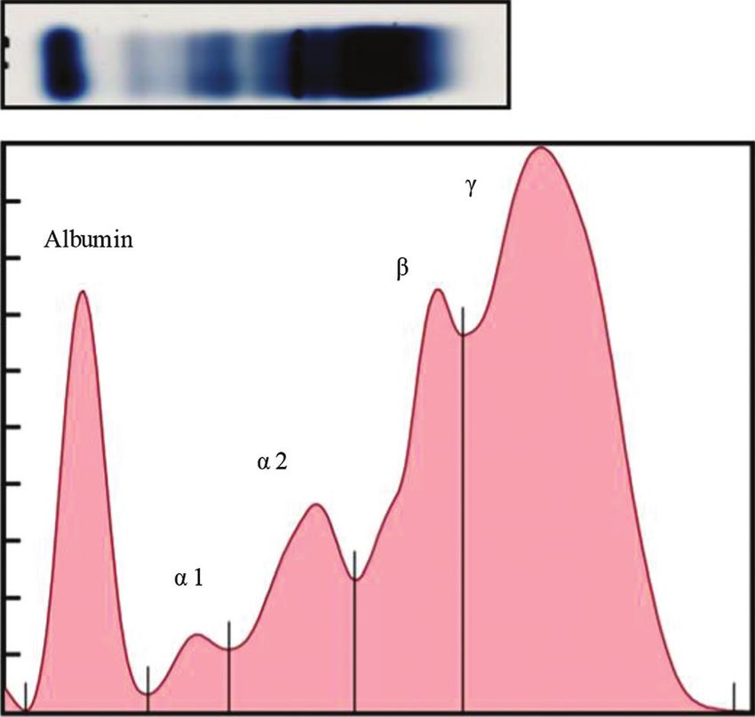
Available at www.veterinaryworld.org/Vol.14/April-2021/7.pdf
disease staging and accurate prognoses. Serum pro- tubes. All serum samples were kept at –20ºC until
tein profiles and inflammatory biomarkers are suitable analyses.
tools to monitor infected animals. Serum protein pro- Determination of the serum protein profile by
files are useful for veterinarians to monitor the health electrophoresis
status of animals. Major canine serum proteins are Total protein concentrations in the 40 positive
separated into five or six bands, including albumin, serum samples were determined using a photometric
α1 globulin, α2 globulin, β globulin, and γ globulin. colorimetric test or a biuret method test kit (Human®,
The β globulin fraction can separate into the β1 glob- Wiesbaden, Germany). Proteins were separated by
ulin and β2 globulin fractions. Serum protein profiles agarose gel electrophoresis (SPIFE® Split Beta SPE
have been used widely as a diagnostic tool to moni- kit, Helena Laboratories, Beaumont, Texas, USA)
tor the status of infectious and other diseases, such as to examine the serum protein profiles. 1.3 milligram
multiple myeloma in dogs [6-10]. protein of each serum sample were electrophoresed at
The acute-phase response is an early defense 400 V for 6 min. The gels were pre-dried at 53°C for
of the body in response to trauma, inflammation, or 12 min, stained with acid blue staining solution, and
infection. The acute-phase response is part of the destained with citric acid using an automated machine
innate host defense system and systemic effects, (Spife® 3000, Helena Laboratories). The density
including fever, leukocytosis, and increased blood of protein bands was analyzed using the Quickscan
cortisol. C-reactive protein (CRP) is one of the acute- Touch program (Helena Laboratories). The specimen
phase proteins. CRP was first described as an acute- electrophoresis protein serum (SPEP) data from dogs
phase protein in 1930 and named for its ability to infected with D. immitis or B. pahangi were compared
bind to C-polysaccharide from Pneumococcus pneu- to the reference ranges of normal, uninfected dogs
monia [11,12]. Pro-inflammatory cytokines, such as reported by Kaneko [13].
interleukin 6 (IL-6) and tumor necrosis factor-alpha Measurement of CRP concentration
(TNF-α), stimulate the liver to produce CRP and The CRP concentrations were measured using
release it into the bloodstream. CRP is a major acute- a fluorescent immunoassay (Vcheck Canine CRP
phase protein in dogs; however, information on serum 2.0 Test Kit, Bionote, Gyeonggi-do, South Korea).
protein profiles and CRP levels in dogs infected with The CRP concentrations were determined in serum
D. immitis (i.e., canine heartworm disease) or B. pah- samples from dogs infected with D. immitis (n=6) or
angi is limited. B. pahangi (n=6) or both parasites (n=1). Five microli-
The aim of this study was to determine and to ters of each sample were diluted with the diluent buffer
compare the serum protein profiles and CRP level in (to 100 µL), mixed, and added to the test device (V200
dogs infected with D. immitis or B. pahangi with nor- Analyzer, Bionote, South Korea). A CRP concentration
mal reference values from healthy dogs, with the goal of more than 30 mg/L represented an abnormal value.
of utilizing these parameters to monitor disease status.
Statistical analysis
Materials and Methods SPEP data from infected dogs were analyzed
Ethical approval using a general linear model (GLM) with the online
The research protocol was approved by SAS version 9.4 (SAS Inst. Inc., Cary, NC, USA)
Chulalongkorn University Animal Committee or SAS University Edition (https://www.sas.com/
(approval no. 1931052). en_us/software/university-edition.html). The GLM
Study period and location
was y=trt+method+trt*method+e, where y was the
Samples were collected from private Small serum protein value from each filarial worm species
Animal Hospitals and Veterinary Clinic in Bangkok and method of use, trt is a factor from each filarial
worm species, method is a factor from the labora-
from August 2019 to July 2020.
tory technique used to measure serum protein pro-
Sample collection files, trt*method is the interaction term between the
Blood samples were collected from 980 canines two factors, and e is the residual value from each
(Canis familiaris). Some dogs exhibited the clinical observation.
signs of filariasis, including exercise intolerance,
coughing, and ascites. However, most dogs did not Results
exhibit any clinical signs of filariasis. All samples Dogs infected with D. immitis and B. pah-
were tested to determine the presence of microfi- angi showed significantly lower albumin levels and
laria using buffy coat blood smears and staining with albumin-to-globulin (A/G) ratios and significantly
Wright–Giemsa. Sheathed and unsheathed microfi- elevated total protein, β2 globulin, and γ globulin
laria were identified to determine the filarial species levels compared with reference values of normal
using acid phosphatase staining [1]. The number of dogs (Table-1). The serum protein electropherograms
dogs positive for D. immitis or B. pahangi or both are shown in Figures-1-4. The serum protein electro-
parasites was 24, 15, and 1, respectively. All canine phoretogram for dogs infected with both is shown in
blood samples were collected in serum collection Figure-5.
Veterinary World, EISSN: 2231-0916 861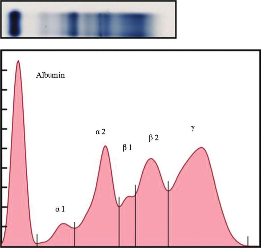
Available at www.veterinaryworld.org/Vol.14/April-2021/7.pdf
Table-1: The total protein, albumin, α1 globulin, α2
globulin, β1 globulin, β2 globulin, and γ globulin levels
and albumin-globulin (A/G) ratio.
Variable Dirofilaria Brugia pahangi Reference
(g/dL) immitis positive positive range [13]
(mean±SD) (mean±SD)
(n=24) (n=15)
Total 9.22±2.40 8.50±1.75 5.40-7.10
protein
Albumin 2.07±0.70 2.28±0.50 2.60-3.30
Alpha-1 0.34±0.17 0.43±0.18 0.20-0.50
Alpha-2 0.66±0.55 0.52±0.45 0.30-1.10
Beta-1 1.15±0.53 0.87±0.46 0.70-1.30
Beta-2 2.23±1.02 1.79±0.06 0.60-1.40
Gamma 2.77±1.84 2.63±1.17 0.90-2.20
A/G ratio 0.33±0.12 0.41±0.15 0.59-1.11
15.00
9.22
Concentration (g/dL)
10.00
Figure-3: A representative electrophoretogram (top) of an
infected with Dirofilaria immitis. The serum protein profile
(bottom) shows the reduced albumin peak, increased β
2.77
5.00 globulin, and γ globulin peaks (hyperglobulinemia).
2.23
2.07
1.15
0.66
0.34 0.33
0.00
Serum protein electrophoresis
Albumin Alpha-1 Alpha-2 Beta-1 Beta-2 Gamma total protein A/G
Figure-1: The albumin, α1 globulin, α2 globulin, β1 globulin,
β2 globulin, γ globulin, and total protein concentrations
(mean±standard deviation in g/dL) and albumin-globulin
(A/G) ratio of dogs infected with Dirofilaria immitis. The
numbers indicate the average values.
15.00
Concentration (g/dL)
8.50
10.00
5.00
2.63
2.28 1.79
0.52 0.87
0.43 0.41
0.00
Serum protein electrophoresis
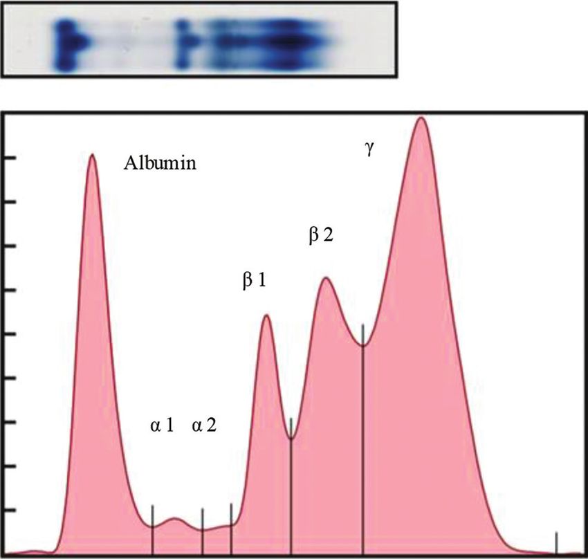
Albumin Alpha-1 Alpha-2 Beta-1 Beta-2 Gamma total protein A/G Figure-4: A representative electrophoretogram (top) of a
dog infected with Brugia pahangi. The serum protein profile
Figure-2: The albumin, α1 globulin, α2 globulin, β1 globulin, (bottom) shows the elevated β globulin and γ globulin
β2 globulin, γ globulin, and total protein concentrations peaks.
(mean±standard deviation in g/dL) and albumin-globulin
(A/G) ratio of dogs infected with Brugia pahangi. The concentration was 12.9 mg/L in dogs infected with
numbers indicate the average values.
B. pahangi. The CRP concentration in the dog infected
with both D. immitis and B. pahangi was >200 mg/L.
The CRP concentrations of dogs infected with only
D. immitis (n=6) were between 13.6 and 116.9 mg/L, Discussion
with an average of 69.6 mg/L. In addition, dogs had Determining the serum protein profile is a basic
high CRP concentration; they may have to monitor test used in animal hematology and clinical chemis-
health status during the treatment process. There were try to monitor the patient’s health and disease status.
four samples with CRP concentrations above 30 mg/L Variations in serum protein profiles commonly occur
and two samples below 30 mg/L. The CRP concen- as secondary symptoms in numerous diseases and may
trations of dogs infected with B. pahangi (n=6) were be the primary symptom of certain conditions. Several
in the normal range (< 30 mg/L):Available at www.veterinaryworld.org/Vol.14/April-2021/7.pdf
of various classes of Igs, and CRP also migrates in
this fraction [19,20]. Moreover, the elevated γ globu-
lin level in dogs infected with only D. immitis might
be related to the high CRP concentration. Similar to
dogs infected with D. immitis, dogs infected with
only B. pahangi showed a reduced albumin level and
A/G ratio, and elevated total protein, β2 globulin, and
γ globulin levels compared with reference levels in
healthy dogs [13]. In humans infected with Loa loa,
total serum protein β globulin and γ globulin frac-
tions with monoclonal gammopathy are elevated. The
albumin, A/G ratio, and α-globulin were all normal in
dogs with B. pahangi [21].
CRP is a major canine acute-phase protein that
increases rapidly in a wide range of inflammatory con-
ditions; CRP is especially high in chronic conditions.
From our results, the average CRP concentration in
dogs infected with only D. immitis was higher than the
reference range, but the CRP concentration was normal
Figure-5: A representative electrophoretogram (top) of a
in dogs infected with only B. pahangi. The CRP con-
dog infected with Dirofilaria immitis and Brugia pahangi.
centration in dogs infected with both D. immitis and
B. pahangi was markedly elevated (> 200 mg/L). A
to detect acute or chronic inflammation and stimulate
striking increase in CRP and decrease in albumin and
humoral immune response [6,14].
paraoxonase-1 activity have been observed in canine
The Spilt Beta SPE Kit separates serum proteins heartworm disease [22]. CRP levels are elevated in
into five or six bands: Albumin, α1 globulin, α2 glob- canine heartworm disease because of inflammation
ulin, β globulin, and γ globulin. The β globulin peak involved in the pathogenesis [23]. Heartworm disease
can also be split into β1 and β2 globulin peaks. Each leads to pulmonary hypertension and, in the late stage,
globulin fraction consists of acute-phase proteins or may induce right-side cardiac insufficiency. Adult
antibodies, and sometimes both [15]. In dogs infected worms are localized in the pulmonary arteries, which
with D. immitis or B. pahangi, albumin levels and induces endothelial damage or proliferative endocar-
the A/G ratio were reduced compared with normal ditis. The CRP concentration can be used as a marker
reference dog values [13]. Reduced albumin levels of endothelial arteritis and pulmonary hypertension
are usually caused by starvation, liver insufficiency, in dogs infected with D. immitis [24]. However, the
kidney disease, congestive heart failure, or parasitic CRP concentration in dogs infected with B. pahangi
disease [6,8,10]. Canine filariasis leads to marked was in the normal range; those dogs were mostly
changes in the parameters used to evaluate liver and asymptomatic. In humans, the CRP concentration in
kidney functions [16]. A reduced A/G ratio is caused patients with asymptomatic microfilaremia was lower
by the overproduction of globulins. In this study, total than in patients with chronic lymphatic pathology. The
protein and γ globulin levels were elevated in dogs patients with chronic lymphatic obstruction caused by
infected with D. immitis or B. pahangi compared with the filarial parasite Wuchereria bancrofti had elevated
the levels in healthy dogs [13]. The high level of total serum CRP [25]. The extremely high CRP concentra-
proteins is indicative of hyperglobulinemia because tion in the coinfected dogs indicates that the inflamma-
of the humoral response induced by D. immitis. The tory response may be caused by D. immitis infection.
high level of γ globulin indicates the high production
of immunoglobulins (Igs), such as IgG, in response Conclusion
to chronic inflammation. Hyperglobulinemia can be The serum protein profiles and CRP concentra-
caused by chronic infection, inflammation, neoplasia, tions in canine filariasis can reflect the health status
and parasitic diseases, such as dirofilariasis, scabies, of infected dogs. The total protein and γ globulin lev-
and Ehrlichia infection [17]. Clinical signs of heart- els increased in canine filariasis compared with the
worm disease appear as chronic infection and marked normal reference range. The CRP concentrations in
allergic response to the adult worms and microfilariae. dogs infected with D. immitis were extremely high,
Hyperglobulinemia is an expected feature of canine whereas those in dogs infected with B. pahangi were
heartworm disease and can be either polyclonal or normal. This information can be utilized by veterinar-
monoclonal gammopathy. We noted clonal gammopa- ians to monitor infected dogs during treatment.
thy using the SPE kit, which identified the monoclonal
Authors’ Contributions
protein IgG. Dirofilariasis has been hypothesized to
elicit the atypical benign clonal proliferation of plasma SA and PT designed the experiments. NK, SS,
cells [18]. In dogs, γ globulin fractions are composed and PK contributed to the analysis and interpretation
Veterinary World, EISSN: 2231-0916 863Available at www.veterinaryworld.org/Vol.14/April-2021/7.pdf
of data. SA and PT wrote the manuscript. NK, SS, (2009) Serum protein electrophoresis: An underused but
and PK assisted in writing and revision of the man- very useful test. Digestion, 79(4): 203-210.
11. Carreton, E., Morchon, R. and Montaya-Alonso, J.A.
uscript. All authors have read and approved the final (2017) Cardiopulmonary and inflammatory biomarkers in
manuscript. heartworm disease. Parasit. Vectors, 10(2): 534.
12. Sproston, N.R. and Ashworth, J.J. (2018) Role of C-reactive
Acknowledgments
protein at sites of inflammation and infection. Front.
The authors would like to thank Assist. Prof. Immunol., 9:754
13. Kaneko, J.J. (1997) Serum proteins and the dysprotein-
Chatree Khatiworavage for statistical analysis. This emias. In: Kaneko, J.J., editor. Clinical Biochemistry of
study was supported by grants from Ratchadapisek, Domestic Animals. 5th ed. Academic Press, San Diego, CA.
Chulalongkorn University, Thailand (Grant no. CU_ p117-138.
GR_63_42_31_03). 14. Histrova, J. and Genova, M. (2015) Serum protein electro-
phoresis by agarose gel M spike screening and beyond-re-
Competing Interests view. Int. J. Sci. Res., 6(6): 1463-1466.
15. Moore, A.R. and Avery, P.R. (2019) Protein characterization
The authors declare that they have no competing using electrophoresis and immunofixation; a case‐based
interests. review of dogs and cats. Vet. Clin. Pathol., 48(1): 29-44.
16. Heshem, M. and Badawy, A. (2007) Hematological and bio-
Publisher’s Note chemical studies on filariasis of dogs. Internet J. Vet. Med.,
Veterinary World remains neutral with regard 4(2): 1-7.
17. Capraiis, D., Sassanelli, M., Paradies, P., Otranto, D. and
to jurisdictional claims in published institutional Lia, R. (2009) Monoclonal gammopathy associated with
affiliation. heartworm disease in a dog. J. Am. Anim. Hosp. Assoc.,
45(6): 295-300.
References
18. Wahed, A. and Dasgupta, A. (2015) Monoclonal gammop-
1. Chungpivat, S. and Taweethavornsawat, P. (2008) The dif- athy and its detection. In: Hematology and Coagulation: A
ferentiation of microfilariae in dogs and cats using Giemsa’s Comprehensive Review for Board Preparation, Certification
staining and the detection of acid phosphatase activity. and Clinical Practice. Elsevier, Amsterdam. p117-123.
J. Thai. Vet. Pract., 20(1): 47-55. 19. Jarensky, A.K., Bondzio, A., Murugaiyan, J., Siebert, U.,
2. Kramer, L., Grandi, G., Leoni, M., Passeri, B., McCall, J., Roesler, U., Kohn, B. and Einspanier, R. (2014)
Genchi, C., Montarino, M. and Bazzocchi, C. (2008) Characterization of native C-reactive protein (CRP) and the
Wolbachia and its influence on the pathology and immunol- corresponding liver mRNA in dogs. Biochem. Biophys. Res.
ogy of Dirofilaria immitis infection. Vet. Parasitol., 158(3): Commun., 452(3): 462-467.
191-195. 20. Yamamoto, S. (1992) Isolation of canine C-reactive pro-
3. McCall, J.W., Genchi, C., Kramer, L.H., Gurrero, J. and tein and characterization of its properties. Vet. Immunol.
Venco, L. (2008) Heartworm disease in animals and Immunopathol., 30(4): 329-339.
humans. Adv. Parasitol., 66:193-285. 21. Laskar, D.B., Rose, M., Gupta, R., Tanowitz, HB. and
4. Kaikuntod, M., Thongkorn, K., Tiwananthagorn, S. Haseeb, MA. (2018) Case report: Monoclonal gammopa-
and Boonyapakorn, C. (2018) Filarial worms in dogs in thy of undetermined significance is associated with Loa loa
Southeast Asia. Vet. Integr. Sci., 16(2): 1-17. infection. Am. J. Trop. Med. Hyg., 99(5): 1206-1210.
5. Kobasa, T., Thammapalo, S., Suvannalabha, S., 22. Mendez, J.C., Carreton, E., Martinez, S., Tvarijonaviciute, A.,
Armesombun, A., Loymak, S., Sawat, L. and Choochite, W. Ceron, J.J. and Montoya-Alonso, JA. (2015) Acute phase
(2004) Identification of Brugia malayi like microfilaria in response in heartworm infected dogs after adulticide treat-
naturally infected cats from Narathiwat province, South ment. Vet. Parasitol., 209(3-4): 197-201.
Thailand. J. Trop. Med. Parasitol., 27(1): 21-25. 23. Yoon, W.K., Kim, Y.W., Suh, S., Choi, R., Lee, S.G. and
6. Jania, B. and Andraszek, K. (2016) Application of native Hyun, C. (2017) Evaluation of cardiopulmonary and inflam-
agarose gel electrophoresis of serum proteins in veterinary matory markers in dogs with heartworm infection during
diagnostics. J. Vet. Res., 60(4): 501-508. treatment with the 2014 American Heartworm society rec-
7. McGrotty, Y. and Knottenbelt, C. (2002) Significance of ommended treatment protocol. Parasit. Vectors, 10(2): 535.
plasma protein abnormalities in dogs and cats. Practice, 24. Venco, L., Bertazzolo, W., Giordano, G. and Paltrinieri, S.
24(9): 512-517. (2014) Evaluation of C-reactive protein as a clinical bio-
8. O’Connell, T.X., Horita, T.J. and Kasravi, B. (2005) marker in naturally heartworm-infected dogs: A field study.
Understanding and interpreting serum protein electrophore- Vet. Parasitol., 206(1-2): 48-54.
sis. Am. Fam. Physician, 71(1): 105-112. 25. Lal, RB., Dhawan, RR., Ramzy, RM., Farris, RM. and
9. Thotova, C., Nagy, O. and Kovac, G. (2016) Serum proteins Gad, AA. (1991) C-reactive protein in patients with lym-
and their diagnostic utility in veterinary medicine: A review. phatic filariasis: Increased expression on lymphocytes in
Vet. Med., 61(9): 475-496. chronic lymphatic obstruction. J. Clin. Immunol., 11(1):
10. Vavricka, S.R., Burri, E., Beglinger, C. and Degen, L. 46-53.
********
Veterinary World, EISSN: 2231-0916 864You can also read