Characterization Of Phospholipase A2 Enzyme Activity In Serum Of The Komodo Dragon (Varanus Komodoensis) - Cirworld
←
→
Page content transcription
If your browser does not render page correctly, please read the page content below
ISSN 2347-6893
                                                                                     Volume 11 Number1
                                                                          Journal of Advances in Biology

 Characterization Of Phospholipase A2 Enzyme Activity In Serum Of The
                Komodo Dragon (Varanus Komodoensis)
      Mark Merchant*1, Danyell Henry1, Rodolfo Falconi1, Becky Musher2, Judith Bryja3
       1Department of Chemistry, McNeese State University, Lake Charles, LA, USA

                                mmerchant@mcneese.edu
   2 Department of Conservation and Research, San Antonio Zoo, San Antonio, TX, USA

                           bekky.muscher-hodges@sazoo.org
   3 Department of Herpetology, Houston Zoo, Houston, TX, USA jbryja@houstonzoo.org

ABSTRACT
Soluble phospholipase A2 (sPLA2) is an enzyme found in the peripheral circulation of vertebrates which has significant
immunological activity. This enzyme exerts immune activity by the hydrolysis of fatty acids from the sn-2 position of
membrane glycerophospholipids of microbes, thus compromising membrane integrity and casing eventual lysis. We
utilized membrane fatty acids labeled with a fluorescent probe (BODIPY) at the sn-2 position fatty acid to label the
membranes. Incubation of different volumes of serum from Komodo dragons with BODIPY-labeled bacteria resulted in
liberation of labeled fatty acid in a serum volume-dependent manner. This cleavage of fatty acid occurred rapidly, with a
biphasic production of fluorescent product. An immediate accumulation of product was noted, which increased steadily for
a 30-minute period, followed by a slower hydrolysis between 30 and 180 min. The activity was temperature-dependent,
with low activities observed at 5oC and a linear increase up to 40oC. The liberation for fatty acid was inhibited by p-Bromo
phenacyl bromide, a specific phospholipase A2 (PLA2) inhibitor, in a concentration-dependent manner, indicating that the
activity was due to the presence of sPLA2.

Indexing terms/Keywords
innate immunity, reptile, varanid.

Academic Discipline And Sub-Disciplines
Biology; Biochemistry; Conservation; Ecology

SUBJECT CLASSIFICATION
Class Q: Science; subclass QL: Zoology, Herpetology

TYPE (METHOD/APPROACH)
Experimental. Laboratory Research

INTRODUCTION
Phospholipase A2 (PLA2) is an enzyme that cleaves fatty acids from the sn-2 position of membrane structural lipids. This
enzyme serves a variety of important biological and cellular functions including remodeling of membranes [1]. Among its
most important functions is the cleavage of intracellular arachidonic acid from membranes, which then serves as a
precursor for eicosanoid synthesis [2-3]. However, another form of PLA2 (sPLA2), first described by Vadas et al. [4], which
circulates as a free enzyme in the serum. This enzyme is found in the peripheral circulation and cleaves Sn-2 fatty acids
from the membranes of microbes, thus compromising membrane integrity and function. As a result, sPLA2 exhibits
important immune activity [5-7] and is thought to be a critical component of innate immunity.
The Komodo dragon (Varanus komodoensis) is a critically-endangered species that is native to only three islands on the
Indonesian archipelago. The largest lizard in the world, Komodo dragons are fierce predators that have been known to
feed on prey more than ten times their own size [8-9]. These animals have saliva which contains large loads of pathogenic
bacteria. They deliver a lethal bite, and follow their prey for days, or even weeks, until it succumbs to systemic infection.
For this reason, they are able to feed on prey items much larger than themselves [8-9]. In addition, it has been suggested
that these reptiles deliver toxins in their bite, based on skull morphology [10]. Although these toxins have not been
isolated or described to date, they are reported to induce systemic shock and anticoagulant effect. Komodo dragons are
territorial, and often engage in territorial battles with members of their own species. In addition, these animals often inflict
wounds on each other during feeding frenzies in which multiple dragons feed on large prey items. However, despite the
fact that their saliva contains large loads of infectious bacteria, Komodo dragons do not seem to suffer the same fate as
their prey. This would seem to indicate that these animals have evolved an effective immune system to combat these
potentially lethal wounds.
Little is known concerning the immune system of Varanids. Several recent studies have characterized antibacterial
activity [11], serum complement [12], and dipeptidyl peptidase [13] activities of serum from the Komodo dragon. In
addition, these animals have been reported to express antimicrobial peptides [14]. Because Komodo dragons are
endangered, it would be useful to further understand their mechanisms of immunity to disease and infection. This study
was conducted to characterize the expression of circulating PLA 2 enzyme activity in the serum of the Komodo dragon.

2163 | P a g e
March 2018                                                                           https://cirworld.com/
ISSN 2347-6893
Volume 11 Number1
Journal of Advances in Biology
MATERIALS AND METHODS
Chemicals and Biochemicals
4,4-difluoro-5,7-dimethyl-4-bora-3a,4a-diaza-s-indacene-3-hexadecanoic acid (BODIPY™ FL C16) was purchased from
Invitrogen (Carlsbad, CA, USA). Ethylene glycol tetraacetate (EGTA), p-bromophenacyl bromide (BPAB), CaCl2, nutrient
broth, sodium hydroxide, and tris HCl were purchased from Sigma Chemical Co. (St. Louis, MO, USA).
Treatment of Animals
Blood samples were collected from Komodo dragons at the Houston and San Antonio zoos. Blood was drawn from the
caudal vein, transferred to Vaccutainer™ tubes, and allowed to clot for at least five h before the serum was collected be
centrifugation. The amount of blood collected from each individual depended on the size of the animal, and was at the
discretion of the attending veterinarian at each institution. Blood was collected from the tail caudal veins three adults (20 -
81.5 kg) and five juveniles (1.5 - 6.2 kg), transferred to Vaccutainer™ tubes, and allowed to clot for at least five hr before
serum was collected by centrifugation. The serum was pooled so that average antibacterial values for this species could
be generated. The collection of blood from these animals was conducted in accordance with the Animal Care and Use
institutional policies of the Houston and San Antonio Zoos.
Preparation of PLA2 substrate
Escherichia coli (ATCC 11105 strain) was used to inoculate 1 L of nutrient broth. Subsequently, 1 mg of BODIPY ® FL C16
(4,4-difluoro-5, 7-dimethyl-4-bora-3a, 4a-diaza-s-indacene-3-hexadecanoic acid; Invitrogen, Carlsbad, CA, USA) dissolved
in 1 ml of DMSO (dimethyl sulfoxide) was added to this culture and incubated at 37 °C for 18 h. The culture was then
centrifuged at 8000 g for 15 min. The supernatant was discarded and the bacterial pellet resuspended in a solution of
0.9% NaCl (w/v). This process was carried out twice. Finally, the pellet was resuspended in 30 ml assay buffer (1 mM
Ca2+ in 100 mM tris-HCl, pH 7.4).
PLA2 assays
This assay was developed to measure the secretory PLA2 enzyme activity in serum. We used fluorescent BODIPY® to
label a fatty acid that binds occupies the sn-2 position of phospholipids. Bacterial cultures add these macromolecules as
component of the outer membrane during growth and proliferation. When bacteria are exposed to the activity of the PLA 2
enzyme, the labeled sn-2 fatty acid is hydrolyzed from the membrane and released from the into the assay buffer. After a
brief centrifugation, the pellet formed contains bacteria with the labeled fatty acid in the membrane, while the supernatant
has free labeled fatty acid that can be measured by spectrofluorometry. PLA 2 activity was measured in a
spectrofluorometer at an excitation of 500 nm and an emission of 510 nm, and excitation and emission slit widths of 1
nm.
To evaluate the kinetic parameters of the PLA2 enzyme activity in Komodo dragon serum, 3.5 ml of serum, 1.75 mL of the
solution containing the fluorescent marked bacteria and 33.25 ml of assay buffer were mixed. At different time intervals (0,
5, 10, 15, 20, 30, 60, 90 min), 1.1 mL of the mixture was removed and added to 900 l of stop buffer (100 mM tris-HCl, 50
mM EDTA, pH 8.0), centrifuged, and the supernatant was measured spectrofluorometric ally as previously described.
To determine the effect of serum titer on PLA 2 activity, different amounts of serum (0, 1, 2, 5, 10, 20, 50, 100, 200 and 500
l) derived from Komodo dragons were added and then balanced to a final volume of 750 L with assay buffer. Then, 100
L of the solution containing labeled E. coli were added and incubated for 30 min. After incubation time, the reaction was
terminated with the addition of 750 L stop buffer, then centrifuged, and the fluorescence intensity of supernatant
determined spectrofluorometric ally.
Statistics and controls
The concentrations of each sample were compared to that of a standard curve developed for pure product, and the nmol
of product formed for each assay was calculated. The fluorescent intensities for each sample were corrected for
background by the subtraction of florescence measured in the absence of alligator serum. Each data point represents the
mean ± standard deviation of four independent determinations
RESULTS AND DISCUSSION
Eukaryotic organisms must constantly protect against colonization by potentially pathogenic microbes. Host defense can
be accomplished by two different, but highly interrelated immunological systems of protection, innate and adaptive
immunity. While adaptive immunity affords high specificity, it typically requires previous exposure and can take several
days to develop following initial infection. In contrast, the innate immune system, while less specific in activity, is based on
microbial molecular pattern recognition [15-16], is available immediately upon infection, and acts as a first line of defense
against infection. Secretory PLA2 act as a significant component of innate immunity, hydrolyzing fatty acids from
glycerophospholipids of microbial membranes, thereby compromising membrane integrity and function.
Phospholipase A2 has been found in a few reptiles, but has been primarily characterized in snake venoms [reviewed in
17]. More recently, circulating PLA2 has been identified in a variety of crocodilian species. Nevalainen et al. [18] detected
PLA2 in the serum of both saltwater (Crocodylus porosus) and Siamese crocodiles (C. siamensis) using radiolabeled fatty
2164 | P a g e
March 2018 https://cirworld.com/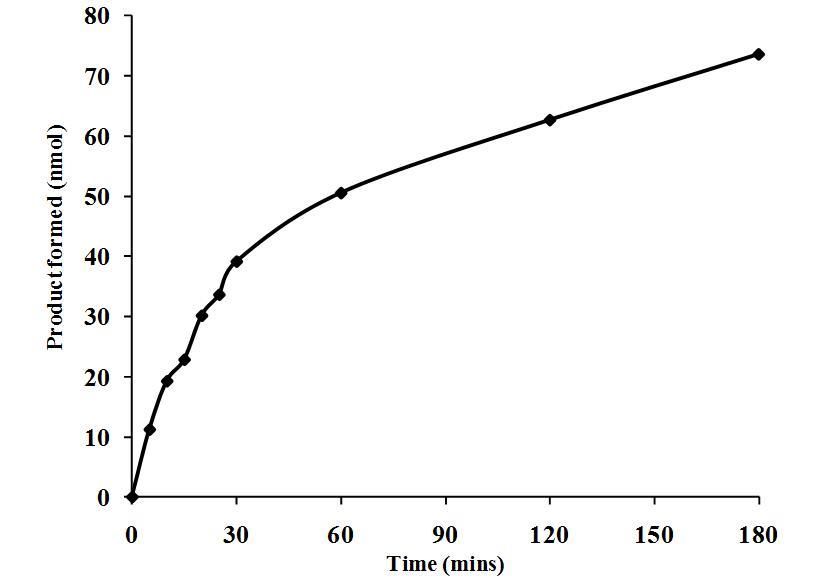
ISSN 2347-6893
                                                                                    Volume 11 Number1
                                                                         Journal of Advances in Biology

acids. More recent studies in our laboratory have identified circulating sPLA 2 in the American alligator (Alligator
mississippiensis) [19], broad snouted-caiman (Caiman latirostris) and yacare caiman (Caiman yacare) [20]. In addition,
sPLA2 activity has been identified three species of African crocodiles, the Nile crocodile (C. niloticus), dwarf crocodile,
dwarf crocodile (Osteolaemus tetraspis), and the slender-snout crocodile (Mecistops cataphractus) [21].
The data displayed in Figure 1 illustrate the titer-dependent PLA2 activity in Komodo dragon serum. Inclusion of only one
µL of serum resulted in the formation of 9.7 nmol of product after 30 min of incubation with substrate. The activity
increased dramatically when 2-50 µL of serum were used, and then increased less dramatically from 100-500 µL. The
curve appeared to be asymptotic in nature, approaching a maximum product formation of approximately 85-90 nmol. This
strong activity is not surprising considering the potent and broad-acting antibacterial activity of the Komodo dragon serum
[11], and also the effective serum complement activity [12] previously described for these animals. These animals are
known to have a broad spectrum of both gram positive and negative bacterial species in their saliva [22]. Therefore, the
strong PLA2 activity shown in Figure 1 might help defend against infection of injuries due to interspecies fighting during
territorial disputes, feeding frenzies, and during mating conflicts.

                Figure 1. Serum volume-dependent PLA2 activity in serum from the Komodo dragon
Different volumes (1-500 µL) were incubated with 100 µL of BODIPY-labeled E. coli bacteria in a 750 mL reaction. After
one hour, the samples were centrifuges and the fluorescent activity of the resulting supernatant was determined as
described in the Materials and Methods section. The results are presented as the means standard deviations of four
independent determinations and are expressed as nmol of product formed/30 min reaction incubation.
The data in Figure 2 demonstrate the kinetic curve of Komodo dragon serum PLA 2 activity. The response was biphasic
and showed the high rate of conversion of substrate to product. The accumulation of enzymatic product occurred very
rapidly, with 11.2 nmol of product formed only five minutes after the addition of substrate. The response was linear for 30
minutes, and then a strong change in slope occurred, and the response exhibited another linear portion from 30-180 min.
The initial rate of product formation during the first thirty minutes of incubation, was approximately 1.3 nmol/min. Since a
ten-fold dilution of plasma was used for this assay, we can assume a 10-fold increase of product formation to 13 nmol/min
in undiluted plasma. This is a very high rate of product formation for PLA2. These data indicate that serum from Komodo
dragons might potentially have a high capacity to kill bacteria due to the PLA 2 activity. This rapid activity would be useful
to suppress microbial growth and proliferation within a short time after an aseptic injury or infection occurs.

2165 | P a g e
March 2018                                                                          https://cirworld.com/
ISSN 2347-6893
Volume 11 Number1
Journal of Advances in Biology
Figure 2. Time-dependent PLA2 activity in serum from Komodo dragons
Aliquots (1.1 mL) of a 38.5 mL reaction (3.5 mL serum, 1.75 mL of BODIPY-labeled E. coli bacteria, and 33.25 mL assay
buffer) were stopped at different time intervals and the fluorescent product was measured as described in the
Methodology section. The results are presented as the means standard deviations of four independent determinations
and are expressed as nmol of product formed/30 min reaction incubation.
Figure 3 shows the temperature-dependent PLA2 activity of Komodo dragon serum. At lower temperature ranges, the
Komodo dragon serum exhibited relatively low activities. However, the PLA 2 activity increased in a near-linear fashion
from 10 to 40oC (y = 8.5579x + 46.071, R² = 0.9451). The production of product at 10oC (61.0 nmol) was only half of that
produced at 40oC (122.7 nmol). The slope of the linear increase suggests an approximate increase of 2.1 nmol of product
per 1oC of temperature increase. Harlow et al. [23] showed that the preferred body temperature of Komodo dragons is
34.0-35.6oC, and thus these animals have a relatively high PLA2 activity (111.1 – 114.3 nmol/30 min) in this temperature
range. The PLA2 thermal profile presented in Figure 3 exhibits different characteristics than the temperature dependent
serum complement activity reported by Merchant et al. [12], but was almost identical to the dipeptidyl peptidase IV activity
[13]. These data would seem to indicate that an innate immune response in Komodo dragons would be optimal at 35-
40oC. It would be reasonable to expect that these animals would need optimal immune activity during warmer weather
when they are more active, and might be more likely to engage in territorial disputes.
Figure 3. Temperature-dependent PLA2 activity in Komodo dragon serum
2166 | P a g e
March 2018 https://cirworld.com/ISSN 2347-6893
Volume 11 Number1
Journal of Advances in Biology
Serum from pooled serum from Komodo dragons were incubated at different temperatures with BODIPY-labeled E. coli
bacteria in a 750 µL reaction. After one hour, the samples were centrifuges and the fluorescent activity of the resulting
supernatant was determined as described in the Materials and Methods section. The results are presented as the means
standard deviations of four independent determinations and are expressed as nmol of product formed/30 min reaction
incubation.
Longo et al. [24] showed that BPAB is a specific inhibitor of both secretory and intracellular PLA 2. This compound acts as
a suicide substrate for PLA2, and alkylates a specific histidine residue in the active site [25]. Figure 4 shows the
concentration-dependent effects of BPAB on fluorescent product formation by serum from Komodo dragons. Treatment
with 50 µ M BPAB produced no discernable change in activity, but inclusion of 100 µ M BPAB resulted in a 30.4%
reduction in accumulation of fluorescent product, compared to the plasma-mediated reaction in the absence of BPAB.
Further treatment with 500 and 1000 µ M BPAB produced stepwise decreases of 76.5 and 85.3% in activity (pISSN 2347-6893
Volume 11 Number1
Journal of Advances in Biology
REFERENCES
1. Brown, W. J., Chambers, K., and Doody, A. 2003. Phospholipase A2 (PLA2) enzymes in membrane trafficking:
mediators of membrane shape and function. Traffic 4, 214-221.
2. Fonteh, A., N., Bass, D. A., Marshal, L. A., Seeds, M., Samet, J. M., and Chilton, F. H. 1994. Evidence that
secretory phospholipase A2 plays a role in arachidonic acid release and eicosanoid biosynthesis by mast cells.
J. Immunol. 152, 5438-5446.
3. Balsinde, J., Winstead, M. V., and Dennis, E. A. 2002. Phospholipase A2 regulation of arachidonic acid
mobilization. FEBS Lett. 531, 2-6.
4. Vadas, P., Browning, J., Edelson, J. and Pruzanski, W. 1993. Extracellular phospholipase A2 expression and
inflammation: the relationship with associated disease states. J. Lipid Mediat. Cell Signal. 8, 1–30.
5. Dominiecki, M. E., and Weiss, J. 1999. Antibacterial action of extracellular mammalian group IIA phospholipase
A2 against grossly clumped Staphylococcus aureus. Infection and Immunity, 67(2), 2299–2305.
6. Beers, S. A., Buckland, A. G., Koduri, R. S., Cho, W., Gelb, M. H. and Wilton, D. C. 2002. The antibacterial
properties of secreted phospholipases A2,” J. Biol. Chem. 277, 1788–1793.
7. Huhtinen, H. T., Gronroos, J. O., and Gronroos J. M. 2003. Antibacterial effects of human group IIA and group
XIIA phospholipase A2 against Helicobacter pylori in vitro. Acta Pathol. Microbiol. Scand. 114, 127– 130.
8. Jessop, T. S., Madsen, T., Sumner, J., Rudiharto, H., Phillips, J. A., and Ciofi, C. 2006. Maximum body size
among insular Komodo dragon populations covaries with large prey density. Oikos 112, 422-429.
doi:10.1111/j.0030-1299.2006.14371
9. Bull, J. J., Jessop, T. S., and Whiteley, M. 2010. Deathly drool: evolutionary and ecological basis of septic
bacteria in Komodo dragon mouths. PLoS ONE 5(6): e11097. doi:10.1371/journal.pone.0011097.
10. Fry B. G., Wroe S., Teeuwisse, W., van Osch, M. J. P., Moreno, K., Ingle, J., McHenry, C., Ferrara T., Clausen
P., Scheib H., Winter K. L., Greisman L., Roelants K., van der Weerd L., Clemente C. J., Giannakis E., Hodgson
W. C., Luz S., Martelli P., Krishnasamy K., Kochva E., Kwok H. F., Scanlon D., Karas J., Citron D. M., Goldstein
E. J. C., McNaughtan, J. E., Normalna, J. A. 2009. A central role for venom in predation by Varanus
komodoensis (Komodo Dragon) and the extinct giant Varanus (Megalania) priscus. Proc. Natl. Acad.Sci. USA,
106, 8969-8974.
11. Merchant, M., Henry, D., Falconi, R., Muscher, B., and Bryja, J. 2013. Antibacterial activity of serum from the
Komodo dragon (Varanus komodoensis). Microbiol. Res. 4, 16-20.
12. Merchant, M., Falconi, R., Muscher, B., and Bryja, J. 2013. Characterization of serum complement activity in
serum of the Komodo dragon (Varanus komodoensis). Adv. Biol. Chem. 2, 353-359.
13. Merchant, M., Henry, D., Falconi, R., Musher, B., and Bryja, J. 2015. Characterization of dipeptidyl peptidase
enzyme activity in serum of the Komodo dragon (Varanus komodoensis). Int. J. Biochem. Res. Rev. 5(2), 145-
152.
14. Bishop, B., Juba, M., Russo, P., Barksdale, S., Scott, S., Settlage, R., Michalak, P., Gupta, K., Vliet, K., Schnur,
J., and Van Hoek, M. 2017. Discovery of Novel Antimicrobial Peptides from Varanus komodoensis (Komodo
Dragon) by Large-Scale Analyses and De-Novo-Assisted Sequencing Using Electron-Transfer Dissociation Mass
Spectrometry. J. Proteom. Res. 16, 1470-1482.
15. Ozinsky, A., Underhill, D. M., Fontenot, J. D., Hajjar, A. M., Smith, K. D., Wilson, C. B., Schroeder, L., and Adrem,
A. 2000. The repertoire for pattern recognition of pathogens by the innate immune system is defined by
cooperation between Toll-like receptors. Proc. Natl. Acad.Sci. USA 97, 13766–13771.
16. Kawai, T., and Akira, S. 2010. The role of pattern-recognition receptors in innate immunity: update on toll-like
receptors. Nature Immunol. 11, 373-384.
17. Fraenkel-Conrat, H. 2008. Snake Venom Neurotoxins Related to Phospholipase A2. J. Toxicol. 1, 205-221.
18. Nevalainen, T. J. Kanchanapangka, S., Youngprapakorn, P., Webb, G. J. W., Manolis, S. C. and Scott, K. F.,
Phospholipase A2 activity of crocodile serum. Reptilia-Amphibia 30, 119–125.
19. Merchant, M., Heard, R., and Monroe, C. 2009. Characterization of phospholipase A2 activity in serum of the
American alligator (Alligator mississippiensis). J. Exp. Zool. A 311, 662-666.
20. Siroski, P., Merchant, M., Poletta, G., Larriera, A., and Ortega, H. 2013. Detection and characterization of
phospholipase A2 (PLA2) in Caiman latirostris and Caiman yacare plasma. Zool. Sci. 30, 35-41.
21. Merchant, M., Royer, A., Broussard, Q., Gilbert, S., and Shirley, M.H. 2011. Characterization of serum
phospholipase A2 activity in three diverse species of West African crocodilians. Biochem Res. Int. 1-7.
2168 | P a g e
March 2018 https://cirworld.com/ISSN 2347-6893
Volume 11 Number1
Journal of Advances in Biology
22. Montgomery, J. M., Gillespie, D., Sastrawan, P., Freeking, T. M., and Stewart, G. L. 2002. Aerobic salivary
bacteria in wild and captive Komodo dragons. J. Wildl. Dis. 38, 545-551.
23. Harlow, H., Purwandana, D., Jessop, T., and Phillips. J. 2010. Size-Related Differences in the Thermoregulatory
Habits of Free-Ranging Komodo Dragons. Int. J. Zool. http://dx.doi.org/10.1155/2010/921371.
24. Longo, W. E., Grossman, E; M;, Erickson, B., Panesar, N., Mazuki, A. E., Kaminski, D. L. 1999. The effect of
phospholipase A2 inhibitors on proliferation and apoptosis of murine intestinal cells. J. Surg. Res. 84, 51–56.
25. Volwerk, J.J., Pieterson, W.A., de Haas, G.H., Histidine at the active site of phospholipase. Biochem. 13, 1446-
1454.
2169 | P a g e
March 2018 https://cirworld.com/You can also read