HADRON THERAPY IN CHILDREN - AN UPDATE OF THE SCIENTIFIC EVIDENCE FOR 15 PAEDIATRIC CANCERS - KCE
←
→
Page content transcription
If your browser does not render page correctly, please read the page content below

KCE REPORT 235

HADRON THERAPY IN CHILDREN
AN UPDATE OF THE SCIENTIFIC EVIDENCE FOR 15 PAEDIATRIC CANCERS

2015                                                             www.kce.fgov.be


KCE REPORT 235
HEALTH TECHNOLOGY ASSESSMENT

HADRON THERAPY IN CHILDREN
AN UPDATE OF THE SCIENTIFIC EVIDENCE FOR 15 PAEDIATRIC CANCERS

ROOS LEROY, NADIA BENAHMED, FRANK HULSTAERT, FRANÇOISE MAMBOURG, NICOLAS FAIRON, LIESBET VAN EYCKEN, DIRK DE RUYSSCHER

2015                                                                                                    www.kce.fgov.be
COLOPHON
Title: Hadron therapy in children – an update of the scientific evidence for 15 paediatric cancers
Authors: Roos Leroy (KCE), Nadia Benahmed (KCE), Frank Hulstaert (KCE), Françoise Mambourg (KCE), Nicolas Fairon
(KCE), Liesbet Van Eycken (Stichting Kankerregister – Fondation Registre du Cancer), Dirk De Ruysscher (KU
Leuven)
Project coordinator: Marijke Eyssen (KCE)
Reviewers: Raf Mertens (KCE), Sabine Stordeur (KCE), Geneviève Veereman (KCE)
External experts: Edward Baert (UGent), Yves Benoit (UGent), Sylviane Carbonnelle (AFCN – FANC), Olivier de Witte (Erasme;
ULB), Bart Depreitere (KU Leuven), Lorraine Donnay (Clinique & Maternité Sainte-Elisabeth, Namur), Hilde Engels
(RIZIV – INAMI), Nancy Van Damme (Stichting Kankerregister – Fondation Registre du Cancer), Paul Van Houtte
(Institut Jules Bordet; ULB), Claudia Wild (Ludwig Boltzmann Institute, Austria)
External validators: Gudrun Goitein (Since September 2014 retired from Paul Scherrer Institute, Villigen, Switzerland), Edward C.
Halperin (New York Medical Centre, US), Stefaan Van Gool (KU Leuven)
Acknowledgements: Kris Henau (Stichting Kankerregister – Fondation Registre du Cancer), Mattias Neyt (KCE), Jo Robays (KCE),
Chris Segaert (RIZIV – INAMI), Beate Timmerman (Westdeutsches Protonentherapiezentrum Essen, Germany),
Leen Verleye (KCE)
Other reported interests: None declared
Layout: Ine Verhulst
Coverpictures: The left cover image is copyrighted by Sage Publications, Inc.
The right cover image is copyrighted by Eric Bouvet / Institut Curie (ref. 4487)
Disclaimer: The external experts were consulted about a (preliminary) version of the scientific report. Their
comments were discussed during meetings. They did not co-author the scientific report and did not
necessarily agree with its content.
Subsequently, a (final) version was submitted to the validators. The validation of the report results
from a consensus or a voting process between the validators. The validators did not co-author the
scientific report and did not necessarily all three agree with its content.
Finally, this report has been approved by common assent by the Executive Board.
Only the KCE is responsible for errors or omissions that could persist. The policy recommendations
are also under the full responsibility of the KCE.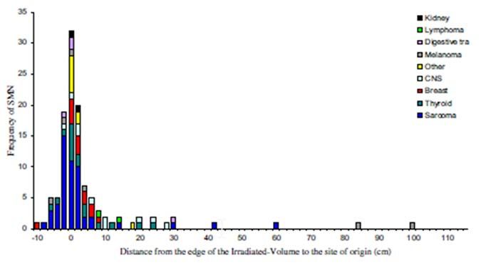
Publication date:                08 January 2015
Domain:                          Health Technology Assessment (HTA)
MeSH:                            Proton therapy; Heavy ions; Radiotherapy; Review [Publication type]
NLM Classification:              WN 250.5.P7
Language:                        English
Format:                          Adobe® PDF™ (A4)
Legal depot:                     D/2015/10.273/04

Copyright:                       KCE reports are published under a “by/nc/nd” Creative Commons Licence
                                 http://kce.fgov.be/content/about-copyrights-for-kce-reports.

How to refer to this document?   Leroy R, Benahmed N, Hulstaert F, Mambourg F, Fairon N, Van Eycken L, De Ruysscher D. Hadron therapy in
                                 children – an update of the scientific evidence for 15 paediatric cancers. Health Technology Assessment (HTA)
                                 Brussels: Belgian Health Care Knowledge Centre (KCE). 2015. KCE Reports 235. D/2015/10.273/04.
                                 This document is available on the website of the Belgian Health Care Knowledge Centre.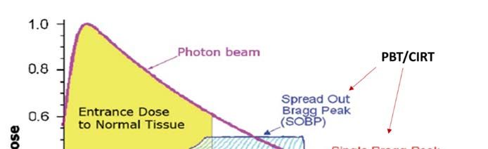
KCE Report 235                                          Hadron therapy                                                                                                                 1

■ TABLE OF CONTENTS
                 LIST OF FIGURES ...............................................................................................................................................4
                 LIST OF TABLES .................................................................................................................................................4
                 LIST OF ABBREVIATIONS & ACRONYMS........................................................................................................5
                 ■       SCIENTIFIC REPORT............................................................................................................................9
                 1            INTRODUCTION ....................................................................................................................................9
                 1.1          CANCER IN CHILDREN AND ADOLESCENTS ....................................................................................9
                              1.1.1  The burden of cancer in children and adolescents ..................................................................9
                              1.1.2  Prognosis ...............................................................................................................................10
                              1.1.3  Sequelae of cancer (therapy) in paediatric cancer survivors .................................................12
                 1.2          HADRON THERAPY ............................................................................................................................14
                              1.2.1  Radiotherapy and radiation effects ........................................................................................14
                              1.2.2  History of hadron therapy .......................................................................................................14
                              1.2.3  Photon versus hadron (charged particle) therapy ..................................................................15
                              1.2.4  Radioprotection ......................................................................................................................17
                              1.2.5  Carbon ion therapy.................................................................................................................17
                              1.2.6  Proton beam therapy..............................................................................................................18
                              1.2.7  Conclusions ............................................................................................................................23
                 1.3          CONCLUSIONS OF THE 2007 KCE REPORT ON HADRON THERAPY ..........................................24
                 1.4          HADRON THERAPY IN BELGIUM ......................................................................................................24
                 1.5          OBJECTIVE OF THIS STUDY .............................................................................................................25
                 2            METHODS ............................................................................................................................................26
                 2.1          LITERATURE SEARCH .......................................................................................................................26
                 2.2          QUALITY APPRAISAL .........................................................................................................................27
                 2.3          DATA EXTRACTION ............................................................................................................................27
                 2.4          STATISTICAL ANALYSIS ....................................................................................................................27
                 2.5          GRADING EVIDENCE .........................................................................................................................27
                 2.6          VALIDATION ........................................................................................................................................29
2 Hadron therapy KCE Report 235
3 RESULTS .............................................................................................................................................30
3.1 NUMBER OF (POTENTIAL) PATIENTS PER INDICATION UNDER STUDY ....................................30
3.2 CHORDOMA & CHONDROSARCOMA ...............................................................................................32
3.2.1 Background ............................................................................................................................32
3.2.2 What is the clinical effectiveness of proton beam therapy in children with skull base and
(para)spinal chordoma or with skull base chondrosarcoma? ................................................33
3.3 CRANIOPHARYNGIOMA.....................................................................................................................35
3.3.1 Background ............................................................................................................................35
3.3.2 What is the clinical effectiveness of proton beam therapy in children with
craniopharyngioma?...............................................................................................................36
3.4 EPENDYMOMA....................................................................................................................................38
3.4.1 Background ............................................................................................................................38
3.4.2 What is the clinical effectiveness of proton beam therapy in children with ependymoma? ...38
3.5 ESTHESIONEUROBLASTOMA ...........................................................................................................40
3.5.1 Background ............................................................................................................................40
3.5.2 What is the clinical effectiveness of proton beam therapy in children with
esthesioneuroblastoma? ........................................................................................................40
3.6 EWING SARCOMA ..............................................................................................................................42
3.6.1 Background ............................................................................................................................42
3.6.2 What is the clinical effectiveness of proton beam therapy in children with Ewing sarcoma? 43
3.7 CNS GERMINOMA ..............................................................................................................................44
3.7.1 Background ............................................................................................................................44
3.7.2 What is the clinical effectiveness of proton beam therapy in children with CNS
germinoma? ...........................................................................................................................44
3.8 LOW-GRADE GLIOMA (INCL. OPTIC PATHWAY GLIOMA)..............................................................45
3.8.1 Background ............................................................................................................................45
3.8.2 What is the clinical effectiveness of proton beam therapy in children with low-grade
glioma? ...................................................................................................................................46
3.9 MEDULLOBLASTOMA & OTHER PRIMITIVE NEUROECTODERMAL TUMOURS (PNET).............48
3.9.1 Background ............................................................................................................................48KCE Report 235 Hadron therapy 3
3.9.2 What is the clinical effectiveness of proton beam therapy in children with medulloblastoma
and PNET? .............................................................................................................................49
3.10 NON-RESECTABLE OSTEOSARCOMA.............................................................................................51
3.10.1 Background ............................................................................................................................51
3.10.2 What is the clinical effectiveness of proton beam therapy in children with non-resectable
osteosarcoma? .......................................................................................................................52
3.10.3 What is the clinical effectiveness of carbon ion radiotherapy (CIRT) in children with non-
resectable or incompletely resected high-grade osteosarcoma? ..........................................53
3.11 PELVIC SARCOMAS ...........................................................................................................................54
3.11.1 Background ............................................................................................................................54
3.11.2 What is the clinical effectiveness of proton beam therapy in children with pelvic
sarcomas? ..............................................................................................................................55
3.12 PINEAL PARENCHYMAL TUMOURS .................................................................................................55
3.12.1 Background ............................................................................................................................55
3.12.2 What is the clinical effectiveness of proton beam therapy in children with pineal
parenchymal tumours?...........................................................................................................56
3.13 RETINOBLASTOMA ............................................................................................................................56
3.13.1 Background ............................................................................................................................56
3.13.2 What is the clinical effectiveness of proton beam therapy in children with retinoblastoma? .57
3.14 RHABDOMYOSARCOMA ....................................................................................................................58
3.14.1 Background ............................................................................................................................58
3.14.2 What is the clinical effectiveness of proton beam therapy in children with
rhabdomyosarcoma? .............................................................................................................59
3.15 (PARA-)SPINAL ‘ADULT TYPE’ SOFT TISSUE SARCOMA ..............................................................61
3.15.1 Background ............................................................................................................................61
3.15.2 What is the clinical effectiveness of proton beam therapy in children with (para)spinal adult-
type soft tissue sarcomas?.....................................................................................................62
3.16 SUMMARY OF SELECTED STUDIES ................................................................................................63
4 DISCUSSION & CONCLUSIONS ........................................................................................................69
5 RECOMMENDATIONS ........................................................................................................................72
■ REFERENCES .....................................................................................................................................734 Hadron therapy KCE Report 235
LIST OF FIGURES Figure 1 – Cancer in children and adolescents: new diagnoses by tumour type and age group,
Belgium 2004-2009 ...............................................................................................................................................9
Figure 2 – Cancer in children and adolescents by tumour type, Belgium 2004-2009 ........................................10
Figure 3 – Frequency distribution of the distances from the edge of the irradiated volume to the
site where solid second neoplasms developed among 115 patients. ................................................................13
Figure 4 – Radiation dose profiles: photons vs. protons ....................................................................................15
Figure 5 – Physical effectivity and radiobiological differential effect of different RT types .................................16
Figure 6 – Passive scattering vs. pencil beam (active) scanning .......................................................................19
Figure 7 – Measured depth–dose curves of an approximately 130 MeV proton beam......................................22
LIST OF TABLES Table 1 – Country-weighted 5-year overall survival (95% CI) by ICCC diagnostic category, sex and age,
for cases diagnosed between 2000 and 2007 ....................................................................................................11
Table 2 – Indications under study .......................................................................................................................25
Table 3 – PICO table and selection criteria ........................................................................................................26
Table 4 – A summary of the GRADE approach to grading the quality of evidence for each outcome ..............28
Table 5 – Levels of evidence according to the GRADE system .........................................................................28
Table 6 – Downgrading the quality rating of evidence using GRADE ................................................................29
Table 7 – Number of (potential) patients per indication under study ..................................................................31KCE Report 235 Hadron therapy 5
LIST OF ABBREVIATION DEFINITION
2D-EBRT Two-dimensional external beam radiotherapy
ABBREVIATIONS &
3D-CPT Three-dimensional conformal proton therapy
ACRONYMS ALARA As low as reasonably achievable
ARMS Alveolar rhabdomyosarcoma
BCR Belgian Cancer Registry
BRIEF Behaviour Rating Inventory of Executive Function
cCR Clinically assessed complete response
CEBAM Belgian Centre for Evidence-Based Medicine
CE-PET/CT Contrast enhanced positron emission tomography - computed tomography
CFFS Cystic failure-free survival
CGE Cobalt gray equivalent
CH Chordoma
CI Confidence interval
CIn Cumulative incidence
CIRT Carbon ion radiotherapy
CND Comprehensive neck dissection
CNS Central Nervous System
CoR Complete remission
CRT Chemoradiotherapy
CS Chondrosarcoma
CSI Craniospinal irradiation
CT Computed tomography
CTC or CTC AE Common terminology for adverse events
CTV Clinical target volume
DFS Disease free survival
DNA Deoxyribonucleic acid
DR Distal recurrence rate6 Hadron therapy KCE Report 235
DSS Disease specific survival
EBRT External beam radiotherapy
EFS Event free survival
ENB Esthesioneuroblastoma
ERMS Embryonal rhabdomyosarcoma
ESMO European Society for Medical Oncology
FDG-PET/CT Fluorodeoxyglucose Positron emission tomography - computed tomography
FFS Failure free survival
FU Follow-up
GBM Glioblastoma multiforme
GCT Germ cell tumour
GH Growth hormones
GRADE Grading of Recommendations Assessment, Development and Evaluation
GSI Gesellschaft für Schwerionenforschung
GTR Gross total resection
GTV Gross tumour volume
Gy Gray, International System of Units (SI) unit of absorbed radiation
GyE Gray equivalent
HIT Heavy ion therapy
HIT Heidelberg Ion Therapy Center
HR Hazard ratio
HRQoL Health-related quality of life
HTA Health technology assessment
ICCC International Classification of Childhood Cancers
IGF-I Insulin-like growth factor 1
IMPT Intensity modulated proton beam therapy
IMRT Intensity modulated radiotherapy
Incl. IncludingKCE Report 235 Hadron therapy 7
IORT Intraoperative radiotherapy
IQ Intelligence Quotient
KCE Belgian Health Care Knowledge Centre
KPS Karnofsky Performance Scale
LCR Local control rate
LET Linear energy transfer
LR Local recurrence rate
MDI Mental development index (inventory)
MRI Magnetic resonance imaging
NFFS Nodular failure-free survival
NGGCT Nongerminomatous germ cell tumour
NIRS National Institute for Radiation Science
NR Not reported
OAR Organs at risk
OER Oxygen enhancement ratio
OR Odds ratio
OS Overall survival
PB Pineoblastoma
PBT Proton beam therapy
PC Pineocytoma
PET Positron emission tomography
PET-CT Positron emission tomography - computed tomography
PICO Participants–Interventions–Comparator–Outcomes
PFS Progression-free survival
PNET Primitive neuroectodermal tumours
PP Pseudo progression
PPT Pineal parenchymal tumours
PPTID PPT with intermediate differentiation8 Hadron therapy KCE Report 235
PR Partial remission
PSI Paul Scherrer Institute
PTV Planning target volume
QoL Quality of life
RANO Response assessment in neuro-oncology
RBE Relative biological effectiveness
RcR Recurrence rate
RCT Randomised controlled trial
RIZIV-INAMI National Institute for Health and Disability Insurance (Rijksinstituut voor Ziekte- en
Invaliditeitsverzekering - Institut National d’Assurance Maladie-Invalidité)
RMS Rhabdomyosarcoma
RpR Response rate
RR Risk ratio/ relative risk
RT Radiotherapy
RT-ind Radiotherapy induced
SD Standard deviation
SE Standard error
SEER Surveillance, Epidemiology and End Results (database)
SIB-R Scales of Independent Behaviour-Revised
SM Secondary malignancy
SOBP Spread Out Bragg Peak
SRT Stereotactic radiotherapy
SSF Special solidarity fund
StD Stable disease
STS Soft tissue sarcomas
TNM Classification (of T describes the size of the primary tumour and whether it has invaded nearby
Malignant Tumours tissue; N describes nearby (regional) lymph nodes that are involved; M describes
distant metastasis
WHO World Health Organisation
WVRT Whole ventricular radiation therapyKCE Report 235 Hadron therapy 9
■ SCIENTIFIC REPORT 1 INTRODUCTION
1.1 Cancer in children and adolescents
1.1.1 The burden of cancer in children and adolescents
In Belgium, childhood cancer comprises less than 1% of the total cancer
burden. Every year, about 320 children (0-14 years) and 175 adolescents
(15-19 years) are diagnosed with cancer1. Leukaemias, brain tumours,
lymphomas and carcinomas are the most frequent malignancies in children
and adolescents (Figure 1). Yet, the proportion of each tumour type varies
by age group. In the age category 0-4 years, leukaemias comprise 31% of
all tumour diagnoses; after that age, the incidence decreases. In
adolescents, lymphomas (25%) and carcinomas (22%) are the most
common tumour types; they are less common at younger age (4% and 1%
respectively in the age group 0-4 years)1(Figure 2).
Figure 1 – Cancer in children and adolescents: new diagnoses by
tumour type and age group, Belgium 2004-2009
[Figure – Source: BCR report “Cancer Incidence in Belgium. Special Issue: Cancer
in children and adolescents. 2013” p141]10 Hadron therapy KCE Report 235 The highest incidence rates for neuroblastomas and other peripheral nerve 1.1.2 Prognosis cell tumours (IV), retinoblastomas (V), renal tumours (VI) and hepatic tumours (VII) are observed in infants (i.e. age
KCE Report 235                                                               Hadron therapy                                                                               11

Table 1 – Country-weighted 5-year overall survival (95% CI) by ICCC diagnostic category, sex and age, for cases diagnosed between 2000 and 2007
                                     N       All children       Girls              Boys               Age < 1 year       Age 1-4 years      Age 5-9 years      Age 10-14
                                                                                                                                                               years

All cancers                          57956   77.9 (77.4-78.3)   78.3 (77.6-79.0)   77.5 (76.9-78.2)   77.9 (76.4-79.4)   79.3 (78.4-80.0)   77.6 (76.6-78.5)   76.6 (75.7-77.5)
Ia: acute lymphoid leukaemia         15860   86.3 (85.5-87.1)   87.6 (86.4-88.6)   85.3 (84.1-86.4)   61.8 (56.0-67.1)   90.6 (89.5-91.7)   88.1 (86.8-89.3)   77.7 (75.5-79.7)
Ib: acute myeloid leukaemia          3094    62.7 (60.5-64.9)   62.6 (59.3-65.7)   62.6 (59.4-65.6)   53.5 (47.0-59.6)   65.1 (61.4-68.5)   67.9 (63.5-71.9)   59.5 (55.1-63.5)
IIa: Hodgkin’s lymphoma              3142    95.4 (94.1-96.5)   94.3 (92.0-96.0)   96.6 (95.5-97.4)   -                  95.5 (91.1-97.8)   94.1 (89.9-96.6)   95.8 (94.5-96.8)
IIb: non-Hodgkin lymphoma            2544    84.0 (82.0-85.8)   84.0 (80.7-86.7)   84.0 (81.5-86.2)   63.3 (49.8-74.0)   78.1 (72.7-82.5)   87.0 (83.8-89.6)   85.4 (82.7-87.8)
(except Burkitt’s lymphoma)
IIc: Burkitt’s lymphoma              1443    90.2 (88.5-91.7)   85.4 (80.0-89.4)   90.7 (88.8-92.3)   40.1 (40.1-40.1)   89.3 (85.3-92.3)   91.1 (88.8-93.0)   87.2 (84.0-89.8)
III: CNS and miscellaneous           9277    57.5 (56.1-58.8)   56.8 (54.7-58.9)   58.0 (56.2-59.7)   48.3 (43.8-52.7)   57.4 (55.0-59.8)   57.0 (54.6-59.3)   60.3 (57.8-62.7)
intracranial and intraspinal
neoplasms
IIIa: ependymomas and choroid        1233    62.8 (58.4-66.8)   61.6 (55.1-67.4)   62.5 (56.5-67.9)   42.4 (30.0-54.3)   55.3 (50.6-59.8)   74.7 (66.5-81.1)   76.2 (68.6-82.2)
plexus tumour
IIIb: astrocytomas                   2714    61.5 (59.0-63.9)   62.1 (58.7-65.3)   60.7 (57.1-64.1)   64.1 (56.3-70.9)   79.4 (75.6-82.7)   55.6 (51.1-60.0)   49.3 (45.0-53.5)
IIIc: intracranial and intraspinal   3119    57.1 (54.6-59.6)   57.1 (53.0-60.9)   57.1 (53.9-60.2)   33.3 (26.6-40.2)   46.5 (42.3-50.5)   67.3 (63.3-71.0)   67.3 (62.2-71.9)
embryonal tumors
Iva: neuroblastoma and               4588    70.6 (68.4-72.6)   71.7 (68.3-74.8)   69.5 (66.7-72.1)   91.1 (89.6-92.5)   58.7 (54.8-62.5)   52.1 (45.8-58.0)   55.7 (45.5-64.6)
ganglioneuroblastoma
V: retinoblastoma                    1627    96.4 (94.6-97.6)   96.1 (93.3-97.8)   97.2 (95.5-98.2)   98.3 (96.6-99.1)   94.6 (89.9-97.2)   96.4 (79.9-99.4)   -
VIa: nephroblastoma and other        3554    89.4 (88.0-90.7)   89.7 (87.7-91.3)   89.2 (87.0-91.0)   84.3 (80.2-87.6)   91.4 (89.7-92.9)   88.2 (85.4-90.4)   76.7 (66.0-84.5)
nonepithelial renal tumours
VIIIa: osteosarcomas                 1500    69.3 (66.2-72.3)   72.8 (68.3-76.8)   66.4 (62.1-70.4)   -                  59.8 (47.6-70.1)   72.1 (66.7-76.8)   68.5 (64.9-71.9)
VIIIc: Ewing’s sarcoma and           1397    67.9 (64.2-71.2)   66.7 (61.4-71.4)   68.7 (63.7-73.1)   70.6 (58.8-79.6)   73.7 (64.6-80.7)   76.3 (71.5-80.4)   62.1 (57.1-66.6)
related sarcomas of bone
IX: rhabdomyosarcomas                2197    67.7 (64.7-70.6)   64.7 (59.4-69.4)   69.7 (66.1-73.0)   61.0 (49.7-70.5)   71.2 (66.2-75.5)   70.6 (65.5-75.2)   62.3 (56.6-67.5)
ICCC=International Classification of Childhood Cancers
[Table – Source: Gatta et al., 2014, p392]
12 Hadron therapy KCE Report 235
1.1.3 Sequelae of cancer (therapy) in paediatric cancer survivors In particular, radiation to the brain has been associated with neurocognitive
deficits, neuroendocrine dysfunction, and hearing loss, with children
Thanks to the substantial improvements in treatment outcomes, the number younger than 7 years old being the most profoundly affected.8, 11 However,
of survivors after cancer in childhood and adolescence is substantial. these side effects may also be caused by other factors, including the brain
According to Olsen et al. (2009) one person in 1000 in the general population tumour itself, hydrocephalus, chemotherapy, surgical morbidity and peri-
in high-income countries is a survivor of childhood cancer.3 Hence, the operative complications.8, 11 Craniospinal irradiation, e.g. used as part of
greatest challenge is now to maintain a sound balance between cure and medulloblastoma treatment, can further lead to primary thyroid dysfunction,
long-term morbidity in those who survive. spinal growth impairment and damage to the lung, heart and intestinal
In fact, in childhood cancer survivors the burden of chronic health problems tract.11, 12 The effect of radiation on spinal growth and development is a
is huge: 30 years after cancer diagnosis the cumulative incidence of chronic complex problem and depends not only on the age and sex of the patient
health conditions reached 73.4% (95% CI: 69.0 to 77.9), with a cumulative and the radiation dose. Also, not all spinal vertebrae respond equally to
incidence of 42.4% (95% CI, 33.7 to 51.2) for severe, disabling, or life- radiation.12, 13 The age of the paediatric patient plays a major role in the
threatening conditions or death due to a chronic condition.4 Survivors face design of the treatment plan. New developments aim at avoiding and/or
major health risks, as much due to sequelae of their treatment as due to the postponing radiotherapy in children, e.g. by altering the chemotherapy
risk of recurrence of the original tumour5. These risks include a continuing regimen.
excess risk of mortality, second primary neoplasms, neurocognitive defects,
cardiovascular disease, other organ dysfunction, endocrine disturbances, 1.1.3.2 Secondary malignancies
growth retardation and disturbance, social and mental impairments and the Among childhood cancer survivors, subsequent malignancies are the
psychosocial effects of their disease and treatment on the patients as well leading cause of non-relapse mortality.14, 15 The Childhood Cancer Survivor
as their families.4-6 Study reported a 30-year cumulative incidence of 7.9% (95% CI: 7.2-8.5) for
1.1.3.1 Impact of radiotherapy second malignant neoplasms (excluding non-melanoma skin cancer).16 This
represents a 6-fold increased risk of subsequent malignancies (occurring at
Radiation therapy is an inherent component of the curative treatment of least 5 years after treatment) relative to the general population.15 Childhood
many childhood tumours. Sadly, radiation exposure entails a major risk of cancer survivors are likely to experience more cancers as adults than are
late morbidity in long-term cancer survivors. Children are particularly individuals of similar age in the general population as a result of host-related
susceptible to the late effects of radiation, even at low doses, as factors (e.g. deletion of the RB1 gene, which causes heritable
demonstrated in epidemiologic studies of exposed populations.7,8 The retinoblastoma) and/or as a result of certain aspects of the anticancer
reasons for this high susceptibility include the sensitivity of developing and therapy they received.17
growing tissues, more proliferating cells in young patients, the longer life In radiotherapy patients, the induced tumours include carcinomas, which
expectancy resulting in a larger window of opportunity for expressing may arise in sites adjacent to or remote from the treated area.9 Their number
radiation damage, and the large number of long-term survivors8-10. In is relatively large, but the relative risk is small. Sarcomas may appear in
addition, younger patients face dosimetric disadvantages as they tend to heavily irradiated tissues, either within or close to the treatment field. The
receive on average higher secondary organ doses than adults due to absolute number of sarcomas is small, but their relative risk after radiation
geometrical factors.10 large.9KCE Report 235 Hadron therapy 13 Further, second cancers are more likely to arise in the intermediate than in Radiation-related solid malignancies develop a minimum of at least 5 to 10 the high-dose area, suggesting a bell-shaped dose-response curve or a years after treatment.15 Hence, solid cancer events within 5 years after levelling off at increasing dose due to the competing factors of cell kill and therapeutic radiation are not plausibly attributed to radiotherapy.15 Using cell mutation.18, 19 In a study analysing 115 cases of secondary cancers after current technology, it is very well possible to calculate for each part of the radiation therapy in childhood, Diallo et al. (2009) observed that 66% of body/organ the dose (incl. secondary neutrons) received. secondary tumours had occurred in the beam-bordering region (i.e., the area The knowledge on secondary malignancies in cancer survivors is surrounding the planning target volume), 22% in regions located more than constrained by the (limited) follow-up period (25 to 30 years) of the existing 5 cm from the irradiated volume and 12% in the central area of the irradiated survivor cohorts.17 Also, most studies have a too short follow-up to permit volume (which corresponds to the planning target volume)(Figure 3). Of sound conclusions. However, the Childhood Cancer Survivor Study (which interest, the peak frequency (36/115 cases, 31%) was observed for volumes follows over 14 000 children with a sibling control group) demonstrated that that had received a dose of
14                                                                                 Hadron therapy                                                         KCE Report 235

1.1.3.3      Conclusion                                                                       For cancer treatment X-rays (photon therapy, conventional radiotherapy),
                                                                                              gamma rays, and charged particles are used. Charged particle radiation
In paediatric radiation oncology, the ultimate goal is to treat the disease                   therapy or hadron therapy uses beams of protons or other charged particles,
while reducing the (late) effects of radiation on growth and development,                     such as carbon, helium, neon, or silicon. At present only protons and carbon
cognition, neuroendocrine function and the induction of secondary tumours.                    ions are in clinical use.27
Reducing the exposure of normal tissues to therapeutic radiation would
presumably decrease the risk of subsequent malignancies.15 Here, the                          1.2.2   History of hadron therapy
option of hadron therapy, particularly proton beam therapy, comes in. Based
                                                                                              In 1954 the very first human patient was treated with proton beam therapy
on the comparison of treatment plans, it was demonstrated that the use of
                                                                                              at Berkeley Radiation Laboratory (California),28 in Europe the first patient
scanned protons could lead to a reduction of the integral dose by a factor
                                                                                              was treated in Uppsala (Sweden) in 1957. Hadron therapy developed at
two to three and to a reduction of the mean and mid-to-low doses to critical
                                                                                              scientific accelerator laboratories and was initially established as a niche
structures when compared to intensity modulated photon plans and
                                                                                              within radiation oncology by pioneering clinicians and scientists working in
conventional photon techniques.23 These and other aspects of hadron
                                                                                              partnership with accelerator physicists.29 In Massachusetts General Hospital
therapy are further explored in the following paragraphs.
                                                                                              (Boston, US) for instance, the first patient was treated with proton beam
1.2     Hadron therapy                                                                        therapy in 1961 at Harvard University’s physics laboratory; it lasted another
                                                                                              40 years before patients could be treated in a clinical setting.30 In most
1.2.1     Radiotherapy and radiation effects                                                  facilities the technical equipment was limited, either regarding the usable
Among the treatment options for cancer, radiation therapy is widely used.                     energy (with low energy only superficially located tumours as tumours of the
Based upon indications for radiotherapy stated in evidence-based                              eye could be treated) or regarding the mobility of the therapy nozzle, as most
guidelines, it was estimated that 52% of cancer patients should receive                       of the centres had fixed beam lines only (allowing treatment for eye and base
radiotherapy at some point during the treatment course.24 Radiotherapy                        of skull site or prostate). Therefore, the longest and most extensive
uses high-energy radiation to kill cancer cells. Radiation therapy kills cancer               experience was made with eye or base of skull tumours as well as with
cells mostly by damaging their DNA; this damage is either direct or indirecta                 prostate cancer.6 Nowadays, proton centres implement rotating beam
ionization of the atoms which make up the DNA chain. Cancer cells whose                       application systems (gantries) to further enlarge the freedom of beam
DNA is damaged beyond repair, stop dividing and die. When the doomed                          arrangements and thus enlarge the list of possible indications.6
cells die, they are broken down and eliminated by the body’s natural                          Worldwide, more than 120 000 patients have been treated with particle
processes.25 Radiation oncology is confronted with a dichotomy: on the one                    therapy: more than 13 000 with carbon ions and more than 105 000 with
hand the tumour’s location and extent has to be defined precisely and the                     proton therapy.27
radiation dose conformed to it so that the probability of local tumour control                It is important to underscore that hadron therapy did not enter clinical
(or palliation) can be maximised while on the other hand ill effects of                       practice through an incremental series of carefully designed hypothesis-
radiation should be avoided by minimising the dose to uninvolved healthy                      driven clinical trials,29 as opposed to e.g. medicinal products which cannot
tissues.26                                                                                    enter the market (and thus clinical practice) without clinical trials.

a       Indirect ionization happens as a result of the ionization of water, forming free
        radicals within the cells that can in turn damage the DNA.
KCE Report 235 Hadron therapy 15
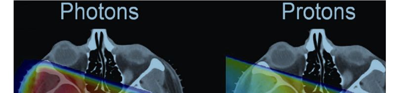
1.2.3 Photon versus hadron (charged particle) therapy to the tumour, the entrance dose usually remains substantially lower than
that of photon radiotherapy.11
1.2.3.1 Physical properties
An additional advantage is that the lateral penumbrac is generally smaller for
Photon radiation consists of high-energy electromagnetic waves. It deposits proton than for photon beams, resulting in higher conformity of the former.34
most of its energy below the skin surface and in normal tissue going in There remains however some uncertainty about the exact extent of the
(‘proximal dose’), hits the target site (the tumour) and still deposits energy lateral penumbra.35 This aspect and some other issues of concern will be
and thus affects normal tissues when coming out past the target (‘distal further explored in paragraph 1.2.6.3.
dose’) (Figure 4). Radiation oncologists try to minimize the effects on healthy
tissues as much as possible by varying the delivery paths, shaping the beam
and/or modulating the beam intensity (e.g. IMRT). Figure 4 – Radiation dose profiles: photons vs. protons
In contrast, charged particles deposit a low dose near the surface and a
large fraction of their energy at or around the target, at the end of the rangeb
of beam penetration. Tissues beyond the tumour location receive very little
of the dose. This peak energy delivery is known as the Bragg Peak (Figure
4).32 The absence of radiation distal to the target is one of the major
advantages of proton radiotherapy, allowing for substantial tissue sparing,
which is of utmost importance in nearly all anatomic sites (e.g. head and
neck, chest, spinal cord, pelvis, central nervous system).
The initial energy of the charged particles determines how deep in the body
the Bragg Peak will form. The intensity of the beam—that is, how many
particles traverse a particular area in unit time—determines the dose that
will be deposited to the tissues. By adjusting the energy of the charged
particles and the intensity of the beam, one can deliver pre-specified doses
anywhere in the body with high precision.33 In this way the proton beam can
be adjusted to match the depth and extent of the target volume and excellent
conformity can be achieved. Because the Bragg Peak of a mono-energetic
proton beam is narrow, several beams with closely spaced penetration
depths are used to treat the entirety of the tumour. This area of uniform dose
over the entirety of the tumour is termed a Spread Out Bragg Peak [Figure – Source: Cotter et al., 2012 p26911]
(SOBP)(Figure 4). While the SOBP does increase dose deposition proximal
b In passing through matter, charged particles ionize and thus lose energy in particle and the inverse of the density of the medium, and is a function of the
many steps, until their energy is (almost) zero. The distance to this point is initial velocity of the particle31.
c
called the range of the particle. The range depends on the type of particle, on The penumbra is the space in the periphery of the main target of radiation
its initial energy and on the material through which it passes. The range of a therapy; it is the volume that receives between 80% and 20% of the isodose.
heavy charged particle is approximately proportional to the mass of the16 Hadron therapy KCE Report 235
1.2.3.2 Radiobiological effect When comparing the relative merits of different types of radiations used in
therapy, two factors have to be considered: the physical selectivity and the
Because charged particles damage cell DNA in qualitatively different ways radiation quality (i.e. high-LET versus low-LET) (Figure 5).38 Improving the
than photons, the same amount of physical radiation can have much more physical selectivity (on the ordinate) should be per se always an advantage.
pronounced biological effects, resulting in larger cellular damage.33 The Selection between low- and high-LET radiation (in abscissa) depends on the
concept of relative biological effectiveness (RBE) has been introduced to tumour characteristics, i.e. histology, grade, doubling time, etc. This is thus
account for this increased efficiency of cell killing. RBE is defined as the ratio a pure radiobiological and medical issue (not technique or machine
of a dose of photons to a dose of any particle to produce the same biological dependent).38
effect. The RBE of protons is approximately 1.1, indicating that protons
result in approximately 10% more biological damage per unit dose than Figure 5 – Physical effectivity and radiobiological differential effect of
photons.33 As such, a proton dose is described as a cobalt Gray equivalent different RT types
(CGE), which translates to an equivalent photon dose measured in Gray
(Gy).11 For example, a proton dose of 50.4 CGE represents an energy
deposit equivalent to 45.8 Gy of photon radiation but has an in vivo effect of
50.4 Gy, attributable to the increased RBE. Carbon ions have a similar RBE
to protons along the particle path but have a markedly increased RBE
(estimated at 3-4) at their maximum depth of penetration. As a result, the
deleterious effects on normal tissues proximal to the tumour are expected to
be similar to proton radiotherapy, while tumour killing is enhanced at
maximum depth.11
Charged particles have a more pronounced biological effectiveness than x-
rays because they have a high rate of energy deposition (high linear energy
transfer (LET)) over a portion of the particle track which is targeted to the
tumour volume. Roughly, the higher the linear energy transfer of the
radiation, the greater the relative ability to damage cellular DNA.26, 33 Protons
have a higher linear energy transfer (LET) than photons, but their
radiobiological properties do not differ substantially.36 The LET of carbon ion
beams increases steadily with increasing the depth to reach the maximum
in the peak region.37
An additional advantage of high linear energy transfer radiation is that it can
affect hypoxic cells within a tumour, which are generally more resistant to
low linear energy transfer radiation, such as photons and electrons.33
[Figure – Source: Wambersie et al., 200438]KCE Report 235 Hadron therapy 17
1.2.4 Radioprotectiond The principle of dose limitation is applicable to exposed workers and the
public, but not to patients, should it be for diagnostic procedures or
Any practitioner treating a child with hadron therapy should be aware that it therapeutic procedures.39 However, deterministic effects will appear in
should be done in the most optimal way, as it should for any therapy. This is patients treated by hadron therapy if a certain limit is exceeded and the
especially the case for children: they are growing and their tissues are in full probability of stochastic effects increases with radiation dose to healthy
development, therefore making them more sensitive to ionizing radiation tissues.
than adults. They also have a longer life expectancy and thus more time to
develop long-term radiation-induced health effects like cancer. When considering available options for disease management, the
practitioner takes his/her decision in a multidisciplinary team, taking several
The three fundamental principles of radioprotection should always be kept factors in consideration like the age of the child, the type of tumour, in order
in mind: justification, optimization and dose limitation.39 The first principle, to offer the best treatment to his/her paediatric patient. When opting for
justification, refers to the fact that the use of ionizing radiation and, more hadron therapy, as for any other therapy using ionizing radiation, the
specifically, of this kind of therapy should be justified, i.e. the benefits of practitioner should keep radioprotection in mind.
treating a patient with ionizing radiation should outweigh the risks associated
with the use of ionizing radiation. Justification is needed on the generic level 1.2.5 Carbon ion therapy
as well as on the individual patient level.40
Heavy ions (e.g. carbon ions) differ from protons in their radiobiological
The second principle, optimization, refers to the fact that the dose of properties; the enhanced RBE is a result of the much higher ionization
radiation should be kept as low as reasonably achievable (ALARA principle). density (high LET). These differences constitute a two-edged sword: some
In this case, we should understand that the dose of radiation to healthy, non- may be advantageous while other may be disadvantageous.44 For instance,
targeted tissues should be kept as low as reasonably achievable, the dose carbon ion beams are attributed an enhanced oxygen enhancement ratio
to the target – besides being as homogenous as possible – remaining of (OER)e.44 Tumours with low radioresponsiveness against low-LET
course therapeutic.41 radiations are assumed to have a high proportion of hypoxic cells, poor
The process of optimization of medical exposure also includes – among reoxygenation pattern and high intrinsic repair capacity. It is assumed that
others – choice of equipment, quality assurance including quality control, such tumours could benefit from high-LET radiations because the reduction
evaluation of doses or activities administered to the patient.42 Concerning in the OER is achieved with increasing LET.37 If reduced OER is aimed at,
equipment choice, taking the example of delivery system, the pencil beam neon ions perform better than carbon ions.44 Heavy ions also exhibit a lesser
(cf. infra) is in most cases to be preferred to the double scattering beam (cf. dependence of radiosensitivity on the position in the cell cycle. This is one
infra) because of the sparing of normal surrounding tissues in the first of the rationales for introducing high-LET carbon ions in cancer therapy.37
case.43 More specifically in this kind of treatment, justification and However, this may also be disadvantageous, as normal cells tend to spend
optimization also imply selecting the most optimal treatment planning. less time in the X-radiosensitive phases of the cell-cycle than malignant
cells, which may result in less healthy cell protection with carbon ions than
with photons.44
d Paragraph written by S Carbonnelle, Federal Agency for Nuclear Control approximately 3 for low-LET photons, electrons, or protons, but is diminished
e with the use of high-LET irradiations to approximately 1.6 to 1.7 at LET values
The oxygen enhancement ratio (OER) is the ratio of radiation dose needed of about 100 keV/µm. The degree to which the OER is reduced is an
to inactivate severely hypoxic or anoxic tumour cells relative to the dose indication of high-LET biologic effectiveness
needed to inactivate well-oxygenated tumour cells. It has a value of (http://www.expertconsultbook.com/).18 Hadron therapy KCE Report 235
It may be argued that the lack of repair of normal tissues when exposed to 21) treated for skull base tumours (chordoma or low-grade chondrosarcoma)
high LET radiation is not relevant, because the high LET region is confined at Gesellschaft für Schwerionenforschung (GSI) in Darmstadt, Germany.11,
49
to the tumour and the normal tissues, lying outside the tumour, are only
exposed to low LET radiation.44 In reality, however, this is not the case: (1) For the present report, the majority of research questions concerned the use
the treated volume always extends the gross tumour volume, (2) tumours of proton beam therapy, with one exception, the use of carbon ion therapy
can be intertwined or embedded in a substrate of normal tissue and (3) in children with non-resectable or incompletely resected high-grade
recent analyses suggested that the LET in a treatment with carbon ions was osteosarcoma with or without metastases. The rationale for this question is
quite high even well outside the target volume.45 Hence, the risk of normal the OSCAR trial (OSteosarcoma – CArbon Ion Radiotherapy Trial to
tissue damage with carbon ions may be a larger problem than anticipated in determine the safety and efficacy of heavy ion radiotherapy in patients with
many cases.44 osteosarcoma) that is presently running at the Heidelberg Ion Therapy
Lastly, when comparing dose distributions between carbon ion and proton Center (HIT)). Enrolment is scheduled until 15 July 2015, but this may be
beams, the lateral fall-off around the target volume is more rapid in carbon alteredf.
ion beams than proton beams. However, in the region beyond the distal end
of the peak, almost no dose is deposited in protons while a certain dose is 1.2.6 Proton beam therapy
deposited in carbon ions. This distal dose is caused as the primary carbon 1.2.6.1 Types of proton therapy
ions undergo nuclear interactions and fragment into particles with lower
atomic number, which produces a fragmentation tail beyond the peak.37 The The protons emerging from a cyclotrong or synchrotronh form a narrow pencil
distal tail and the associated potentially increased toxicity to organs close to beam; in order to cover a treatment field of the size of a tumour and hence
the tail of high LET particles is a major concern about its use in children.46 produce a Spread Out Bragg Peak, the pencil beam must be either scattered
by a foil or scanned. Currently, both passive and active scanning beam
The largest experience in carbon ion beam therapy comes from the National
delivery systems are in use, though in the majority of centres the passive
Institute for Radiation Science (NIRS) in Chiba, Japan, where since 1994
scattering system is used.
more than 8000 patients with different tumour types have been treated with
carbon ion beams.27, 47 In neither of the two references, special attention is Passive scattering technique (or scatter foil technique)
given to carbon ion radiotherapy in children. Only a retrospective case series
Passive scattering is the simplest technique: a proton beam hits the scatter
on carbon ion therapy in non-resectable osteosarcoma treated at NIRS in
foil and is spread laterally (Figure 6). The beam is further shaped via brass
Chiba48 with a patient population comprising a mixture of children and adults,
apertures that are placed in the gantry head. Beam depth is manipulated via
was published. To the best of our knowledge, the paediatric experience with
a modulation wheel, which produces the varying energies needed to treat
carbon therapy is extremely limited and includes 17 patients (age range 5-
the entire target under the SOBP. The beams are further shaped to conform
f http://www.klinikum.uni-heidelberg.de/OSCAR.129200.0.html?&L=1 into a closed path) is time-dependent, being synchronized to a particle beam
g of increasing kinetic energy. The synchrotron is one of the first accelerator
A cyclotron is a type of particle accelerator in which charged particles concepts to enable the construction of large-scale facilities, since bending,
accelerate outwards from the centre along a spiral path. The particles are beam focusing and acceleration can be separated into different
held to a spiral trajectory by a static magnetic field and accelerated by a components.31
rapidly varying (radio frequency) electric field.31
h A synchrotron is a particular type of cyclic particle accelerator, descended
from the cyclotron, in which the guiding magnetic field (bending the particlesKCE Report 235 Hadron therapy 19
to the distal edge of the tumour with Lucite compensators that account for The pencil beam technology has two main advantages over the passive
both tissue inhomogeneity and tumour shape.11 This technique has been in scattering technique. First, it allows for shaping of both the proximal and
use since the 1950ies and is currently the most common proton beam distal edges of the treatment field: decreasing the entry dose while
technique employed.6, 11 maintaining a lack of exit dose. Second, the neutron scatter, which is of
There are several disadvantages associated with the passive scattering concern regarding secondary cancer induction, is reduced significantly
technique: (1) for each beam individual hardware (compensators and thanks to the lack of shielding and blocks in the gantry head, an advantage
collimators) is required; (2) there is little control of the proximal edge of the that will be particularly important for the paediatric patient.6, 11 On top of that,
beam and hence conformity is often less than with IMRT and, last but not pencil beam scanning offers the possibility of intensity modulated proton
least, (3) as the proton beam is always larger than the patient-specific beam therapy (IMPT).
aperture shaped to match the target, the protons will bombard the brass
collimator and produce secondary neutrons.6, 50, 51 During passive scattered Figure 6 – Passive scattering vs. pencil beam (active) scanning
proton therapy neutrons are generated both in the treatment head (external
neutrons) and inside the patient (internal neutrons) through proton-nuclear
interactions.10 It is estimated that these external neutrons deliver a total-
body equivalent dose that is even larger than the leakage radiation from
conventional linear accelerators.9 Upon Hall’s 2006 publication,9 animated
discussions on the extent and impact of the secondary neutrons have been
held in the literature.52-54 As this issue is so important, it will be further
discussed in paragraph 1.2.6.3. Yet, the passive scattering technique may
be indicated in those cases where the target has a regular, not too complex
shape (G Goitein, personal communication).
Active scanning technique
There are two categories of active scanning systems: spot-scanning or
pencil beam scanning on the one hand and uniform beam scanning on the
other.
Spot-scanning or pencil beam scanning
This technique was first introduced at the Paul Scherrer Institute in
Switzerland. At present it is employed in only a couple of centres worldwide.
In this technique, magnets steer a small pencil beam of protons to specific
positions within a tumour target without the need for brass apertures or
compensators (Figure 6).11 Hence, by magnetic control, directing of range
shifter plates into the beam and table movements during treatment, the
proton beam gets adjusted to the target volume.6, 55 The depth of the beam
[Figure – Source: Hall 2006 p69]
is varied in the accelerator itself; this process is called active modulation.1120 Hadron therapy KCE Report 235
Intensity modulated proton beam therapy enhances the ability of the pencil Based on (in silico) planning comparative studies, it has been concluded that
beam to treat the tumour from multiple angles, at various depths and proton beams have the potential for a similar or even improved coverage of
degrees of intensity. While each treatment field generated in pencil beam the planning target volume while keeping the dose at the organs at risk
covers the entire area of the tumour, true IMPT patches together fields that significantly lower.23,58 It has been suggested that in the near future a large
treat one portion of the target area at a time until the entire tumour is treated. fraction of definitive radiation treatment would be based on particle beams
This distinction allows for more precise dose distribution in which very and 4D image guiding.59
specific amounts of radiation can be given across the peaks and valleys of An additional advantage of proton beam therapy is that it leaves its
the tumour. It’s especially well-suited for patients with complicated tumour “signature” in the body for some time as irradiated tissue emits gamma rays;
shapes nestled in the head and neck region where you want to retain key different isotopes (of carbon, nitrogen, or oxygen) have different half-lives,
functions such as vision, speech, swallowing and taste on the order of minutes.60 Real-time positron emission tomographic (PET)
(http://www.mdanderson.org/patient-and-cancer-information/proton- scans are therefore possible: the radiation dose delivered to the tissue can
therapy-center/for-patients/videos-articles-and-podcasts/impt- be assessed and compared with the planned dose. In this way, subsequent
background.pdf). doses and areas can be planned.60
Yet, pencil beam is more sensitive to any misalignment or density change. Another method to verify the position of the end-of-range of the proton beam
Uniform beam scanning is prompt gamma imaging. While primary protons normally stop inside the
In the uniform scanning technique, the system uses a range modulator, patient, protons produce secondary prompt and delayed radiation that may
patient collimator and range compensator similar to the passive scattering be used for in vivo range verification.61 Prompt gamma-rays are emitted
technique, but it utilizes magnets instead of scattering foils to spread the almost instantaneously during the decay of the excited nuclear reaction
beam laterally.56 With this system, the beams are scanned in a fixed pattern products to their ground state. Therefore, they can be used for an immediate
with a uniform intensity for each layer, while in the pencil beam scanning and more accurate proton range verification than PET scanners. A number
system, beams are scanned with variable intensity and pattern.57 Overall, of prompt gamma-ray measurements along the path of proton (and carbon
the uniform scanning system uses less material in the beam path compared ion) pencil-beams have been reported; several detectors are under
to the passive delivery system and therefore is supposed to produce fewer development by different groups.61
neutrons.56 Some experts are less enthusiastic about proton beam therapy and have
expressed major concerns. Tepper (2008) notified that radiation dose
1.2.6.2 Proton beam therapy – the holy grail in paediatric distributions are a model for clinical reality, but they are not clinical reality.62
radiation oncology? Models usually predict outcomes effectively when they are used to predict
As was explained above, the principal motivation for using proton beams situations within the realm from which the models were derived (i.e.
instead of photons is the avoidance of high local doses to sensitive tissues interpolated from the data), but the use of those models to predict outcomes
(“organs at risk”, OAR), in order to reduce acute and late effects (e.g. extrapolated beyond the range of the initial data, produces answers that are
secondary malignancies, growth disturbances, neurocognitive deficits, much more suspect.62
neuroendocrine dysfunction, hearing loss). This is especially the case in Merchant (2013) argued that the differences in radiobiological effect when
children. Brain tumours, metastases in the spinal cord or tumours in the comparing photons with protons imply that we are comparing a known entity
pelvic region may be extremely difficult to treat with surgical interventions, with an unknown entity: the dose-volume histogram for proton therapy might
and radiation therapy (e.g. in case of incomplete resection) may have a mean something substantially different from the dose-volume histogram for
tremendous acute and/or late impact (see higher). photon therapy.12 He underscores that the multifaceted difference betweenYou can also read