NEW TARGETED THERAPIES FOR MALIGNANT NEURAL TUMORS - DIVA
←
→
Page content transcription
If your browser does not render page correctly, please read the page content below
Digital Comprehensive Summaries of Uppsala Dissertations
                from the Faculty of Medicine 1632

                New targeted therapies for
                malignant neural tumors
                From systematic discovery to zebrafish models

                ELIN ALMSTEDT

    ACTA
UNIVERSITATIS
 UPSALIENSIS    ISSN 1651-6206
                ISBN 978-91-513-0857-9
  UPPSALA       urn:nbn:se:uu:diva-402542
    2020
Dissertation presented at Uppsala University to be publicly examined in Rudbecksalen,
Rudbecklaboratoriet, Dag Hammarskjölds väg 20, Uppsala, Friday, 6 March 2020 at 13:00
for the degree of Doctor of Philosophy (Faculty of Medicine). The examination will be
conducted in English. Faculty examiner: Professor Rogier Versteeg (Head of Department of
Oncogenomics, Academic Medical Center, University of Amsterdam).

Abstract
Almstedt, E. 2020. New targeted therapies for malignant neural tumors. From systematic
discovery to zebrafish models. Digital Comprehensive Summaries of Uppsala Dissertations
from the Faculty of Medicine 1632. 61 pp. Uppsala: Acta Universitatis Upsaliensis.
ISBN 978-91-513-0857-9.

Cancers in the neural system presents a major health challenge. The most aggressive brain tumor
in adults, glioblastoma, has a median survival of 15 months and few therapeutic options. High-
risk neuroblastoma, a childhood tumor originating in the sympathetic nervous system, has a 5-
year survival under 50%, despite extensive therapy. Molecular characterization of these tumors
has had some, but so far limited, clinical impact. In neuroblastoma, patients with ALK mutated
tumors can benefit from treatment with ALK inhibitors. In glioblastoma, molecular subgroups
have not yet revealed any subgroup-specific gene dependencies due to tumor heterogeneity
and plasticity. In this thesis, we identify novel treatment candidates for neuroblastoma and
glioblastoma.
   In paper I, we discover novel drug targets for high-risk neuroblastoma by integrating
patient data, large-scale pharmacogenomic profiles, and drug-protein interaction maps. Using
a novel algorithm, TargetTranslator, we identify more than 80 targets for this patient group.
Activation of cannabinoid receptor 2 (CNR2) or inhibition of mitogen-activated protein kinase
8 (MAPK8) reduces tumor growth in zebrafish and mice models of neuroblastoma, establishing
TargetTranslator as a useful tool for target discovery in cancer.
   In paper II, we screen approximately 1500 compounds across 100 molecularly characterized
cell lines from patients to uncover heterogeneous responses to drugs in glioblastoma. We
identify several connections between pathway activities and drug response. Sensitivity to
proteasome inhibition is linked to oxidative stress response and p53 activity in cells, and can
be predicted using a gene signature. We also discover sigma receptors as novel drug targets for
glioblastoma and find a synergistic vulnerability in targeting cholesterol homeostasis.
   In paper III, we systematically explore novel targets for glioblastoma using an siRNA
screen. Downregulation of ZBTB16 decreases cell cycle-related proteins and transcripts in
patient-derived glioblastoma cells. Using a zebrafish assay, we find that ZBTB16 promotes
glioblastoma invasion in vivo.
   In paper IV, we characterized the growth of seven patient-derived glioblastoma cell lines
in orthotopic zebrafish xenografts. Using automated longitudinal imaging, we find that tumor
engraftment strongly correlates with tumor initiation capacity in mice xenografts and that the
heterogeneous response to proteasome inhibitors is maintained in vivo.
   In summary, this thesis identifies novel targets for glioblastoma and neuroblastoma using
systematic approaches. Treatment candidates are evaluated in novel zebrafish xenograft models
that are developed for high-throughput glioblastoma and neuroblastoma drug evaluation.
Together, this thesis provides promising evidence of new therapeutic options for malignant
neural tumors.

Keywords: neuroblastoma, glioblastoma, data integration, zebrafish models, precision
medicine

Elin Almstedt, Department of Immunology, Genetics and Pathology, Neuro-Oncology,
Rudbecklaboratoriet, Uppsala University, SE-751 85 Uppsala, Sweden.

© Elin Almstedt 2020

ISSN 1651-6206
ISBN 978-91-513-0857-9
urn:nbn:se:uu:diva-402542 (http://urn.kb.se/resolve?urn=urn:nbn:se:uu:diva-402542)
To my family
List of Papers

This thesis is based on the following papers, which are referred to in the text
by their Roman numerals.

      I      Almstedt, E., Elgendy, R., Hekmati, N., Rosén, E., Wärn,
             C., Olsen,     T.K., Dyberg,     C., Doroszko,      M., Larsson,
             I., Sundström,    A., Arsenian     Henriksson,      M., Påhlman,
             S., Bexell, D., Vanlandewijck, M., Kogner, P., Jörnsten,
             R., Krona, C., Nelander, S. (2020) Integrative discovery of treat-
             ments for high-risk neuroblastoma. Nature Communications,
             11(1):71.

      II     Johansson, P., Almstedt, E., Doroszko, M., Kundu, S., Vinel, C.,
             Schmidt, L., Baskaran, S., Lundsten, S., Rosén, E., Elgendy, R.,
             Elfineh, L., Häggblad, M., Martens, U., Lundgren, B., Frigault,
             M.M., Lane, D.P., Nestor, M., Marino, S., Krona, C., Nelander,
             S. A drug association map of glioblastoma informs precision tar-
             geting of p53-dependent metabolic states. Cell Reports, in revi-
             sion.

      III    Baskaran, S., Almstedt, E., Hansson, C., Kalushkova, A., Ati-
             enza Párraga, A., Spyrou, A., Forsberg Nilsson, K., Jernberg
             Wiklund, H., Elfineh, L., Weishaupt, H., Kundu, S., Krona, C.,
             Nelander, S. ZBTB16 orchestrates growth and invasion in glio-
             blastoma. Manuscript

      IV     Almstedt, E., Rosén, E., Gloger, M., Hekmati, N., Koltowska,
             K., Krona, C., Nelander, S. Real-time evaluation of glioblastoma
             treatments in patient-specific zebrafish xenografts. Manuscript

Reprints were made with permission from the respective publishers.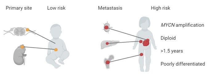
Contents

Cancers............................................................................................................ 9
  Cellular processes involved in cancer development and maintenance ....... 9
  Treating cancer using conventional and targeted therapies ...................... 11
  Nervous system tumors ............................................................................ 11
Neuroblastoma .............................................................................................. 13
  Risk groups define treatment stratifications ............................................. 13
  Molecular characterization of neuroblastoma .......................................... 14
  Neuroblastoma evolution and heterogeneity ............................................ 18
  Emerging targeted therapies for neuroblastoma ....................................... 18
Glioblastoma ................................................................................................. 20
   Current treatment of glioblastoma ............................................................ 21
   Molecular characteristics of glioblastoma ................................................ 22
   Glioblastoma subgroups and heterogeneity ............................................. 22
   Challenges for glioblastoma treatment ..................................................... 24
Strategies to identify targeted therapies ........................................................ 26
   Mapping genomic alterations in patient tumors ....................................... 26
   Mapping cancer cell line vulnerabilities .................................................. 27
   Mapping transcriptional effects of drugs.................................................. 28
   Data integration reveals possible targets and mechanisms....................... 28
   Mapping synergistic drug pairs ................................................................ 29
Experimental models of neural cancers ........................................................ 31
  Cell based models of neural cancers ........................................................ 31
  In vivo models of neural cancers .............................................................. 31
  Using zebrafish for drug evaluation ......................................................... 32
Present investigations.................................................................................... 35
   Paper I Integrative discovery of treatments for high-risk
   neuroblastoma .......................................................................................... 35
   Paper II A drug association map of glioblastoma informs precision
   targeting of p53-dependent metabolic states ............................................ 36
   Paper III ZBTB16 orchestrates growth and invasion in glioblastoma.... 36
   Paper IV Real-time evaluation of glioblastoma treatments in patient-
   specific zebrafish xenografts .................................................................... 37
Discussion and future perspectives ............................................................... 38
  Data mining as a strategy for target identification ................................... 38
  Novel targets for neuroblastoma .............................................................. 38
  Novel targets for glioblastoma ................................................................. 39
  New in vivo models for glioblastoma treatment evaluation ..................... 40
  Future perspectives ................................................................................... 41
  Concluding remarks ................................................................................. 41
Populärvetenskaplig sammanfattning ........................................................... 42
Acknowledgements ....................................................................................... 44
References ..................................................................................................... 47
Cancers

Cancer is one of the leading causes of death in the world with approximately
18 million new cases and 9.6 million deaths in 2018 (Bray et al., 2018). Com-
prising more than 100 separate entities, cancer diseases are usually classified
by the primary site tissue or the cell-of-origin. Clinically, cancer is divided
into different stages, from localized disease to invasive or metastatic disease,
which in most cases is associated with a worse prognosis.
    Cancer diseases are thought to be caused by genomic alterations (muta-
tions), leading to unrestrained cell proliferation. The genetic alterations of
cancer cells are observed at different levels, including point mutations, gene
truncations, deletions, translocations, copy number alterations, overexpres-
sion, or by epigenetic regulation. Cancer-associated gene alterations can ap-
pear randomly during cell division, as the number of stem cell divisions re-
quired to form a tissue is associated with an increased risk of developing can-
cer in that organ (Tomasetti and Vogelstein, 2015), or by external stimuli, e.g.
exposure to mutagens such as UV light or tobacco smoke (Alexandrov et al.,
2013). Age is one of the leading risk factors for developing cancer, likely due
to the accumulation of acquired mutations during life (Alexandrov et al.,
2013). Generally, childhood tumors have a lower mutational burden than adult
tumors (Alexandrov et al., 2013; Lawrence et al., 2013). In children, 7-8% of
patients carry germline gene variants that predispose for cancer (Gröbner et
al., 2018).

Cellular processes involved in cancer development and
maintenance
The mutations that cause cancer affect several dimensions of cellular func-
tions, sometimes referred to as Hallmarks of cancer (Figure 1) (Hanahan and
Weinberg, 2011). First, cancer is characterized by uncontrolled proliferation
as a result of oncogene activation, reduced ability to induce cell death, inacti-
vation of tumor suppressor pathways, and replication immortality. Second, to
sustain the elevated need of energy and oxygen as a result of cell growth and
proliferation, cancer cells alter their metabolism and induce angiogenesis
(Hanahan and Weinberg, 2011). Third, malignant cells commonly activate mi-
gratory and invasive pathways to invade into surrounding tissue. Forth, cancer

                                                                               9
cells induce a proinflammatory microenvironment, which promotes tumor growth and lacks an effective anti-tumor immune response. Underlying the mutation diversity is an increased genomic instability, which leads to an in- creased mutation rate and intratumoral heterogeneity (Hanahan and Weinberg, 2011). Cancer-associated genetic alterations are particularly frequent in a set of cellular pathways that regulate oncogenic signaling and such alterations are shared between many tumor types (Bailey et al., 2018; Sanchez-Vega et al., 2018). Commonly altered pathways include cell cycle regulators, such as the cyclin-dependent kinase (CDK) family of proteins, receptor tyrosine kinases (RTKs; e.g. ALK, and EGFR), and developmental transcription factors, such as Myc (Sanchez-Vega et al., 2018; Schaub et al., 2018). Activation of down- stream signaling pathways, e.g. PI3K-AKT-mTOR and RAS-MAPK path- ways, or alternatively, by the inactivation of pathway inhibitors, such as CDKN2A/B/C, Rb1, PTEN, and NF1, are also frequent in cancer (Sanchez- Vega et al., 2018). To avoid regulated cell death through apoptosis, tumor cells often inactivate the tumor suppressor p53 (Sanchez-Vega et al., 2018). During replication in normal somatic cells, telomeres are shortened, leading to a lim- ited number of successive cell divisions for a cell. Replication immortality require activation of telomere maintenance pathways, typically by activating telomerase reverse transcriptase (TERT) or through the alternative lengthen- ing of telomeres (ALT) pathway (Hanahan and Weinberg, 2011). Together, these alterations give a survival benefit for the cancer, but can also affect the cells negatively and induce cellular stress. To cope with this, the cell activates compensatory mechanisms, which can be exploited for therapies. Figure 1. Hallmarks of cancer. 10
Treating cancer using conventional and targeted
therapies
The current treatment regimen for most solid tumors includes a combination
of surgery, radiotherapy, and chemotherapy. For some tumor types, there are
additional treatment options. These include (i) stem cell transplantation or im-
mune checkpoint blockade that boost the immune response against the tumor,
e.g. PD1 inhibition in malignant melanoma (Robert et al., 2015), (ii) differen-
tiation therapy to limit the proliferative capacity of the tumor cells, e.g. 13-
cis-retinoic acid in neuroblastoma (Matthay et al., 1999, 2009), and (iii) tar-
geted therapy by inhibiting key oncogenes in the tumor cells, e.g. HER2 inhi-
bition in HER2-positive breast cancer (Ryan et al., 2008; Slamon et al., 2001).
Other modes of therapy have shown promising effects in clinical trials, e.g.
tumor treating fields in glioblastoma (Stupp et al., 2017), but have not yet
made it into standard treatment regimens. To an increasing extent, treatments
are tailored to every patient based on clinical or molecular biomarkers, a strat-
egy often referred to as precision medicine. Two common strategies to evalu-
ate precision therapy for cancer patients are umbrella trials (i.e. dissecting out
all possible targets within one disease) and basket trials (i.e. targeting a com-
mon molecular alteration in several different diseases) (Hierro et al., 2019).
The aim of precision medicine is to increase the treatment accuracy in a way
that a patient receives the right treatment, at the right time, and with minimal
effect on non-cancer cells.

Nervous system tumors
Cancer in the nervous system present a major health challenge. In children,
cancers in the nervous system have the second highest cancer-related mortal-
ity, after leukemia (Bray et al., 2018; Ferlay et al., 2019). The most common
malignant diagnosis includes neuroblastoma, a tumor presenting in the sym-
pathetic nervous system, and the brain tumor medulloblastoma. Cancer in the
nervous system is the 12th most common cause of cancer-related death in the
world (Bray et al., 2018; Ferlay et al., 2019) and glioblastoma is the most
common malignant brain tumor (Ostrom et al., 2019). The clinical course of
brain tumors varies drastically, where patients with glioblastoma (grade IV)
have a dismal prognosis of just over a year with the best available treatment,
while low grade lesions might be cured with surgery only (Louis et al., 2016).
In neuroblastoma, the clinical heterogeneity spans from spontaneous regres-
sion of metastatic disease, to high-risk neuroblastoma where only half of the
patients are alive 5 years after their diagnosis, despite extensive therapy (Cohn
et al., 2009). Nervous system tumors, especially brain tumors, are also a major
cause of cancer related morbidity, reduced quality of life, and long-term de-
pendencies on the health care system for survivors (Taphoorn et al., 2010).

                                                                               11
New treatments for high-risk neuroblastoma and glioblastoma are highly warranted. In this thesis, we explore different ways of identifying novel tar- geted treatments for these diagnoses and develop zebrafish xenograft models for treatment evaluation. 12
Neuroblastoma
Neuroblastoma is the most common extracranial solid tumor in children, with
a median age of 1.6 years at diagnosis (London et al., 2005). Neuroblastoma
develops in the peripheral nervous system from the developing sympathetic
ganglia, originating from the neural crest (Brodeur, 2003; Westerman et al.,
2011). The most common primary sites are the adrenal medulla and sympa-
thetic ganglia along the spinal cord, and it spreads primarily to bone marrow,
bone, lymph nodes, and liver (DuBois et al., 1999). Some patients with wide-
spread disease limited to skin, liver, and bone marrow still show spontaneous
regression and excellent survival, while high-risk patients have poor survival,
making neuroblastoma a clinically heterogeneous disease (Cohn et al., 2009).
Familial neuroblastoma is often caused by heritable mutations in the receptor
tyrosine kinase ALK or the transcription factor PHOX2B (Carén et al., 2008;
Krona et al., 2008; Mosse et al., 2004; Mossé et al., 2008; Trochet et al.,
2004).
Risk groups define treatment stratifications
Neuroblastoma patients are treated based on a risk-group system. The risk
groups are based on clinical, molecular and histological features of the tumor
including tumor spreading (stage), age of the patient, histology (proliferation
index and degree of differentiation), amplification of the MYCN gene, chro-
mosomal aberrations of 11q, and DNA ploidy (Figure 2) (Ambros et al., 2009;
Cohn et al., 2009). Staging is defined as localized, with or without invasion
into surrounding structures, or metastatic disease, according to either the In-
ternational Neuroblastoma Staging System (INSS) or the International Neu-
roblastoma Risk-Group (INRG) Staging Systems. Neuroblastoma has a sepa-
rate staging entity, 4S (MS in INRG), for young patients with widespread dis-
ease, as these tumors often regress spontaneously and are considered low-risk
(Nickerson et al., 2000). Patients older than 1.5 years belong to a higher risk
group (London et al., 2005).
The treatment of neuroblastoma varies largely between the risk groups.
Low-risk patients have an excellent survival even without treatment, or with
either surgery or chemotherapy alone (Strother et al., 2012). For the high-risk
patient group, the prognosis is much worse, with only half of the patients sur-
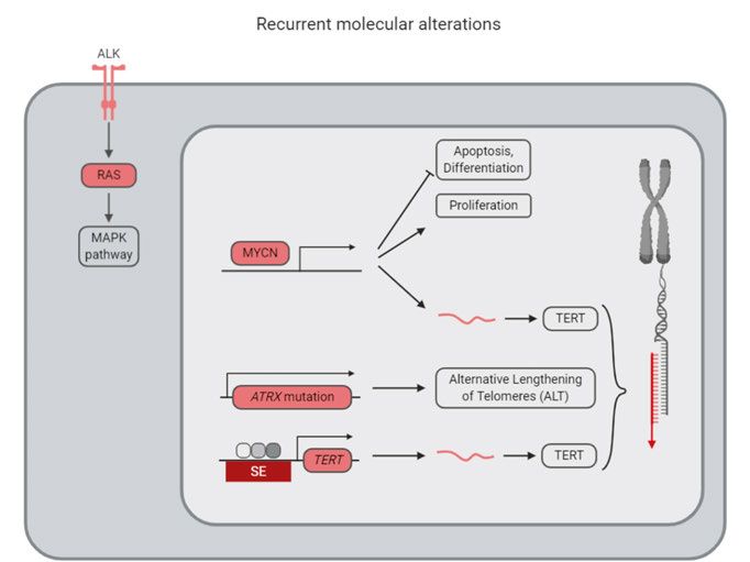
13viving after 5 years despite extensive therapy protocols including chemother- apy, surgery, radiation, immunotherapy, stem cell transplantation, and differ- entiation therapy (Kushner et al., 1994; Matthay et al., 1999; Park et al., 2011; Pinto et al., 2015). During the last decade, large-scale efforts coupling patient clinical and molecular data have started to decipher the molecular character- istics of high-risk neuroblastoma, in an effort to understand more about the mechanisms underlying the disease and to find novel treatments. Figure 2. Neuroblastoma primary and metastatic sites. Low- and high-risk patients are stratified based on clinical and molecular markers. Molecular characterization of neuroblastoma Childhood tumors typically carry fewer mutations than adult cancers. In neu- roblastoma, the average mutation burden is 12 missense somatic mutations, with a higher frequency in high-risk tumors (Molenaar et al., 2012). There are relatively few recurrent mutations between neuroblastoma patients (Figure 3). The most common mutations include activation of the receptor tyrosine kinase ALK and inactivation of the transcription factor ATRX, involved in telomere elongation, affecting approximately 10% of the patients each (Cheung et al., 2012; Molenaar et al., 2012; Pugh et al., 2013). The relative paucity of somatic mutations in targetable proteins makes it difficult to develop targeted therapies for neuroblastoma based on DNA sequencing of the primary tumor alone. Copy number aberrations occur in many high-risk patients, including gain of the MYCN locus on chromosome 2p24 (Schwab et al., 1983), loss of 1p36 (Brodeur et al., 1981; Maris et al., 2000), loss of 11q (Guo et al., 1999), and gain of 17q (Bown et al., 1999; Caron et al., 1996). Chromothripsis (localized chromosomal shredding and subsequent random assembly, leading to struc- tural variations in the genome) have been found to be associated with poor survival, affecting 18% of stage 3 and 4 patients (Molenaar et al., 2012). Struc- tural rearrangements have been associated with alterations in the DNA dam- age response pathway such as FANCM and FAN1, which are part of the Fan- coni anemia pathway (Molenaar et al., 2012). 14
MYCN
Amplification of the transcription factor MYCN, encoding the N-Myc protein,
is the most common genetic alteration in neuroblastoma and is associated with
a worse prognosis (Ambros et al., 2009). In normal cells, MYCN is expressed
during development (Zimmerman et al., 1986). Targeted expression of MYCN
in tyrosine hydroxylase (TH) positive neural crest cells is sufficient for neuro-
blastoma formation in mice (Weiss et al., 1997). Loss of N-Myc induces cell
death and differentiation (Kang et al., 2006). It has been proposed that N-Myc
regulates differentiation through miRNA control of nuclear hormone recep-
tors (Ribeiro et al., 2016). Unfortunately, N-Myc is a difficult target for drug
development and consequently many studies focus on indirect targeting of N-
Myc through reduced MYCN transcription, increased degradation of N-Myc
by targeting N-Myc phosphorylating proteins, or targeting of downstream ef-
fector pathways (Pinto et al., 2015).
ALK
Familial cases of neuroblastoma constitute 1% of the cases and 50% of these
cases are associated with activating ALK mutations (Devoto et al., 2011;
Mossé et al., 2008). In sporadic neuroblastoma, ALK is mutated in 8-12% of
the patients (Bresler et al., 2014; Carén et al., 2008; Mossé et al., 2008), and
relapse patients tend to acquire ALK mutations (Schleiermacher et al., 2014).
ALK mutation is predictive of survival (Pugh et al., 2013). ALK inhibitors are
approved for non-small cell lung cancer and targeting of ALK is under clinical
investigation, see below.
Telomere maintenance
Recently, replication immortality through telomere maintenance has been pro-
posed to be one of the main risk-determinants in neuroblastoma, along with
p53 and RAS pathway alterations (Ackermann et al., 2018). In high-risk tu-
mors, telomere maintenance is obtained through either MYCN amplification,
TERT rearrangements, or ATRX mutations, while tumors with spontaneous re-
gression lack these alterations (Ackermann et al., 2018). Further alterations in
RAS and/or p53 pathways increase the risk for the patients to very high-risk
(Ackermann et al., 2018).
Whole genome sequencing of high and low-risk patients have identified
genomic translocation in the 5p15.13 region in 21-23% of the tumors, leading
to increased TERT expression by placing an active enhancer in proximity to
the TERT gene (Peifer et al., 2015; Valentijn et al., 2015). Cases with TERT
rearrangements show increased telomere length (Valentijn et al., 2015) and
the alteration is almost mutually exclusive to MYCN amplification and ATRX
mutations (Peifer et al., 2015; Valentijn et al., 2015). However, MYCN ampli-
fied tumors upregulate TERT mRNA expression (Peifer et al., 2015). No pa-
tient samples showed TERT promoter mutations (Lindner et al., 2015), which
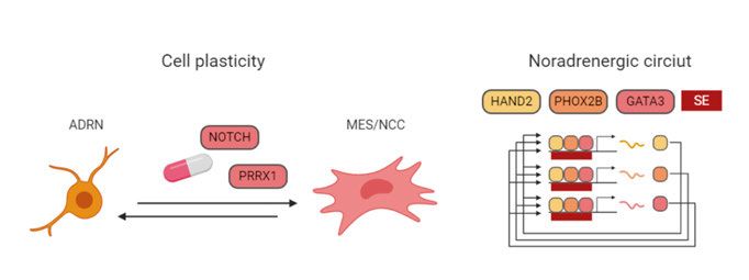
15is common in glioblastoma. In high-risk tumors without TERT upregulation, alternative lengthening of telomeres (ALT) was activated (Peifer et al., 2015). TERT in itself is a difficult drug target, but the region upstream of TERT shows Polycomb modification characteristics (Valentijn et al., 2015) and conse- quently, the TERT expression could possibly be regulated by inhibition of the Polycomb repressive complex. Loss-of-function mutations in ATRX are associated with older patients and they appear in all patients older than 12 years (Cheung et al., 2012). Structural variation in the ATRX gene leads to decreased expression (Molenaar et al., 2012). N-terminal deletions in the ATRX gene activate REST, a transcription factor which repress neuronal differentiation programs and recruit Polycomb Repressive Complex 2 (PRC2), and can possibly be targeted with EZH2 inhi- bition, a subunit of the PRC2 (Qadeer et al., 2019). Super-enhancers in neuroblastoma Due to the low frequency of mutations in neuroblastoma, there has been a recent interest in understanding the neuroblastoma epigenetic regulation. It has long been known that neuroblastoma cells can switch between epithelial- like and neuroblast-like cell states (Ross et al., 1983), indicating an epigenet- ically regulated phenotype switch. Recent studies have shown that isogenic neuroblastoma cells can adopt either a neural crest cell/mesenchymal-like state (MES/NCC) or an adrenergic/noradrenergic cell state (ADRN; Figure 3), which can be separated based on CD133 expression levels (Boeva et al., 2017; van Groningen et al., 2017). Sorted cell populations can switch phenotype both in vitro and in vivo (van Groningen et al., 2017). The cell identity is de- termined by core transcriptional regulatory networks in which a set of tran- scription factors increase their expression through a feed-forward loop, mainly driven by the activation of super-enhancers, and which are identified by an increased density of histone 3 lysine 27 acetylation and trimethylation (H3K27ac, H3K27me3) epigenetic marks (Boeva et al., 2017; van Groningen et al., 2017). The transcription factors regulating the ADRN identity include GATA3 and PHOX2B, while MES/NCC-cells are driven by AP-1 transcrip- tion factors, consisting of heterodimers of FOS and JUN family members (Boeva et al., 2017; van Groningen et al., 2017). NOTCH and PRRX1 expres- sion reprogrammed cells into a more MES/NCC state (van Groningen et al., 2017, 2019). MES/NCC cells were more resistant to chemotherapy, suggest- ing that targeting of MES/NCC cells or transforming them into ADRN cells might be a possible treatment strategy (Boeva et al., 2017; van Groningen et al., 2017). Recurrent neuroblastoma Neuroblastoma patients succumb to refractory or relapsed disease. In relapsed neuroblastoma, the mutational burden increases (Eleveld et al., 2015; 16
Schramm et al., 2015). Many mutated genes are involved in epithelial to mes-
enchymal transition (EMT) processes (Schramm et al., 2015) or activation of
the RAS-MAPK pathway, including ALK, KRAS, HRAS, PTPN11, or NF1
mutations (Eleveld et al., 2015). Patients also acquire chromosomal aberra-
tions characteristic of aggressive and metastatic disease, such as chromosome
9 deletions harboring the DOCK8 locus (Schramm et al., 2015), partial loss of
chromosome 6, 1p, or 11q, or homozygous deletion of CDKN2A (Eleveld et
al., 2015). Interestingly, the transcriptional landscape is altered in relapsed
neuroblastoma without major changes in the epigenetic profile (Schramm et
al., 2015).

Figure 3. Recurrent genomic alterations and cell plasticity in neuroblastoma. SE:
Super Enhancer.

                                                                                    17
Neuroblastoma evolution and heterogeneity
Despite the higher frequency of mutations in recurrent neuroblastoma, re-
lapsed tumors have a lower heterogeneity of chromosomal aberrations than
primary tumors (Schramm et al., 2015). Many mutations in relapse samples
are shared with the primary tumor (Eleveld et al., 2015), though the spectrum
of mutations differs between locoregional and metastatic relapses. Locore-
gional relapses show a broader spectrum of acquired mutations than metastatic
relapses (Schramm et al., 2015), suggesting that several local subclones could
be responsible for the relapse and that tumor evolution occur both temporally
and spatially.
Analysis of chromosomal rearrangements has revealed four different evo-
lutionary trajectories in neuroblastoma, with high regional heterogeneity indi-
cating a worse prognosis (Karlsson et al., 2018). Specifically, clonal sweeps,
in which one clone takes over and dominates an anatomical region, and muta-
tional explosions were both predictors of recurrence (Karlsson et al., 2018).
This motivates the collection of biopsies from several locations and that new
biopsies are collected upon recurrence.
Emerging targeted therapies for neuroblastoma
Emerging therapies for neuroblastoma aim to target specific molecular altera-
tions or specifically expressed proteins. In an early phase clinical trial, only
9% of ALK mutated patients responded to treatment with the ALK inhibitor
crizotinib (Mossé et al., 2013), possibly due to activating mutations in the ALK
kinase domain (Bresler et al., 2014). Ongoing clinical trials are trying to over-
rule the crizotinib resistance using second-generation ALK inhibitors such as
ASP3026, loratinib, and LDK-378 (Li et al., 2016)(clinicaltrials.gov,
NCT03107988, NCT01742286). Recent development of an antibody-drug
conjugate targeting ALK has showed promising effects in vivo, hopefully
treating neuroblastoma cells that would otherwise develop a resistance
through mutation of the receptor (Sano et al., 2019). In relapse samples, some
of the mutations in RAS-MAPK pathways might be targeted with MEK in-
hibitors (Eleveld et al., 2015).
MYCN, TERT, and ATRX and are three commonly altered genes in neuro-
blastoma. Unfortunately, targeting of the encoded proteins with small mole-
cule inhibitors has proven difficult. Instead, treatment strategies have aimed
to indirectly target N-Myc protein levels as it has proven difficult to regulate
the protein directly. Current efforts are made to downregulate MYCN tran-
scription by the use of BET bromodomain inhibition (Puissant et al., 2013) or
protein destabilization through PI3K (Chesler et al., 2006), ROCK (Dyberg et
al., 2017), or Aurora A kinase interference (Brockmann et al., 2013; DuBois
18et al., 2018). Inhibition of the N-Myc downstream target ornithine decarbox-
ylase, the rate-limiting enzyme of polyamine biosynthesis, using the inhibitor
DFMO, is also under clinical evaluation (clinicaltrials.gov: NCT01586260,
NCT02030964), though a treatment combination with polyamine uptake
might be needed (Gamble et al., 2019).
Other therapies in clinical studies include targeted radiotherapy using the
radioactive norepinephrine receptor ligand 131I-MIBG (DuBois et al.,
2015)(NCT03126916) and immunotherapy targeting the ganglioside 2 (GD2)
antigen using monoclonal antibodies or genetically engineered T-cells ex-
pressing chimeric antigen receptors (CAR T-cells) (Louis et al., 2011)
(NCT02107963). Clinical evaluation of the use of targeted DNA sequencing
of neuroblastoma for precision medicine is ongoing (NCT02868268).
Despite the extensive effort in identifying targeted therapies, little is known
about therapeutic targets beyond gene mutations and tissue-specific gene ex-
pression. Given the low frequency of mutations in neuroblastoma, researchers
have screened for epigenetic regulators that affect neuroblastoma growth and
differentiation (Lochmann et al., 2018; Veschi et al., 2017). A recent epige-
netically focused drug screen aiming to induce differentiation found that an
H3K27 demethylase inhibitor induced differentiation in neuroblastoma, in-
cluding retinoic-acid resistant cell lines (Lochmann et al., 2018). Another
combined siRNA and chemical screen on epigenetic regulators identified
SETD8, a histone 4 lysine 20 methyl (H4K20me1) transferase, as a possible
target in neuroblastoma (Veschi et al., 2017). SETD8 downregulation or inhi-
bition reactivated p53 pro-apoptotic functions and induced differentiation
(Veschi et al., 2017). Considering the cell plasticity and epigenetic regulation
in neuroblastoma, new ways of identifying targeted therapies based on cell
state and transcriptional programs can hopefully increase the repertoire of pos-
sible treatments for the high-risk disease.
19Glioblastoma
The most common malignant brain tumor is glioblastoma, accounting for al-
most half of all primary malignant brain tumors and 15% of all brain tumors
(Ostrom et al., 2019). Glioblastoma is more or less incurable, with a 6.8% 5-
year survival rate (Ostrom et al., 2019) and a median survival time of 15
months with standard-of-care therapy (Stupp et al., 2005). In the western pop-
ulation, more than 3 of 100.000 individuals are diagnosed with glioblastoma
every year, with a higher incidence in the Caucasian population (Ostrom et
al., 2019). The median age of diagnosis is 65 years, the incidence increasing
with age, and few patients are diagnosed before age 40 (Ostrom et al., 2019).
Age of the patient is highly correlated with disease outcome and younger pa-
tients have a better prognosis (Louis et al., 2016). Symptoms of glioblastoma
are commonly unilateral and are related to the site of the tumor or increased
intracranial pressure and can be manifested as seizures, nausea, neurocogni-
tive changes, a reduction in mobility, or severe headaches (DeAngelis, 2001).
Glioblastoma is a highly invasive, but rarely metastatic, grade IV astrocy-
toma (Louis et al., 2016). Histological features of glioblastoma include high
cellularity, poor differentiation, high mitotic activity, nuclear atypia, hetero-
geneity in cellular morphology, extensive local and distant infiltration, micro-
vascular proliferation, and necrosis (Louis et al., 2016). Glioblastoma is di-
vided into isocitrate dehydrogenase (IDH) 1/2-wild type (primary) glioblas-
toma, accounting for 90% of glioblastoma, and IDH-mutant (secondary) glio-
blastoma, which progresses from lower stage brain tumors (Ceccarelli et al.,
2016; Nobusawa et al., 2009; Ohgaki and Kleihues, 2013). Secondary glio-
blastoma has a slightly better survival prognosis than primary glioblastoma,
suggesting a different biology between the two entities (Parsons et al., 2008).
The IDH-mutant subgroup is also associated with methylated CpG islands in
the DNA, a phenotype called G-CIMP (Ceccarelli et al., 2016; Noushmehr et
al., 2010).
A glioblastoma tumor can occupy a large part of a cerebral lobe already at
presentation, even though the duration of symptoms has been very short. The
most common sites are the subcortical white matter and deep grey matter in
the temporal lobe, parietal lobe, frontal lobe, and occipital lobe (Lai et al.,
2011). Infiltration into the cortex, along the corpus callosum and into the con-
tralateral hemisphere is common (Louis et al., 2016). For IDH-mutant glio-
blastoma, the localization is predominantly the region surrounding the rostral
lateral ventricles (Lai et al., 2011), as compared to the more widespread origin
20of IDH-wild type glioblastoma. Radiologically, the tumor is visible as an ir-
regular, contrast enhancing mass with a dark necrotic center. The infiltration
into surrounding tissues is very rapid and cells can spread along white matter
tracts, or form Scherer structures (Scherer, 1938), including perineuronal sat-
ellitosis, perivascular aggregation and subpial spread. The mechanism behind
glioblastoma infiltration is not completely understood, but involves activation
of cell motility programs, cell-cell and cell-matrix interactions (e.g. through
integrins), remodeling of extracellular matrix (e.g. secretion of proteolytic en-
zymes such as matrix metalloproteinases), as well as cues from the microen-
vironment (Bellail et al., 2004; Demuth and Berens, 2004). Despite extensive
infiltration, glioblastoma does not commonly spread to other organs, although
circulating tumor cells have been found in patient blood, suggesting an im-
mune-surveillance mechanism (Sullivan et al., 2014). CD8+ T-cells might be
more prominent in long term survivors (Yang et al., 2010).
Although the cell-of-origin is still under investigation, studies suggest that
glioblastoma arise from oligodendrocyte precursor cells (Liu et al., 2011) or
neural stem/progenitor cells (Alcantara Llaguno et al., 2009; Lee et al.,
2018a). The cell-of-origin affects both tumorigenicity and drug sensitivity,
with transformed neural stem cells having a higher tumorigenic potential
(Jiang et al., 2017).
Current treatment of glioblastoma
The standard-of-care therapy for glioblastoma is surgery, radiation, and chem-
otherapy, using the DNA-alkylating agent temozolomide (Stupp et al., 2005).
Temozolomide sensitivity is affected by promoter methylation of the DNA
repair gene O-6-methylguanine-DNA methyltransferase (MGMT), causing
gene silencing (Hegi et al., 2004). Inactivation of the gene leads to a decrease
in DNA repair and an increased sensitivity to temozolomide, making MGMT
promoter methylation a biomarker of increased temozolomide sensitivity in
patients (Stupp et al., 2005). However, treatment with alkylating agents, such
as temozolomide, leads to an increased mutation rate due to the lack of func-
tional DNA repair mechanisms, and recurrent tumors commonly show a hy-
permutation phenotype (Cancer Genome Atlas Research Network, 2008).
While the separation of IDH-wild type and -mutant glioblastoma is useful
when evaluating patient prognosis, this molecular separation of glioblastoma
subgroups has not yet resulted in any molecular-guided treatment options for
these different subgroups.
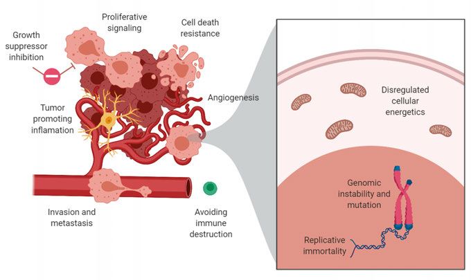
21Molecular characteristics of glioblastoma Compared to neuroblastoma, glioblastoma has a higher mutational burden and more frequently altered pathways. Recurrent genetic aberrations in glioblas- toma activate receptor tyrosine kinases (mainly EGFR, PDGFRA, MET, and FGFR) and downstream signaling pathways (RAS-BRAF and PI3K path- ways), or inhibit their negative regulators (NF1 and PTEN, respectively; Fig- ure 4) (Brennan et al., 2013; Cancer Genome Atlas Research Network, 2008). Glioblastoma cells typically inactivate genes involved in p53 regulation (most common alterations occur in TP53, MDM2, and MDM4), and activate cell cy- cle regulators upstream of RB1 (e.g. CDK4, CDK6, cyclins, and CDKN2A/B/C) (Brennan et al., 2013; Cancer Genome Atlas Research Net- work, 2008) (Figure 4). Increased EGFR signaling can be gained through mu- tation, overexpression, or deletion of exons 2-7 in the extracellular domain (EGFRvIII) leading to constitutively active receptor signaling (Brennan et al., 2013; Huang et al., 1997). EGFR-amplified glioblastoma cells are commonly located in the tumor border, suggesting a role for EGFR in promoting invasion (Snuderl et al., 2011). Glioblastoma cells acquire TERT promoter mutation or mutations in ATRX, involved in telomere maintenance (Ceccarelli et al., 2016; Eckel-Passow et al., 2015). These mutations are mutually exclusive (Cecca- relli et al., 2016). The most common chromosomal aberrations in glioblastoma include gain of chromosome 7, harboring the EGFR, MET, and CDK6 locus (Brennan et al., 2013). Glioblastoma subgroups and heterogeneity Glioblastoma is highly heterogeneous, both between patients and within the same patient. In the last decade, extensive efforts have been focused on sub- grouping different patients based on their molecular features, aiming to under- stand the underlying mechanism of this heterogeneity and to adapt treatments based on different molecular phenotypes. In IDH-wild type glioblastoma, transcriptional and methylation analysis first suggested four molecular sub- groups (Brennan et al., 2013; Sturm et al., 2012; Verhaak et al., 2010), while later studies have confirmed three of these (Ceccarelli et al., 2016; Wang et al., 2017). Named after cellular resemblance, the subgroups are called proneu- ral, mesenchymal, and classical (Verhaak et al., 2010; Wang et al., 2017). The classical subgroup included well-established glioblastoma genomic altera- tions, such as EGFR oncogene amplification or overexpression, loss of the PTEN locus on chromosome 10, downregulation of the CDKN2A/RB1 tumor suppressor pathways, wild type TP53, and an upregulation of neural precursor markers (Verhaak et al., 2010). Cells in the proneural subgroup are often TP53 mutated, have an inactivated CDKN2A pathway, an activation of PDGFRA 22
Figure 4. Glioblastoma heterogeneity and molecular characteristics.

through amplification or elevated expression, and express high levels of mark-
ers from the oligodendrocytic and neural developmental lineage (Verhaak et
al., 2010). Characterization using patient methylation data further divided the
proneural subgroup into two distinct groups, where one showed a hypermeth-
ylated phenotype (G-CIMP) with a better prognosis, tightly linked to IDH1
mutation status (Noushmehr et al., 2010). The mesenchymal group has an in-
activation of NF1 and PTEN tumor suppressor genes and activation of MET,
together with upregulation of markers from the astrocyte lineage (Verhaak et
al., 2010). These subgroups showed different response to treatments, with
more intensive therapy being beneficial for patients in the classical and mes-
enchymal subgroups, but not in the proneural subgroup (Verhaak et al., 2010).
    Further studies have described a more complex model of glioblastoma het-
erogeneity. Sampling multiple regions of a tumor revealed a mix of subgroups
within the same patient (Lee et al., 2017; Sottoriva et al., 2013) and with dif-
ferent driver mutations represented in different spatial regions (Kumar et al.,
2014; Snuderl et al., 2011), in line with glioblastoma being a tumor type with

                                                                             23
great intratumor (within patient) heterogeneity. Phylogenetic trees describing
the evolution of intratumor clones identified sequential copy number altera-
tions, with EGFR and CDKN2A/B being early hits, followed by PDGFRA and
PTEN alterations (Sottoriva et al., 2013). Analysis of single cell RNAseq and
chromosomal aberrations have further confirmed that multiple subgroups are
present within the same patient (Patel et al., 2014; Wang et al., 2017). Drug
screening on patient samples derived from different regions of the same tumor
showed a heterogeneous drug response (Lee et al., 2017). Recent studies of
single cells have found that glioblastoma subgroups might rather be different
cell states and that glioblastoma cells can exist in four independent cell states
(Neftel et al., 2019). Cells from a specific state can repopulate the other states
(Neftel et al., 2019), indicating a level of cell plasticity. The state frequency is
influenced by copy number alterations or mutations in key glioblastoma genes
(CDK4, EGFR, PDGFRA, and NF1), each promoting one of the four states
(Neftel et al., 2019). Glioblastoma heterogeneity and cell plasticity present a
major treatment challenge for glioblastoma.
Challenges for glioblastoma treatment
There are several challenges associated with glioblastoma treatment. First, the
infiltrative nature of glioblastoma cells results in residual tumor cells after
surgery and radiation. Second, the blood-brain barrier and the high intra-
tumoral pressure is a challenge for drug delivery. Third, intratumoral hetero-
geneity, tumor cell plasticity, and genomic instability promotes the outgrowth
of resistant clones (Louis et al., 2016).
The extensive invasiveness of glioblastoma, where cells can spread several
centimeters from the originating tumor mass, is a likely cause of tumor recur-
rence as not all cells will be resectable by surgery. Invading cells will receive
lower doses of radiation and are protected from chemotherapy behind a stable
blood-brain-barrier, making it difficult for many compounds to access the gli-
oblastoma cells (Giese et al., 2003). The infiltrating cells also show a reduced
proliferation rate (Darmanis et al., 2017), indicating that these cells might be
less sensitive to conventional therapy targeting cell proliferation. Single cell
RNAseq of patient material has suggested common mechanisms for tumor in-
filtration, as infiltrating cells from different patients show similar transcrip-
tional profiles (Darmanis et al., 2017). Motility might be induced through ac-
tivation of PI3K pathways, as multifocal tumors have a higher frequency of
PIK3CA mutations than solitary tumors and are more sensitive to
PI3K/AKT/mTOR inhibitors (Lee et al., 2017).
The heterogeneity within the tumor constitutes a major challenge for the
treatment of glioblastoma. At recurrence, 63% of glioblastomas switch the
transcriptional subtype (Wang et al., 2016), adopting a more mesenchymal
phenotype (Wang et al., 2017). Some tumors become hypermutated due to
24the temozolomide treatment (Wang et al., 2016). Studies on clonal rates and
primary-recurrence similarities have shown that clones responsible for patient
relapse exist years before diagnosis (Wang et al., 2016). Only a subset of mu-
tations are shared between the primary and recurrent tumor mutations (Kim et
al., 2015a, 2015b; Wang et al., 2016), suggesting that targeting of early genetic
events, which are shared between a larger proportion of the tumor cell popu-
lation, has been more promising than targeting of subclonal events (Lee et al.,
2017). Interestingly, shared mutations are typically not glioblastoma-associ-
ated driver genes (Wang et al., 2016) and early events might not be necessary
for tumor maintenance, only for tumor initiation, which limits the number of
suitable drug targets (Lee et al., 2017).
Genomic heterogeneity is coupled to the functional heterogeneity of tumor
cells (Meyer et al., 2015). While some patient subclones are sensitive to treat-
ment, other subclones present with a multi-therapy resistance phenotype. This
is a plastic process in the cells as single glioblastoma cells can be expanded to
include both therapy sensitive and resistant subpopulations (Segerman et al.,
2016). Multi-therapy resistance is shifted along a proneural-mesenchymal
axis, with mesenchymal cells being more resistant to treatment (Segerman et
al., 2016). The plasticity of glioblastoma cells makes it difficult to identify
new treatments for glioblastoma patients.
Despite extensive efforts in glioblastoma characterization, targeting of ma-
jor driver events, such as EGFR, have not resulted in any clinical benefit (Lee
et al., 2015; Thiessen et al., 2010; Uhm et al., 2011; Weller et al., 2017). The
resistance is possibly due to dynamic clonal populations of cells harboring
EGFR mutant extrachromosomal DNA (Nathanson et al., 2014) or through
alternative mechanisms such as NGR1 or PI3K/AKT/mTOR pathway upreg-
ulation (Lee et al., 2018b). Most likely, several events need to be targeted
simultaneously to hit different mechanisms of glioblastoma growth and inva-
sion. To identify novel therapies for glioblastoma, we need reliable models
that capture the whole spectrum of heterogeneity and plasticity of glioblas-
toma cells and new methods for stratifying patients based on drug sensitivity.
25Strategies to identify targeted therapies
Finding new specific drug targets for high-risk neuroblastoma and glioblas-
toma remains a matter of crucial importance. During the last decade, extensive
analysis of patient genomic data has revealed druggable molecular alterations
in several tumor types, some which have shown in vivo relevance and been
approved for clinical use, including HER2 overexpressing breast cancer (Ryan
et al., 2008; Slamon et al., 2001) and BRAF V600E mutated melanomas
(Chapman et al., 2011). Targeting of specific alterations has therefore become
a tractable option for cancer drug discovery.
However, inhibition of single targets based exclusively on patient mutation
data is not always fruitful. For example, glioblastoma patients with EGFR al-
terations do not respond to EGFR inhibition (Lee et al., 2015; Thiessen et al.,
2010; Uhm et al., 2011; Weller et al., 2017). With relatively few recurrent
mutations in neuroblastoma, there are limited possibilities for targeted thera-
pies based on mutations and DNA sequencing alone.
Extensive data collection from functional screens in serum-cultured cell
lines and patient-derived cells, using drugs, siRNA, shRNA, or CRISPR tech-
nologies, have found that drug sensitivity does not necessarily correlate with
mutated single targets, but rather with molecular profiles containing many
types of data (Twomey et al., 2017). Future treatments will likely rely on (i)
robust patient stratifications using different data types to identify the patients
that will benefit from a treatment, (ii) innovative data integration methods that
find novel drug targets beyond recurrent mutations, and (iii) drug combina-
tions to overcome resistance mechanisms. Here, I describe different strategies
for drug target identification.
Mapping genomic alterations in patient tumors
The massive amount of available cancer data has led to new opportunities in
cancer drug discovery. Patient clinical and molecular data, typically copy
number variation (CNV), transcriptome, and exome collections, is at the cen-
ter of drug discovery. During the last decade, an increasing amount of molec-
ular and clinical data from cancer patients has been collected and is available
in public data sources. The Cancer Genome Atlas (TCGA) has characterized
the molecular landscape in more than 11 000 patients covering 33 cancer di-
agnosis, including glioblastoma (Bailey et al., 2018; Brennan et al., 2013;
26Ding et al., 2018; Liu et al., 2018; Sanchez-Vega et al., 2018). Neuroblastoma
patients have been extensively characterized in several studies (Cheung et al.,
2012; Molenaar et al., 2012; Pugh et al., 2013; Zhang et al., 2015). Addition-
ally, more than half of the tumors, both adult and pediatric, harbor potentially
druggable events (Bailey et al., 2018; Gröbner et al., 2018). The landscape of
cancer mutations from almost 1.4 million tumor samples have been summa-
rized in the Catalogue Of Somatic Mutations In Cancer (COSMIC) resource,
including information of mutation druggability and impact on protein struc-
ture and function (Tate et al., 2019). Understanding common mechanisms
within each disease and differences between cancer diagnoses have helped to
identify several links between genomic variation, such as oncogene activation,
and patient outcome. Recent data collection extends to include pan-cancer en-
hancer expression (Chen et al., 2018), metabolome (Li et al., 2019; Peng et
al., 2018), immunoprofiling (Thorsson et al., 2018), influence of tumor cell-
of-origin (Hoadley et al., 2018), splicing variations (Jayasinghe et al., 2018;
Seiler et al., 2018), and specific pathway alterations, such as MYC (Schaub et
al., 2018). In coming years, large-scale and pan-cancer collections of prote-
ome, single cell, and spatial influence on patient tumors is expected to emerge.
Together, these data give a solid understanding of cancer genome variations
and map out possible mechanisms for targeted interventions. However, as pre-
viously discussed, not all alterations are possible to target, nor might they be
crucial for tumor maintenance or cause relapse. This has led to the need to
define cancer molecular dependencies.
Mapping cancer cell line vulnerabilities
The sensitivity of cancer cells to drugs or gene interference has been exten-
sively studied in established cell lines covering multiple cancer diagnosis, us-
ing large-scale screening methods. Functional screening can be done using
several technologies, including CRISPR (gene knockout), siRNA/shRNA
(gene expression knockdown), or drug (typically targeting proteins). Readout
should be high-throughput and reflect responses of interest, e.g. reduced cell
viability (measured through metabolic activity with AlamarBlue or MTT), re-
duced proliferation (EdU), altered cell cycle distribution (DNA content anal-
ysis in combination with EdU/BrdU or FUCCI), or increased apoptosis (e.g.
cleaved caspase 3/7). Linking cell line molecular data to drug sensitivity is the
gold standard for identifying biomarkers that can predict cancer vulnerabili-
ties. Databases containing cancer cell line sensitivity to chemical compounds
(Barretina et al., 2012; Garnett et al., 2012; Rees et al., 2016; Yu et al., 2016)
and gene interference (Cowley et al., 2014; Gönen et al., 2017) have been
linked to cancer cell line molecular data (Ghandi et al., 2019), providing a
cancer Dependency Map (Depmap) database (Tsherniak et al., 2017) of asso-
27ciations between cell type, molecular status, and vulnerabilities. These re- sources are valuable tools for cancer drug target and biomarker discovery, but do not provide information about the mechanisms behind target vulnerability, which is necessary for understanding drug resistance mechanisms and how drugs affect cellular pathways involved in cancer maintenance. Another draw- back in these large efforts is the choice of cell models, as established cell lines, which have been cultured in serum over decades, tend to drift away genetically from the original patient tumor and do not resemble the patient tumors as well as primary cell cultures (see next chapter). Functional screens in primary cell cultures will be a necessary extension to screens in traditional cells lines to fully understand the diversity of responses within a disease entity. Mapping transcriptional effects of drugs The need for systematic mapping of molecular responses in cells after external intervention led to the early development of the ConnectivityMap database (Lamb et al., 2006), containing transcriptional response profiles in a set of cancer cell lines, and have successfully been used to identify new targets in neuroblastoma (Preter et al., 2009). More recent developments of the pipeline have resulted in the vastly extended L1000/LINCS dataset, with 1.3 million transcription response profiles after shRNA, drug, and overexpression con- structs (Subramanian et al., 2017). Drugs mimicking or reversing a specific signature are searchable using the characteristic direction signature search en- gine (L1000CDS2) (Duan et al., 2016). With improved data processing, e.g. by the Remove Unwanted Variation (RUV) method, it is possible to remove noise in the L1000/LINCS data, improving its potential for discovery (Lö- nnstedt and Nelander, 2017). Continued mapping of patient and cell line re- sponses to drugs will be of high importance for discovery and mechanism of novel targets in cancer. Ongoing effects in the LINCS project aim to under- stand the drug effect on the proteome. While this type of data might reveal more important drug mechanisms, it is not yet developed to the same extent as transcriptome analysis, thus, most studies are still focused around transcrip- tional analysis. Data integration reveals possible targets and mechanisms Interpretation of large-scale data is becoming one of the central challenges of biomedical research. By combining large-scale databases using integrative analysis, connections between patient data, cell vulnerabilities to drugs, and molecular mechanisms can be understood (Figure 5). For example, analyzing 28
L1000 signatures with gene set enrichment analysis (GSEA) (Subramanian et
al., 2005) can reveal novel functions of drugs or identify altered pathways.
Other databases for data interpretation contain information on drug-protein
interactions (STITCH) (Szklarczyk et al., 2016), protein-protein interactions
(STRING) (Szklarczyk et al., 2015), and protein-target specificity (Chemical
Probes Portal) (Blagg and Workman, 2017) and can also be used to identify
drug target protein networks for a specific cancer. An increasing number of
tools are designed to help biomedical researchers integrate and obtain an over-
view of these different levels of data. For instance, visualization of connec-
tions within large-scale data, e.g. through a network of drug-mutation corre-
lations or drug-target interactions, help researchers to interpret data relations.
Tools for this include Cytoscape (Shannon et al., 2003) or web applications
such as STITCH or STRING. As data collections increase exponentially, the
development of novel tools for data integration and interpretation will become
invaluable for our understanding of biology and identify novel targets.
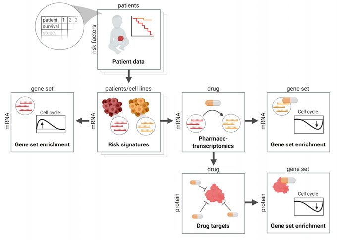
Figure 5. Integration of data resources. Aligning and integration of data matrices
covering different aspects of cancer biology opens up new opportunities for drug tar-
get discovery.
Mapping synergistic drug pairs
Although targeted therapy has shown promising initial results in clinical stud-
ies, many patients suffer from relapse due to low in vivo efficacy of drugs or
29You can also read