Hand Foot and Mouth Disease A Generalized Overview - IAPO
←
→
Page content transcription
If your browser does not render page correctly, please read the page content below
Hand Foot and Mouth Disease
A Generalized Overview
Clifford Chen
Hand foot mouth disease (HFMD) is a common viral illness of childhood
that can occasional affect adults. It can be easily recognized from other viral
infections by its typical distribution of the exanthema over the hands and feet
and oral enanthem1.
HFMD is caused by a virus in the non-polio enterovirus family. These viruses
can present in a wide variety of presentations other than hand foot mouth disease
(Figure 1 A, B and C). Other conditions that have been attributed to these viruses
are meningitis, herpangina, acute gastroenteritis,
A and nonspecific exanthema other than hand foot
mouth disease. The coxsackievirus serotype A16
and enterovirus serotype 71 are the most common
virus of the non-polio enterovirus family to cause
HFMD2. Most recently much attention has been
placed on the enterovirus EV71. While most pa-
tient affected with this serotype has a mild and
typical disease course, there has been increasing
rate of severe sequela involving the central ner-
B vous system or pulmonary edema3. The World
Health Organization reports that countries in the
Western Pacific Region are experiencing a wide-
spread epidemic of the EV714. The infection typ-
ically presents in the warmer climates. Regions
with seasons have the HFMD during the summer
and fall, while topical areas infections can occur
year-round.
HFMD symptoms have many similar fea-
tures with other viral illness. Patients of HFMD
often have the low-grade fever, sore throat, and
C malaise. In infants and toddlers, irritability and
loss of appetite may occur. HFMD is unique by
its unique location of the rash affecting the hand,
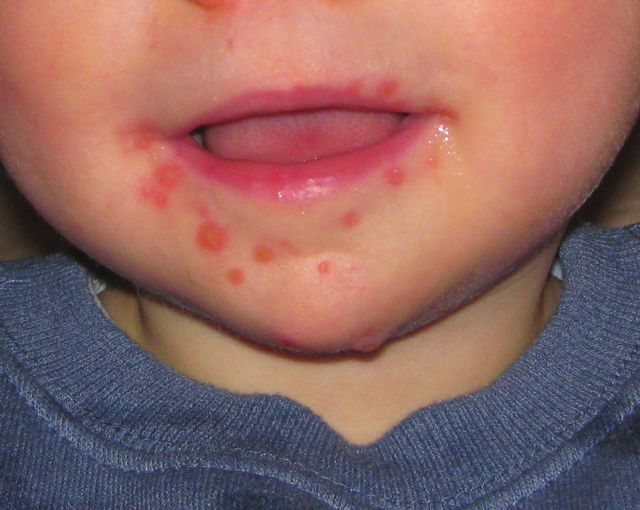
foot, and mouth. The exanthema of HFM disease
itself can be described in multiple morpholo-
gies: vesicular, macular or macular popular5. The
distribution of the erythematous rash classically
covers the palms of the hands and/ or soles of the
Figure 1 A, B and C. Hand foot and feet. In some case, the exanthema affects the but-
mouth disease tocks, knees or elbows. The rash is nonpruritic
138   !   XV IAPO MANUAL OF PEDIATRIC OTORHINOLARYNGOLOGY
and may or may not be painful. Also, patients develop oral enanthem which are
painful erythematous blisters of oral mucous more commonly the tongue and buc-
cal mucosal. Patient affected by the HFM D may have the characteristic rash and
not the oral enanthem and vice visa. Typically the fever, sore throat, and malaise
are the first symptoms of the infection followed by the oral mucosal involvement
in 1 to 2 days after the onset of the fever. The rash over the hands and feet can
develop within 1 to 2 days after that. The total duration of illness varies from 7 to
10 days.
      More recently there has been more atypical infection associated with HFM
disease. The coxsackievirus A6 has been linked to the atypical presentation of
HFMD. The rashes vary in appearance and may be more extensive leading to ve-
siculobullous lesions, bullae, erosions, ulcerations and eschar formation6. One to
3 weeks after the infection, the patient may experience palmar and plantar desqua-
mation. Another feature that may occur is onychomadesis, nail dystrophy about 1
to 2 month after the infection. In Western Pacific Region where they are currently
facing epidemic levels of HFMD caused by enterovirus EV71, there are seeing
high rates of severe illness and complications. Patients are experiencing central
nervous system complications like rhombencephalitis, acute flaccid paralysis, and
aseptic meningitis7. Some patients may develop further automatic nervous system
dysregulation and eventual cardiopulmonary failure8.
      Diagnosis of the disease is made clinically. Characteristic history and physi-
cal exam of fever, sore throat and malaise with the rash of the hand, foot and mouth
will provide enough information to make the diagnosis. Laboratory confirmation
is not necessary. If the presentation is atypical or presentation is another various
disease, it may be beneficial to send the confirmation test. Rapid diagnosis tests are
available if needed in either real time PCR or indirect immunofluorescence assay.
      Since HFMD is due to a virus, it is a self-limiting disease. Typically the in-
fection lasts from 7 to 10 days. Treatment is usually centered around supportive
care, pain management, and disease prevention.
      Patient with this disease often experiences oral discomfort with any intake of
liquids or solids. Because of this, dehydration is a primary concern for infants and
toddlers are most at risk. Assessing the current patient hydration status and sever-
ity of the disease can determine the likelihood of dehydration for the future. Oral
rehydration solution should be the first line for rehydration. If the patient does not
tolerate or refuse to take fluids by mouth, then intravenous hydration should be
started, and hospitalization may be required.
      Pain management is another facet of managing the patient with hand foot
mouth disease. Oral lesions are fairly painful. Most patients can be managed with
non-narcotics like acetaminophen and ibuprofen as needed. Occasionally narcot-
ics may be required to be given. If the patient is unable to tolerate medications by
mouth, intravenous pain medications should be considered.
      The primary reason most patients hospitalized for HFMD is dehydration
associated with the painful mouth lesions. Topical medication for the oral mu-
cositis can be considered, but the evidence for the use of topical medications are
inadequate and mostly anecdotal. One large study looking at topical lidocaine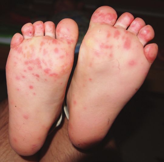
XV IAPO MANUAL OF PEDIATRIC OTORHINOLARYNGOLOGY ! 139
for treatment of mucositis should no benefit from the control group9. Advising
the family to try cool to cold liquids and foods may help soothe the mouth pain.
Examples are frost snacks and cold milk. Avoiding salty, acidic, or spicy foods
or liquids is advised.
      In regions with enterovirus EV71 causing HFMD, patient with severe dis-
ease with neurological or cardiopulmonary disease often receive a trial of intrave-
nous immunoglobulin (IVIG). The anecdotal data indicates if IVIG is given early
that overall outcomes are improved10.
      Prevention is another aspect to needs addressed. Enterovirus is a highly con-
tagious disease that is transmitted by oral ingestion of the virus. Infected people
shed this virus most of the mucous surfaces and secretions. Commonly nasal se-
cretions, saliva, stool, respiratory droplets and fluid from the blister can spread
the virus. If the patient is hospitalized, droplet and contact precautions should be
used to control the spread of the virus. Good hygiene should be used to repress the
spread at home. Hand washing, disinfect common areas and not sharing objects
placed in mucosal areas. Withholding patient with HFM from school, work, and
daycare is strongly advised. Infected people are considered contagious until the
fever has subsided, and the mouth sores are healed and should be able to return to
daily activities afterward
      Hand foot mouth disease is a common pediatric illness caused by various
members of the non-polio enterovirus family. The presentation of the illness of
the classic rash on the palms of the hands and soles of the feet with oral ulceration
of the tongue and buccal mucosa will confirm the diagnosis. General this disease
is a self-limiting that requires supportive care, pain management, and prevention
measures. There are serotypes of this infection in Western Pacific Region that has
been identified that can lead to more severe and life-threatening disease.

References

1.   Robinson, C. R., Doane, F. W., & Rhodes, A. J. (1958). Report of an outbreak of febrile
     illness with pharyngeal lesions and exanthem: Toronto, summer 1957—isolation of group
     A coxsackie virus. Canadian Medical Association Journal, 79(8), 615.
2.   Khetsuriani, N., LaMonte-Fowlkes, A., Oberst, S., Pallansch, M. A., & Centers for Dis-
     ease Control and Prevention. (2006). Enterovirus surveillance—United States, 1970–
     2005. MMWR Surveill Summ, 55(8), 1-20..
3.   Ooi, M. H., Wong, S. C., Lewthwaite, P., Cardosa, M. J., & Solomon, T. (2010). Clinical fea-
     tures, diagnosis, and management of enterovirus 71. The Lancet Neurology, 9(11), 1097-1105.
4.   World Health Organization. (2011). A guide to clinical management and public health re-
     sponse for hand, foot and mouth disease (HFMD).
5.   Miller, G. D., & Tindall, J. P. (1968). Hand-foot-and-mouth disease. JAMA,203(10), 827-830..
6.   Mathes, E. F., Oza, V., Frieden, I. J., Cordoro, K. M., Yagi, S., Howard, R., ... & Bayliss,
     S. (2013). “Eczema coxsackium” and unusual cutaneous findings in an enterovirus out-
     break. Pediatrics, 132(1), e149-e157.
7.   Ooi, M. H., Wong, S. C., Mohan, A., Podin, Y., Perera, D., Clear, D., Solomon, T. (2009).
     Identification and validation of clinical predictors for the risk of neurological involvement
     in children with hand, foot, and mouth disease in Sarawak. BMC infectious diseases, 9(1), 1.
140 ! XV IAPO MANUAL OF PEDIATRIC OTORHINOLARYNGOLOGY
8. Fu, Y. C., Chi, C. S., Chiu, Y. T., Hsu, S. L., Hwang, B., Jan, S. L., ... & Chang, Y. (2004).
Cardiac complications of enterovirus rhombencephalitis.Archives of disease in child-
hood, 89(4), 368-373.
9. Hopper, S. M., McCarthy, M., Tancharoen, C., Lee, K. J., Davidson, A., & Babl, F. E.
(2014). Topical lidocaine to improve oral intake in children with painful infectious mouth
ulcers: a blinded, randomized, placebo-controlled trial. Annals of emergency medi-
cine, 63(3), 292-299.
10. Chang, L. Y., Hsia, S. H., Wu, C. T., Huang, Y. C., Lin, K. L., Fang, T. Y., & Lin, T. Y.
(2004). Outcome of enterovirus 71 infections with or without stage-based management:
1998 to 2002. The Pediatric infectious disease journal,23(4), 327-332.You can also read