A case of haemoptysis in a girl with Noonan syndrome
←
→
Page content transcription
If your browser does not render page correctly, please read the page content below
CASE REPORT
A case of haemoptysis in a girl with Noonan syndrome
R K Mopeli, MB ChB, FCPaed (SA); M Lebea, MB ChB, FCPaed (SA), MMed;
C Verwey, MB ChB, MMed, FCPaed (SA), Cert Paed Pulmonol
Department of Paediatrics and Child Health, Faculty of Health Sciences, University of the Witwatersrand, Johannesburg, South Africa
Corresponding author: C Verwey (charl.verwey@wits.ac.za)
Noonan syndrome (NS) is an autosomal dominant condition affecting 1 in 2 000 live births. It is characterised by distinctive physical features,
congenital heart disease and multiple other comorbidities including haematological abnormalities. Haemoptysis is the expectoration of
blood originating from the lower respiratory tract. It is uncommon in children but can be life threatening. Perfusion of the lower respiratory
system arises from the pulmonary arterial circulation and the bronchial circulation, or bleeding may arise from either. In children, the
most common causes of haemoptysis are respiratory tract infections, aspirated foreign bodies and bronchiectasis. We present a 7-year-old
girl with recurrent haemoptysis.
Afr J Thoracic Crit Care Med 2020;26(3):119-121. https://doi.org/10.7196/AJTCCM.2020.v26i3.023
A 7-year-old girl with Noonan syndrome (NS) presented with a
history of recurrent haemoptysis. She was diagnosed with pulmonary
stenosis at the Paediatric Cardiology Department at Chris Hani
Baragwanath Academic Hospital (CHBAH) and was treated with
a transannular patch at the age of 6. She had severe pulmonary
regurgitation following the surgery and was on treatment with
furosemide, potassium chloride and digoxin.
She presented to her local hospital with a history of coughing up
fresh red blood a year after undergoing surgery. The parents could
not quantify the amount of blood; however, it was thought to be
significant because she had a haemoglobin level of 7.5 g/dL and
required a transfusion of packed red blood cells. It was reported that
she had two other episodes of haemoptysis 11 months prior but did
not seek medical attention. She was airlifted to CHBAH for further
management. On arrival, she was noted to be in respiratory distress
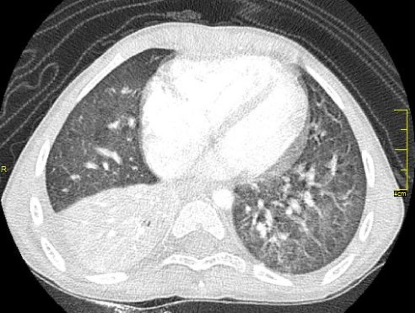
and was put on 60% O2 via a rebreathing mask. She was pink and Figure
Fig. 1.l:Chest
Chest computed
computedtomography scan showing
tomography a right lower
scan showing lobe lower-lobe
a right collapse consolidation
(Indicated by arrow)
well perfused. She had an early diastolic murmur of pulmonary collapse consolidation (indicated by arrow).
regurgitation.
Detailed echocardiography excluded pulmonary hypertension
and pulmonary vein stenosis. Laboratory investigations showed
haemoglobin (11 g/dL), platelets (235 × 109 cells/L), international
normalised ratio (1.02) and prothrombin time (34). She was HIV-
negative and had normal urea and creatinine levels. She had a von
Willebrand factor antigen of 34% (normal range 50 - 160%) and
activity of 88% (normal range 66 - 99%) and her Factor VIII level was
52 IU/dL. Her sputum showed no acid-fast bacilli, a Mycobacterium
tuberculosis culture was negative and no other pathogenic bacteria
were isolated. A computed tomography (CT) scan of the chest
showed a right lower-lobe dense consolidation, which was thought
to be due to lobar pneumonia (Fig. 1). She had a rigid bronchoscopy,
which also showed an inflammatory polyp at the entrance to the
posterior basal segment of the right lower-lobe bronchus (Fig. 2).
The inflammatory polyp and lobar pneumonia were thought to be the
cause of the haemoptysis, which was exacerbated by her underlying
haematological abnormalities. She was treated with antibiotics and Fig. 2. Polyp obstructing bronchial opening.
AJTCCM VOL. 26 NO. 3 2020 119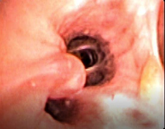
CASE REPORT

discharged home. A repeat bronchoscopy 3 months later showed that
the polyp had significantly decreased in size.
   She presented to the local hospital 2 weeks after the repeat
bronchoscopy with another episode of massive haemoptysis, where she
was resuscitated with fresh frozen plasma and a platelet transfusion.
She was airlifted to CHBAH after stabilisation. A repeat bronchoscopy
was done, but the patient had massive haemoptysis during the
procedure and required resuscitation and intensive care admission for
mechanical ventilation. She was referred to the cardiothoracic team
for lobectomy to control the haemoptysis. She continued to have more
episodes of haemoptysis while awaiting surgery. Rigid bronchoscopy
was done and did not reveal a polyp. She had a cardiac catheterisation
for further investigation of the haemoptysis and an angiogram showed
a torturous right bronchial artery, with extravasation of blood into
the right lower lobe. The patient was referred for embolisation of
the torturous bronchial artery. A descending aorta angiogram in Figure     Fig.3:3.Angiogram
                                                                                     Angiogram    showing
                                                                                             showing         theright
                                                                                                     a torturous  torturous
                                                                                                                      bronchialright
                                                                                                                                arterybronchial
                                                                                                                                      (Indicated byartery
                                                                                                                                                    arrow)
the anterior-posterior view confirmed the findings (Fig. 3). The (indicated by arrow).
prominent bronchial artery formed a confluence with an abnormal
vessel that arises from the right common carotid artery.                   valve stenosis and septal defects being common. [2] Bleeding
   She had a right bronchial artery embolisation and was stable post disorders have been reported in up to 65% of patients with NS.[3,4] A
procedure. That evening she bled again during rigid bronchoscopy. number of coagulation factor deficiencies, von Willebrand disease,
The decision was taken to do a lobectomy to control the bleeding. The thrombocytopenia, and platelet dysfunction have been described.[3,4]
histology of the resected lobe showed normal lung parenchyma, with The most common factor deficiency is factor XI, followed by factors
patchy bronchopneumonia, alveolar haemorrhage and fibrointimal XII and VIII. Our patient was found to have von Willebrand disease.
hyperplasia of the hilar vessels. The bronchus at the resection margin This may have worsened the degree of her blood loss, although it
was normal and patent. There was no granulomatous inflammation was not the primary cause of her bleeding. It was also clear that the
or malignant neoplasm. The patient had an uneventful course post polyp that had been visualised initially was not the primary cause of
lobectomy, and after 6 months follow-up there was no further bleeding bleeding, as she subsequently had further haemoptysis even after it
reported.                                                                  had disappeared. It could be argued that the polyp predisposed her to
                                                                           the lobar pneumonia, which increased vascular pressure in that area
Discussion                                                                 and caused the abnormal vessel to bleed.
The diagnosis of the cause of haemoptysis in children is not easy.            Intracardiac left-to-right shunts can cause pulmonary hypertension
When investigating the aetiology, it may be helpful to divide the in children, resulting in haemoptysis as a complication. Our patient
causes into those arising from parenchymal diseases and those arising had an echocardiography which excluded left-to-right shunting.
from pulmonary vascular disorders.                                         Cardiac catheterisation did not demonstrate pulmonary hypertension
   It is best to begin with a detailed history and physical examination to in this patient, with an invasive mean pulmonary pressure of 8 mmHg.
differentiate between extrapulmonary bleeding such as haematemesis            There is not much in the literature linking NS to primary
and haemoptysis. A history of foreign-body aspiration should be abnormalities of the pulmonary vasculature. The first description of
elicited if present. The respiratory examination may reveal localised an association between NS and primary abnormalities was reported in
wheezing suggestive of a foreign body or crepitations with decreased 1989 in a 19-year-old female who had severe pulmonary hypertension,
breath sounds, which may be caused by pneumonia or bronchiectasis. with the clinical and pathological features that were suggestive of
Radiological investigations such as a chest radiograph or CT scan primary pulmonary hypertension.[5] Our patient was bleeding from a
will provide useful information but may also be normal. Blood tests prominent torturous bronchial artery. This may just be an incidental
should be done in all children to screen for anaemia, raised infective abnormality aggravated by the lobar pneumonia and not necessarily
markers and bleeding abnormalities. Sputum should be evaluated for related to the fact that she has NS. It could also have been part of a
the presence of pathological microorganisms. If all the above does collateral bronchial circulation.
not lead to a diagnosis or the bleeding persists, a bronchoscopy is
indicated.[1]                                                              Conclusion
   Our patient had NS which we had to take into consideration This case illustrates the importance of having a broad-based approach
while investigating the cause of her haemoptysis. NS is a condition when investigating any patient with haemoptysis and that many
inherited in an autosomal dominant pattern and is present in 1 in different pathologies can act together to cause haemoptysis.
2 000 live births. It is characterised by distinctive physical features
such as hypertelorism, low-set, backward rotated ears, a short webbed
neck, short stature and multiple comorbidities including cardiac and Declaration. None.
haematological abnormalities.[2] More than 80% of patients with NS Acknowledgements. None.
have an abnormality of the cardiovascular system, with pulmonary Author contributions. CV, RKM and ML took care of the patient, prepared

120 AJTCCM VOL. 26 NO. 3 2020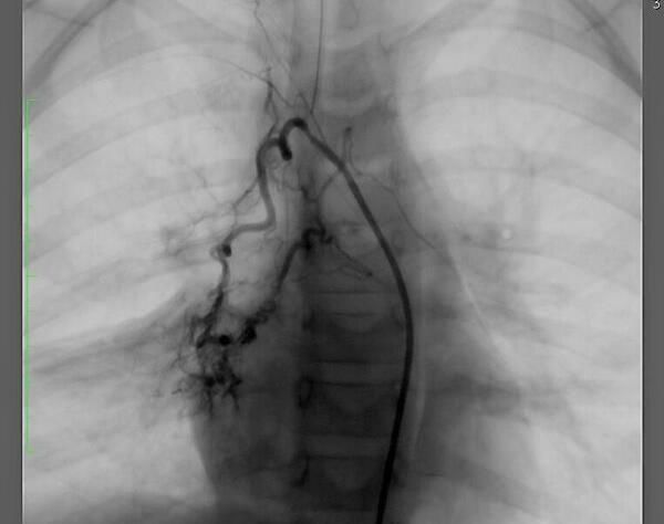
CASE REPORT

the figures, wrote the manuscript, and approved final version of the                    3. Briggs BJ, Dickerman JD. Bleeding disorders in Noonan syndrome. Pediatr Blood
                                                                                           Cancer 2012;58:167-172. https://doi.org/10.1002/pbc.23358
manuscript for publication.
                                                                                        4. Wiedand G, Hofbeck M, Zenker M, Budde U, Rauch R. Bleeding diathesis in
Funding. None.                                                                             Noonan syndrome: Is acquired von Willebrand syndrome the clue? Thromb Res
Conflicts of interest. None.                                                               2012;130(5):251-254. https://doi.org/10.1016/j.thromres.2012.08.314
                                                                                        5. Tinker A, Uren N, Schofield J. Severe pulmonary hypertension in Ullrich-Noonan
                                                                                           syndrome. Br Heart J 1989;62:74-77. https://doi.org/10.1136%2Fhrt.62.1.74
1. Gaude GS. Haemoptysis in children. Indian Pediatr 2010;47(3):245-254. https//doi:
   10.1007/s13312-010-0044-z
2. Romano AA, Allanson JE, Dahlgren J, et al. Noonan syndrome: Clinical features,
   diagnosis and management guidelines. Pediatrics 2010;126:746-749. https://doi.
   org/10.1542/peds.2009-3207                                                          Accepted 29 July 2020.

                                                                                                                   AJTCCM VOL. 26 NO. 3 2020                     121
You can also read