Lessons of the month: Massive gastrointestinal bleeding in a young woman with idiopathic thrombocytopenic purpura (ITP) - RCP Journals
←
→
Page content transcription
If your browser does not render page correctly, please read the page content below
LESSONS OF THE MONTH Clinical Medicine 2021 Vol 21, No 1: e100–2
Lessons of the month: Massive gastrointestinal bleeding
in a young woman with idiopathic thrombocytopenic
purpura (ITP)
Authors: Ksheetij Kothari, A Mallikarjun Patil,B Renuka MalipatelC and Harshad DevarbhaviD
Cytomegalovirus (CMV) is a ubiquitous pathogen, belongs to On postoperative day 16, she developed fresh blood from her
ABSTRACT
the herpes virus family and can infect the gastrointestinal (GI) rectum. There was no history of abdominal pain, diarrhoea, fever
system. The disease is usually noted in immunocompromised nor intake of non-steroidal anti-inflammatory drugs (NSAIDs). There
patients such as solid organ transplant recipients on was no history suggestive of epistaxis, gum bleeding, haematuria
immunosuppressive drugs, patients with malignancy receiving or skin rash. Her medication history included prednisolone 10 mg/
chemotherapy, patients with AIDS, patients on steroids for day and pantoprazole. On clinical examination, she was afebrile,
autoimmune disorders, and is rarely seen in immunocompetent blood pressure of 110/70 mmHg, pulse rate of 80 beats per
individuals. In the GI system, CMV most commonly involves the minute and respiratory rate of 18 breaths per minute. She had
colon, followed by oesophagus, stomach and, rarely, the small mucosal pallor and no petechiae on the skin. Her abdomen was
intestine. The GI manifestation of CMV infection is usually soft and non-tender with normal bowel sounds. A digital rectal
anorexia, diarrhoea, and blood in stools, abdominal pain and examination revealed fresh blood. Laboratory parameters showed
fever. Very rarely, CMV infection may present with a massive GI haemoglobin (Hb) of 8 g/dL (drop by 2 g), platelet count 86 × 109/L
bleed. We report a case of 36-year-old pregnant woman with and total leukocyte count of 7.8 × 10⁹/L. Coagulation profile and
idiopathic thrombocytopenic purpura (ITP) who presented with liver function tests were normal. An upper gastrointestinal (GI)
massive GI bleeding following delivery, attributed to isolated endoscopy appeared normal until the third part of the duodenum
CMV enteritis. and colonoscopy examination showed fresh blood in the ileum
with normal-appearing colonic mucosa suggestive of source
KEYWORDS: cytomegalovirus, enteritis, massive gastrointestinal of bleeding proximal to ileum and distal to duodenum. Post-
bleeding procedure, she developed hypotension with passage of massive
amount of fresh blood. She was transferred to the intensive care
DOI: 10.7861/clinmed.2020-0803 unit and resuscitated with intravenous fluid therapy and packed
cell transfusion. Her blood investigations revealed low Hb (4 g/dL)
and platelet count was 90 × 109/L. Computed tomography (CT)
Case presentation angiography did not reveal a source of bleeding. She continued
to have GI bleeding with haemodynamic instability despite
A 36-year-old woman, 34 weeks pregnant, presented with
resuscitative measures, hence subjected to emergency laparotomy
bleeding from her vagina. She was diagnosed with idiopathic
with intraoperative enteroscopy. Enteroscopy showed a large
thrombocytopenic purpura (ITP) and was taking steroids
amount of fresh blood in the distal jejunum and proximal ileum
(prednisolone 10 mg/day) for the previous 5 months. On
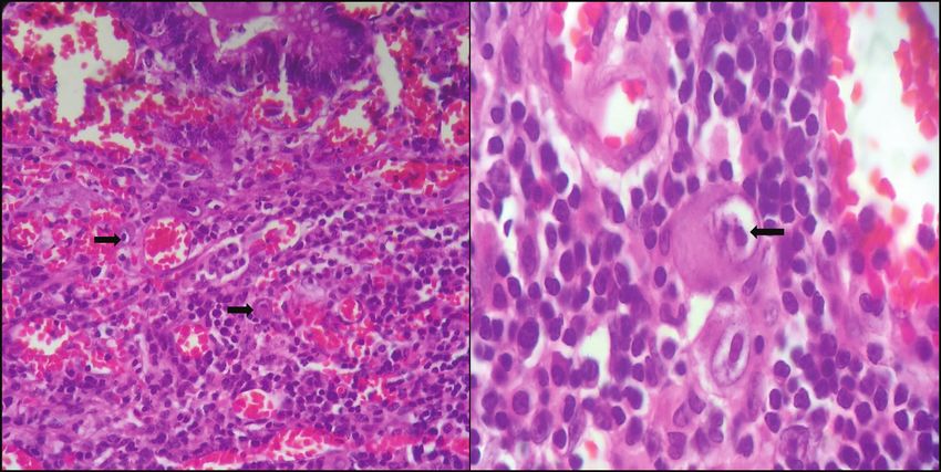
with multiple, discrete, actively bleeding ulcers (Fig 1). The diseased
examination, she was haemodynamically stable and had evidence
segment was resected (80 cm) and end-to-end anastomosis was
of fetal distress, so she was then taken for emergency lower
done.
segment caesarean section. Intraoperatively, she was found to
Gross examination of resected small bowel revealed large
have placenta accreta and required a hysterectomy to control the
punched-out ulcers (Fig 2). On histopathological examination,
bleeding.
characteristic intranuclear owl’s eye inclusion bodies were seen in
endothelial and stromal cells suggesting cytomegalovirus (CMV)
infection (Fig 3). The serum CMV polymerase chain reaction
Authors: A senior resident, St John’s Medical College Hospital, test revealed viraemia (2,000 copies/mL). She was treated with
Bangalore, India; Bprofessor, St John’s Medical College Hospital, intravenous ganciclovir 300 mg every 12 hours and an additional
Bangalore, India; Cassistant professor, St John’s Medical College 3 units of packed cell and platelet transfusion. Although bleeding
Hospital, Bangalore, India; Dprofessor and head of department of was controlled, her general condition deteriorated due the
gastroenterology, St John’s Medical College Hospital, Bangalore, development of multi-organ failure. She died 5 days after surgery
India despite antiviral therapy and other supportive care.
e100 © Royal College of Physicians 2021. All rights reserved.
CMV enteritis

                                                                            a                                   b

                                                                            Fig 3. Haematoxylin and eosin stain showing ulceration, granulation
                                                                            tissue and enlarged stromal cells with characteristic intranuclear owl’s
                                                                            eye inclusion bodies. a) Magnification × 400. b) Magnification × 1,000.

                                                                            enteroscopy in haemodynamically stable individuals. Rarely in
                                                                            haemodynamically unstable patients (as with our patient), when
                                                                            CT doesn’t reveal the cause and the requirement of surgical
                                                                            resection is imminent, intraoperative enteroscopy is required to
Fig 1. Enteroscopy showing multiple, discrete ulcers in jejunum (arrows).   ascertain the source of bleeding and surgical treatment can be
                                                                            done at the same time.3
                                                                              Primary CMV infection is usually acquired in the first 20 years
                                                                            of life, which is either asymptomatic or may have self-limiting
Discussion                                                                  mononucleosis (such as symptoms in immunocompetent
                                                                            individuals).4 Following primary infection, CMV remains dormant
Our patient had obscure overt gastrointestinal bleeding (bleeding           in host tissue in harmony with host immunity and gets reactivated
from unknown source after standard endoscopy, colonoscopy and               with suppressed immune response. The CMV infection of the
radiological imaging).1 The common causes in young individuals              gastrointestinal tract is usually due to reactivation of the dormant
are small bowel ulcers due to inflammatory bowel disease, NSAIDs            CMV due to immunosuppression and rarely due to superinfection
intake and small bowel tumours (such as gastrointestinal tumours            of the diseased GI tract, like in inflammatory bowel disease. CMV
(GIST), lymphoma, neuroendocrinal tumours and carcinoma),                   can infect any part of GI system from oesophagus to rectum, but
Meckel’s diverticulum, Dieulafoy’s lesion and telangiectasia.2 After        commonly involves colon (55%), oesophagus and stomach (40%)
the negative standard endoscopy and colonoscopy examination,                and, rarely, the small intestine (4.3%).5
further investigations to ascertain the source of bleeding include            CMV infects vascular endothelial cells and surface epithelium of
CT angiography to rule out a structural cause. If the CT is                 the small bowel and causes sub-epithelial haemorrhages, erosions,
normal, further evaluation includes capsule endoscopy and push              and superficial and deep ulcers which manifest with persistent
                                                                            diarrhoea, weight loss, obscure GI bleeding, intestinal obstruction,
                                                                            perforation and, rarely, massive GI bleeding.6 The pathogenesis of
                                                                            massive GI bleeding is due to vasculitis, vascular erosion and deep
                                                                            ischaemic ulcers. There are few case reports of massive GI bleeds
                                                                            due to CMV enteritis in literature.7–10 In most of the published cases,
                                                                            patients were immunocompromised. Diagnosis of CMV enteritis
                                                                            is difficult due to non-specific presentation and requirement of
                                                                            enteroscopy for histopathological diagnosis. Presence of CMV
                                                                            inclusion bodies is considered to be diagnostic. In our patient,
                                                                            diagnosis was done on basis of histopathological examination of
                                                                            a surgically resected specimen which showed presence of CMV
                                                                            inclusion bodies. Ganciclovir therapy is the treatment of choice.
                                                                            For massive GI bleeds, surgical resection is to be considered if
                                                                            embolisation therapy is not feasible/fails.

                                                                            Conclusion
                                                                            CMV enteritis can rarely present as massive GI bleed. It needs
                                                                            to be suspected as one of the causes of GI bleed in immune-
                                                                            compromised patients and needs to be confirmed by enteroscopy.
                                                                            Early initiation of anti-viral therapy may avert the need for surgery.
Fig 2. Resected part of small bowel showing punched out ulcers (arrows).    Delayed diagnosis increases morbidity and mortality. ■

© Royal College of Physicians 2021. All rights reserved.                                                                                      e101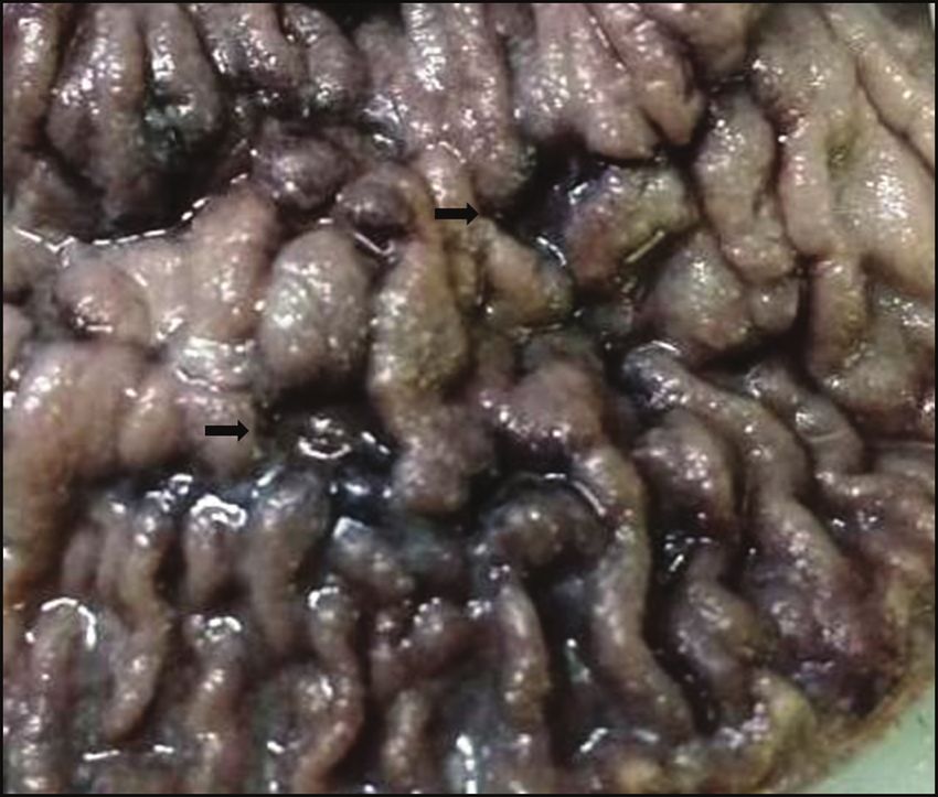
Ksheetij Kothari, Mallikarjun Patil, Renuka Malipatel et al

References                                                                  7 Keates J, Lagahee S, Crilley P, Haber M, Kowalski T. CMV enteritis
                                                                              causing segmental ischemia and massive intestinal hemorrhage.
1 Raju GS, Gerson L, Das A, Lewis B. American Gastroenterological             Gastrointest Endosc 2001;53:355–9.
  Association (AGA) Institute technical review on obscure gastroin-         8 Varma V, Perera MT, Olliff S et al. Cytomegalovirus ileitis causing
  testinal bleeding. Gastroenterology 2007;133:1697–717.                      massive gastrointestinal haemorrhage in a patient following
2 Gerson LB, Fidler JL, Cave DR et al. ACG clinical guideline: Diagnosis      hepatic resection. Trop Gastroenterol 2011;32:145–7.
  and management of small bowel bleeding. Am J Gastroenterol                9 Morunglav M, Theate I, Bertin G, Hantson P. CMV enteritis causing
  2015;110:1265–87.                                                           massive intestinal hemorrhage in an elderly patient. Case Rep Med
3 Zaman A, Sheppard B, Katon RM. Total peroral intraoperative                 2010;2010:385795.
  enteroscopy for obscure GI bleeding using a dedicated push               10 Choi SW, Chung JP, Song YK et al. Lower gastrointestinal bleeding
  enteroscope: diagnostic yield and patient outcome. Gastrointest             due to cytomegalovirus ileal ulcers in an immunocompetent man.
  Endosc 1999;50:506–10.                                                      Yonsei Med J 2001;42:147–51.
4 Godgame RW. Gastrointestinal cytomegalovirus disease. Ann
  Intern Med 1993;119:924–35.
5 Chamberlain RS, Atkins S, Saini N, White JC. Ileal perforation
  caused by cytomegalovirus infection in a critically ill adult. J Clin    Address for correspondence: Prof Mallikarjun Patil, Department
  Gastroenterol 2000;30:432–5.                                             of Gastroenterology, St John’s Medical College Hospital,
6 Iwasaki T. Alimentary tract lesions in cytomegalovirus infection.        Sarjapur Road, Bangalore 34, Karnataka, India.
  Acta Pathol Jpn 1987;37:549–65.                                          Email: drmalli_arjun@yahoo.co.in

e102                                                                                              © Royal College of Physicians 2021. All rights reserved.
You can also read