Hard x-ray photoelectron spectroscopy: a snapshot of the state-of-the-art in 2020 - IOPscience
←
→
Page content transcription
If your browser does not render page correctly, please read the page content below
Journal of Physics: Condensed Matter

TOPICAL REVIEW • OPEN ACCESS

Hard x-ray photoelectron spectroscopy: a snapshot of the state-of-the-art
in 2020
To cite this article: Curran Kalha et al 2021 J. Phys.: Condens. Matter 33 233001

View the article online for updates and enhancements.

                               This content was downloaded from IP address 46.4.80.155 on 31/08/2021 at 18:33
Journal of Physics: Condensed Matter

J. Phys.: Condens. Matter 33 (2021) 233001 (44pp)                                                                 https://doi.org/10.1088/1361-648X/abeacd

Topical Review

Hard x-ray photoelectron spectroscopy: a
snapshot of the state-of-the-art in 2020
Curran Kalha1,13 , Nathalie K Fernando1,13 , Prajna Bhatt1 , Fredrik O L
Johansson2 , Andreas Lindblad2 , Håkan Rensmo2 , León Zendejas
Medina3 , Rebecka Lindblad3 , Sebastian Siol4 , Lars P H Jeurgens4 ,
Claudia Cancellieri4 , Kai Rossnagel5,6 , Katerina Medjanik7 , Gerd
Schönhense7 , Marc Simon8 , Alexander X Gray9 , Slavomír
Nemšák10 , Patrick Lömker11 , Christoph Schlueter11 and Anna
Regoutz1,∗
1
   Department of Chemistry, University College London, 20 Gordon Street, London, WC1H 0AJ, United
Kingdom
2
   Department of Physics and Astronomy, Uppsala University, Box 516, 75120 Uppsala, Sweden
3
   Department of Chemistry—Ångström Laboratory, Uppsala University, Box 538, SE-75121, Uppsala,
Sweden
4
   Empa, Swiss Federal Laboratories for Materials Science and Technology, Laboratory for Joining
Technologies and Corrosion, Dübendorf, Switzerland
5
   Institute of Experimental and Applied Physics, Kiel University, 24098 Kiel, Germany
6
   Ruprecht Haensel Laboratory, Deutsches Elektronen-Synchrotron DESY, 22607 Hamburg, Germany
7
   Johannes Gutenberg Universität, Institut für Physik, 55128 Mainz, Germany
8
   Sorbonne Université, CNRS, Laboratoire de Chimie Physique—Matière et Rayonnement, LCPMR,
F-75005 Paris, France
9
   Department of Physics, Temple University, Philadelphia, PA 19122, United States of America
10
    Advanced Light Source, Lawrence Berkeley National Laboratory, Berkeley, CA 94720, United States of
America
11
    Deutsches Elektronen-Synchrotron DESY, 22607 Hamburg, Germany

E-mail: a.regoutz@ucl.ac.uk

Received 21 December 2020, revised 3 February 2021
Accepted for publication 1 March 2021
Published 13 May 2021

Abstract
Hard x-ray photoelectron spectroscopy (HAXPES) is establishing itself as an essential
technique for the characterisation of materials. The number of specialised photoelectron
spectroscopy techniques making use of hard x-rays is steadily increasing and ever more
complex experimental designs enable truly transformative insights into the chemical,
electronic, magnetic, and structural nature of materials. This paper begins with a short historic
perspective of HAXPES and spans from developments in the early days of photoelectron
spectroscopy to provide an understanding of the origin and initial development of the
technique to state-of-the-art instrumentation and experimental capabilities. The main
motivation for and focus of this paper is to provide a picture of the technique in 2020,
∗
 Author to whom any correspondence should be addressed.
13
  Both authors contributed equally to the manuscript.
                  Original content from this work may be used under the
                  terms of the Creative Commons Attribution 4.0 licence. Any
further distribution of this work must maintain attribution to the author(s)
and the title of the work, journal citation and DOI.

1361-648X/21/233001+44$33.00                                                   1   © 2021 The Author(s). Published by IOP Publishing Ltd   Printed in the UK
J. Phys.: Condens. Matter 33 (2021) 233001                                                                                 Topical Review

including a detailed overview of available experimental systems worldwide and insights into a
range of specific measurement modi and approaches. We also aim to provide a glimpse into
the future of the technique including possible developments and opportunities.
Keywords: photoelectron spectroscopy, hard x-ray photoelectron spectroscopy,
photoemission spectroscopy

S Supplementary material for this article is available online

(Some figures may appear in colour only in the online journal)

1. Introduction and historic perspective                               definition remains in place. A range of abbreviations is in
                                                                       use for the technique with HAXPES, which was first used
All photoelectron spectroscopy (PES) is based on the fun-              in 2005 [15, 16], being the most widely established and oth-
damental principle of the photoelectric effect [1–3], which            ers including HXPS, HXPES, HX-PES, and HIKE. To date,
was explained in terms of the photon concept by Einstein and           HAXPES has been applied in many scientific areas from fun-
Rutherford [4, 5]. The photoelectric effect describes how light        damental atomic, molecular, and condensed matter physics, to
that hits a material gives rise to the emission of electrons,          surface and interface science, and technologically important
i.e. photoelectrons. From this foundation it took decades of           areas like catalysis, electrochemistry, energy materials, includ-
experimental work to develop an experimental technique that            ing batteries, fuel cells, and photovoltaics, and electronic
could capitalise on this effect to obtain physical and chemical        devices.
information. It was only with techniques developed for nuclear            In standard laboratory-based soft x-ray photoelectron spec-
spectroscopy by Siegbahn and his group, that the first steps of        troscopy (SXPS or XPS) monochromated Al Kα and Mg Kα,
PES were taken [6, 7]. The group of Siegbahn initially aimed           at hν = 1486.7 eV and 1253.6 eV, respectively, are most
for correcting nuclear energies from effects of inner shell core       widely used as they can be produced with high intensities and
electron excitations. Such corrections required the develop-           have intrinsically narrow line widths to enable good energy
ment of electron spectrometers for high energies and in fact           resolution necessary for both core and valence state spec-
the first spectrometers were limited to energies of a few keV          troscopy. In the first published paper by Kai Siegbahn on
and above. Their first core photoelectron spectrum showing             electron spectroscopy for chemical analysis (ESCA), which
discrete lines, detected the Cu 1s photoelectrons excited by           we now commonly know as XPS, he points out that for all
Mo Kα1 (hν = 17.479 keV) and Mo Kα2 (hν = 17.374 keV)                  elements of the periodic table ‘one or several narrow atomic
x-rays [8].                                                            levels’ can be found and measured using Al Kα or Mg
    In this context the early development of PES coincides             Kα x-rays. He goes on to state that, if one is interested in
with the emergence of hard x-ray photoelectron spectroscopy            deeper atomic levels, these can be excited with Cr (hν Kα
(HAXPES). From the beginning careful energy calibration,               = 5.415 keV), Cu (hν Kα = 8.046 keV), Ag (hν Lα
linked to the understanding of work function, Fermi level, and         = 2.984 keV), or W (hν Lα = 8.398 keV), but that in his opin-
control over charging, was realised as a very important aspect.        ion Al Kα or Mg Kα are preferable due to their line widths.
Such control and the use of hard x-ray set-ups allowed for             This point of view is understandable as Al and Mg have line
the early indication of the chemical shift [9, 10], the exis-          widths of below 1 eV, while the hard x-ray sources generally
tence of which was firmly established together with quanti-            have widths above 2 eV. Nevertheless, very early HAXPES
tative insights from the use of Cu Kα (hν = 8.046 keV) and             studies were performed using Cu Kα in the 1960s [14, 17].
Cr Kα (hν = 5.415 keV) [11, 12]. During this time the use                 Beyond these initial proof of concept experiments at labora-
of electron spectroscopy as a tool for chemical analysis was           tory sources, it is the advent of synchrotron sources that really
established [13], and started to spread over the world [14],           catalyzed the development of HAXPES. The first reported
with much of this work being performed using hard x-rays.              synchrotron-based HAXPES measurements were performed
Thus, early parts of modern photoelectron spectroscopy were            by Lindau, Pianetta et al at the Stanford Synchrotron Radiation
accomplished using energies that today would use the acronym           Project using synchrotron radiation from the Stanford Positron
HAXPES.                                                                Electron Accelerator Ring (SPEAR) facility [18, 19]. This first
    HAXPES is generally defined as x-ray photoelectron spec-           experiment reported the Au 4f core level measured at 8 keV
troscopy (XPS) which uses x-ray energies above 2 keV, as               and with an impressive energy resolution of 0.25 eV achieved
historically beyond 2 keV crystal monochromators replaced              by combining two Si(220) crystals and a Si(440) channel-cut
grating monochromators used in the soft x-ray regime.                  crystal. Following on from this first experiment developments
Although developments in grating technology have led to some           both in the USA and at HASYLAB, Hamburg, Germany, in
grating monochromators being able to go up to 5 keV, the               the 1980s and 1990s led to a number of core level HAXPES

                                                                   2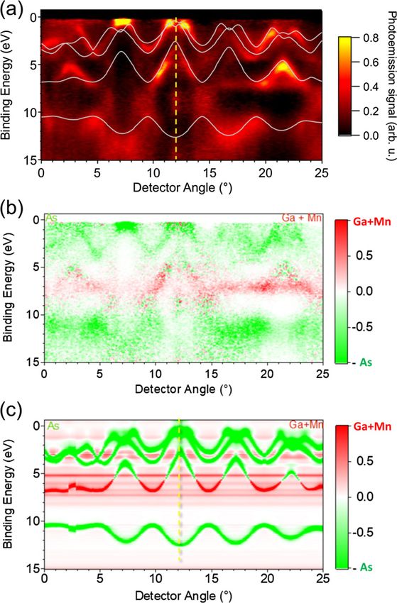
J. Phys.: Condens. Matter 33 (2021) 233001                                                                                                  Topical Review

         Table 1. HAXPES beamlines currently in operation at synchrotrons worldwide. The information given includes the name of
         the beamline, the synchrotron, the host country, the type of source used [insertion device (ID) or bending magnet (BM)], the
         energy range hν available at the beamline, the best energy resolution ΔE and at what energy hν this is accessible (in
         brackets), the x-ray hν spot size at the sample position, and any publications directly relating to the beamlines.
         Beamline          Synchrotron        Country      Source        hν/keV         ΔE (hν)/meV (keV)          hν spot/μm2       References
         BM25 SpLine          ESRF             France        ID           3–40                  80 (5)             100 × 100              [49]
         GALAXIES            SOLEIL            France        ID          2.3–12                 80 (4)               80 × 30            [50–52]
         EMIL               BESSY II          Germany        ID         0.08–10                    a
                                                                                                                    100 × 20              [53]
         KMC-1              BESSY II          Germany        BM           2–12               250 (2–6)              100 × 50            [54, 55]
         P22                PETRA III         Germany        ID           2.4+                   50                  15 × 15            [56, 57]
         X07MB                 SLS           Switzerland     ID           0.3–8                770 (4)            3000 × 3000             [58]
         I09                   DLS               UK          ID         2.1–12+                70 (6)                15 × 30              [59]
         BL09XU              SPring-8          Japan         ID         4.91–100               70 (6)                 10 × 1              [60]
         BL12XUc             SPring-8          Japan         ID           6–12               100–1000               < 40× 40              [61]
         BL15XU              SPring-8          Japan         ID           2–36                 60 (6)                25 × 30              [62]
         BL16XU              SPring-8          Japan         ID          4.5–40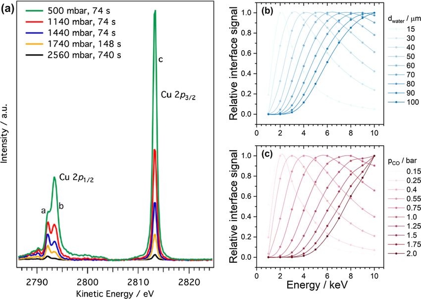
J. Phys.: Condens. Matter 33 (2021) 233001                                                                                      Topical Review

Figure 1. Scientific areas and publication landscape of HAXPES. (a) Number of publications and citations between 2004 and 2020,
(b) number and percentage of publications across different scientific fields between 2004 and 2020, and (c) research areas as identified by
the beamline survey. The publication records in (a) and (b) were extracted from Web of Science based on all publications including the
search terms ‘hard x-ray photoelectron spectroscopy’, ‘hard x-ray photoemission’, ‘HAXPES’, ‘HXPS’, ‘HX-PES’.

underpinning the technique are introduced where most appro-               2.1. Synchrotron-based systems
priate, including photoionisation cross sections (section 2.2),
accessibility of core levels (section 2.2), information depth             As part of this review, we have conducted a survey of the 24
and related concepts (section 3.1.1), recoil (section 3.1.1), and         currently operative HAXPES beamlines worldwide in Octo-
angular asymmetry (section 3.2.2). Ultimately, the motivation             ber and November 2020. The survey was circulated and com-
of the authors has been to provide an unbiased and balanced               pleted by beamline scientists, which are acknowledged below,
overview of the state-of-the-art of HAXPES in 2020.                       and key beamline characteristics are presented in table 1. Fur-
                                                                          ther information on optics and end stations of each beam-
                                                                          line is available in the supplementary information. Beamline
2. Instrumentation
                                                                          X07MB at the SLS in Switzerland, which is primarily an x-ray
This section gives an overview of the three principle sources             absorption beamline, is also included in the overview tables
at which HAXPES experiments can be conducted, includ-                     for completeness, although the HAXPES end station is a non-
ing synchrotrons, laboratory sources, and x-ray free elec-                permanent installation and not in open user operation. Since
tron lasers. Each section provides an up-to-date overview                 the early 2000s the number of beamlines available has con-
of available experimental stations and their characteristic               tinued to grow and figure 2(a) gives an overview of the years
functionalities. The synchrotron section provides a detailed              beamlines came into operation. From the distribution of beam-
overview of all currently operational HAXPES beamlines                    lines worldwide, summarised in figure 2(b), it is clear that
worldwide.                                                                SPring-8 still dominates the field with its sheer number of

                                                                      4
J. Phys.: Condens. Matter 33 (2021) 233001                                                                                    Topical Review

Figure 2. The current worldwide availability of operational HAXPES beamlines. (a) A timeline of the operational starting dates of existing
HAXPES beamlines. The data point labels correspond to the beamline identifiers included in table 1. EMIL (BESSY II) is currently
undergoing commissioning. Beamline P09 at PETRA III was operational between 2010 and 2018, with the capabilities transferred to
beamline P22 in 2019. (b) The number of HAXPES beamlines available within each country.

beamlines available, but strong activities in Europe, the North         them with channel-cut post-monochromators to achieve high
Americas, and the rest of Asia are continuing to provide ever           energy resolution at specific photon energies. Large variations
more improved experimental capabilities and increased user              in both the best experimental energy resolution and the x-ray
access to the technique.                                                spot size at the sample position exist across beamlines and
   As is reflected in the publication track record of the tech-         showcase the importance of choosing the most suitable beam-
nique discussed above and summarised in figures 1(a) and                line for a given scientific question depending on e.g. sample
(b), existing beamlines deliver instrument capability to a wide         size, spectra of interest, and intrinsic line width of features.
range of scientific communities. As part of our survey we also          Further important aspects when selecting a beamline for an
asked beamline staff to identify the top three research areas           experiment are the in situ and operando capabilities of the end
their beamline contributes to. Figure 1(c) provides a sum-              stations. Many end stations provide a variety of in situ sample
mative overview of the scientific areas with over half of the           preparation facilities, such as sputtering, cleaving and anneal-
responses falling in the energy & environment as well as thin           ing, as well as sample deposition options. The majority of end
films & devices categories. The original answers as given in            stations also enables certain operando capabilities, predomi-
the survey, and how they were grouped for the preparation
                                                                        nantly focused on controlling the temperature of the sample
of this figure, are included in the supplementary information.
                                                                        during measurement from cryo-cooling down to as low as 10 K
The information taken from the survey provides an alternative,
                                                                        up to annealing temperatures above 1500 K. Ambient pressure
complementary view on active scientific areas compared to the
                                                                        and electrical biasing capabilities, which will be discussed in
information extracted from publication statistics.
                                                                        detail in sections 3.3.2 and 3.3.3, respectively, are gaining pop-
   The existing HAXPES beamlines do have several aspects
                                                                        ularity due to the ability to conduct dynamic measurements of
in common, but also differ in certain areas, as can be seen
from the survey results presented in table 1 and the further            samples under operation-relevant conditions.
tables in the supplementary information. The vast majority of              Synchrotron-based facilities are continuing to evolve and
the beamlines is based on insertion devices and only four use           new beamlines are being planned and built. After the recent
bending magnets resulting in variation in the available pho-            EBS (Extremely Brilliant Source) upgrade of the ESRF, beam-
ton flux with it varying between 1011 and 1013 photons/s at             line BM25-SpLine is undergoing improvements. BL09XU and
the sample position. While a very high photon flux can be an            BL47XU at SPring-8 will be integrated into BL09XU and will
advantage, it should be noted that lower flux density can be an         be launched as a new dedicated HAXPES beamline with two
asset when studying materials prone to radiation-induced dam-           endstations in 2021. BL09XU will be temporarily shut down
age. The accessible x-ray energy ranges mainly differ in the            in February 2021, followed by an upgrade to the optics and
lowest available energy. There is an almost even split between          equipment. After commissioning, the operation is planned to
beamlines starting at around 2 keV and beamlines starting at            resume in October 2021.
higher energies of 5 keV and above. All beamlines use Si(111)              A new HAXPES beamline is currently under commission-
double crystal monochromators (DCM) and many combine                    ing at the Shanghai Synchrotron Radiation Facility (SSRF) and

                                                                    5
J. Phys.: Condens. Matter 33 (2021) 233001                                                                                       Topical Review

is planned to open to users in 2021. BL20U-Energy Materi-
als (E-line) combines soft and hard x-ray techniques aimed at
investigating fundamental properties of energy materials and
catalysis. The planned beamline specifications include a pho-
ton energy range of 130 eV to 18 keV with a flux of 2 ×10¹²
photons/s at 5 keV and 4 ×10¹² photons/s at 244 eV at x-ray
spot sizes of 80 × 20 μm2 at 5 keV and 90 × 10 μm2 at 244 eV,
respectively. The beamline will house an ambient pressure PES
endstation, which will include a Scienta HIPP-2 analyser and
will operate across a photon energy range from 1.5 to 10 keV.
   The emergence of 4th generation synchrotrons based on
multi-bend achromat lattice designs, enabling higher bright-
ness and coherence, promises new possibilities for HAXPES.
MAX-IV, the first 4th generation synchrotron had first elec-
trons on the 25th of August 2015 and is now operational.
The ESRF upgrade to the Extremely Brilliant Source (EBS) is
also complete and first electrons were injected on the 28th of
                                                                          Figure 3. Theoretical one-electron photoionisation cross sections
November 2019. Further planned new sources include Sirius
                                                                          for selected orbitals of oxygen and vanadium across the photon
in Brasil and the High Energy Photon Source (HEPS) in                     energy range of 1–15 keV. Cross section values taken from [76].
China, and upgrades are planned for a number of existing
synchrotrons, including APS-U, Diamond-II, PETRA IV, and
SPring-8-II.
                                                                          In this section, we give a brief overview of the main com-
                                                                          mercial and custom-built systems currently available and in
2.2. Laboratory-based HAXPES                                              operation.
                                                                              Laboratory-based HAXPES systems have exploited the
Over the past decade a new generation of laboratory-based                 accessibility of additional, deeper core levels to study depth-
HAXPES systems has emerged, which makes use predom-                       dependent phenomena in both bulk as well as multilayer sys-
inantly of Cr (Kα = 5.42 keV) and Ga (Kα = 9.25 keV)                      tems [85–91]. A general advantage not just of laboratory
based x-ray sources. The first monochromated Cr Kα-based                  systems, but any HAXPES experiment, is the ability to access
laboratory HAXPES systems were designed and built in the                  deeper core levels opening up new experimental and analytical
early 2010s by Keisuke Kobayashi, Hiroshi Daimon, Masaaki                 strategies [43, 92–95]. Figure 4 gives a schematic overview of
Kobata and colleagues [73, 74], who realised the impor-                   core levels of transition metals and lanthanides that become
tance of overcoming limitations due to decreasing photoion-               available when moving across the hard x-ray energy range.
isation cross sections to enable laboratory-based HAXPES.                 An example of the usefulness of accessing deeper core lev-
Their approach to address this challenge was to combine a                 els has been shown in recent work on the satellite structures
monochromated, focused, high flux x-ray source with a wide-               of transition metal oxides, where using the transition metal
acceptance objective lens and a Scienta R4000 high-energy                 1s rather than the 2p core levels enables a clearer understand-
hemispherical analyser, achieving a competitive energy reso-              ing of the spectra features present [96, 97]. Another example
lution and speed of acquisition. To date, the majority of HAX-            that has been explored with laboratory HAXPES is in complex
PES systems is still located at synchrotron sources due to                materials with multiple elements present that often have over-
the mentioned decrease in photoionisation cross sections at               lapping core levels in the soft x-ray range, such is the case for
higher x-ray energies, which necessitate high x-ray flux to               LiNi0.8 Co0.2−y Aly O2 layered oxide cathodes for batteries [85].
ensure usable signal counting rates. Figure 3 shows the the-              Probing the Al 1s core level instead of the lower energy Al 2s
oretical one-electron cross sections from Scofield for selected           and 2p states excludes spectral contamination from any of the
orbitals of oxygen and vanadium for energies from 1 to 15 keV             other elements present.
extracted and plotted using the Galore software package [75].                 The ability to excite deep core levels consequently also
    Several sets of theoretical cross sections are available in the       enables the measurement of the associated Auger transitions
literature, including by Scofield [76], Yeh and Lindau [77], and          [90, 100]. For many industrially relevant materials such as Al,
most recently from Trzhaskovskaya and Yarzhemsky [78, 79],                Si, Ti and their corresponding oxides and nitrides, the 1s core
and these are now available as digitised datasets for ease of             level is not accessible using monochromated Al Kα radiation
use [80–84]. The different cross section datasets clearly illus-          (see figure 4). However, access to these lines enables chem-
trate the loss of photoelectrons when moving from the soft                ical state analysis using the Auger parameter concept, which
into the hard x-ray range. The new, high flux laboratory x-ray            is very sensitive to changes in the local chemical environment
sources coupled with highly efficient photoelectron analysers             and often more robust than the observation of core level bind-
and detectors are enabling us to overcome these limitations and           ing energy shifts alone [101, 102]. In many instances the Auger
to move HAXPES back in the laboratory environment, where                  parameter is even sensitive to structural changes such as poly-
it began using Cu sources back in the early days of ESCA.                 morphic phase transitions [103] or the crystallisation of amor-

                                                                      6
J. Phys.: Condens. Matter 33 (2021) 233001                                                                                      Topical Review

Figure 4. Core levels of the transition metals (L) and lanthanides (R), including the 1s to 4s orbitals. The colors indicate at what minimum
excitation energy the core levels can be accessed in photoelectron spectroscopy experiments. All core levels beyond 4s are not included but
can all be accessed using excitation energies below 2 keV. Information taken from references [98, 99].

phous materials [104]. It is worth noting that Auger parameter                In contrast to synchrotron- or FEL-based HAXPES, the
studies were commonly employed in the 1970s and 1980s and                 excitation energy in laboratory-based HAXPES systems is
reference values for many materials are reported in literature            fixed to the characteristic x-ray emission of the anode mate-
[102]. This is due to the fact, that older spectrometers often            rial. This has important implications for the information depth
featured non-monochromated x-ray sources, which provided                  (see general discussion on this topic in section 3.1.1. Depend-
high-energy Bremsstrahlung which could be used to excite                  ing on the core level which is probed, the binding energy and
deep core levels and the associated Auger lines [105]. Due to             consequently the kinetic energy of the photoelectrons can vary
the dominance of monochromated soft x-ray sources in today’s              over a wide range. The largest information depth occurs at
commercial laboratory XPS systems such measurements are                   the highest kinetic energy and therefore in the valence region,
not possible anymore.                                                     whereas deep core levels occur at lower kinetic energy and
   Although laboratory HAXPES systems have a lower energy                 thus have smaller information depth. For TiO2 , measured with
resolution compared to many synchrotron endstations, they                 Cr Kα radiation the difference in information depth (3λ) can
have been successfully used to understand the electronic                  reach up 20 nm [90], and may be even more pronounced
structure of the occupied states (valence bands) of materials             for higher excitation energies. One strategy to obtain near-
[100, 106]. Meaningful valence band spectra have been col-                constant probing depth at one excitation energy it to use the
lected for established materials, such as rutile TiO2 [100], as           angular dependence of the escape depth of the detected photo-
well as for novel energy materials, including Pb and Sn incor-
                                                                          electrons (see detailed discussion in section 3.1.5). This is par-
porating V2 O5 nanowires [106], and direct comparison with
                                                                          ticularly relevant for the quantification of the depth-dependent
projected densities of states from density functional theory
                                                                          composition, as well as depth-dependent chemical state(s) and
(DFT) aids the interpretation of specific aspects of their elec-
                                                                          electronic structure, of heterogeneous materials and thin film
tronic structure. A recent review paper by Razek et al also
                                                                          systems [107].
emphasised the benefits of laboratory HAXPES to accelerate
                                                                              Many laboratory-based HAXPES systems are equipped
material screening, in particular for energy applications [106].
Finally, laboratory HAXPES systems have been employed to                  with an additional soft x-ray source, which can provide com-
study buried layers and interfaces in device-relevant multi-              plementary information on e.g. the surface chemistry and
layer samples. Results on Si-based and post-transition metal              composition, with known sensitivity factor values facilitating
oxide device multilayers and transistor structures have demon-            quantification efforts. Analysis and quantification of PES data
strated the ability to probe the core level spectra of buried             has to take into account not only the photoionisation cross
layers and extract information on their chemical state in situ            sections, but also the angular asymmetry of photoelectrons
without the need of sputter depth profiling as is necessary in            (see detailed discussion in section 3.2.2). If the angles between
SXPS studies [87, 88, 100]. A recent study on SiC/SiO2 device             the soft and hard x-ray sources are not the same, then suitable
multilayers, which are used for power electronic applica-                 corrections have to be applied. One strategy to circumvent this
tions, has also demonstrated the opportunity to target specific           is implemented in the Al/Ag combination of the Kratos AXIS
interface states related to defect states at semiconduc-                  Supra+ system, where the same monochromator is used for
tor–dielectric interfaces [89]. Several of the studies mentioned          both hard and soft x-rays, and therefore both x-ray beams are
here also exemplify the complementarity of laboratory- and                delivered at the sample angle relative to the sample surface.
synchrotron-based HAXPES systems to enable a compre-                      In an ideal setup, the chosen angle is close to the magic angle
hensive and efficient exploration of samples of interest                  at about 54◦ , where the effects of angular anisotropy cancel
[85, 87, 89, 106].                                                        out. A complementary soft x-ray source is in addition useful in

                                                                      7
J. Phys.: Condens. Matter 33 (2021) 233001                                                                                  Topical Review

calibrating the linearity of the kinetic energy scale of the anal-       2.2.2. Scienta Omicron HAXPES lab. In 2018 Scienta
yser. The use of dual source designs in laboratory-based HAX-            Omicron brought their first integrated HAXPES Lab system
PES has additional advantages. The fixed excitation energy in            onto the market. It uses an Excillum MetalJet-D2+ 70 kV
laboratory-based HAXPES can represent a challenge for the                x-ray source, which is based on a Ga metal-jet anode, and
calibration of the linearity of the energy scale of the analyser         together with a Si monochromator delivers Ga Kα x-rays at
due to a lack of well-defined calibration lines at high binding          an energy of 9.25 keV [110–112]. The liquid Ga jet enables
energies (i.e. low kinetic energies). The combination of soft            the use of a small spot, high intensity electron beam to gen-
and hard x-ray sources provides well-defined reference val-              erate electrons without the anode degeneration often suffered
ues of the kinetic energies of e.g. Ag and Au core lines across          by solid anodes at high power loads. This in turn gives a high
the kinetic energy scale. In single-source HAXPES systems                photon flux, of 6.8 ± 0.2 ×10⁸ photons/s at the sample posi-
comparatively sharp Auger lines, such as the Cu L2,3 M4,5 M4,5 ,         tion, necessary for HAXPES experiments in the laboratory.
can provide alternative reference lines at the lower limit of the        The Excillum x-ray source can operate across a range of power
kinetic energy scale [90].                                               settings, from 50 to 250 W, giving tunability of the x-ray flux
                                                                         produced while keeping the energy resolution constant. The
2.2.1. PHI Quantes. The first beta HAXPES system from                    x-ray spot size is 30 × 45 μm2 when it enters the analysis
Physical Electronics (PHI Quantes) was shipped in February               chamber. The rotational movement of the four-axis manipu-
2016. The following year, in February 2017, the system was               lator in the analysis chamber allows the angle between the
officially introduced to the market. The Quantes is a fully              incoming x-rays and the sample surface to be finely controlled,
automated photoelectron spectroscopy instrument, specifi-                including the use of reproducible, grazing incidence geome-
cally designed for spatially resolved chemical analysis and              try. The high energy Scienta Omicron EW4000 hemispheri-
HAXPES, which uses scanned, focused, monochromated                       cal electron energy analyser (HEA) with its large acceptance
Al Kα and Cr Kα x-rays. The Cr Kα source has an exci-                    angle of 60◦ coupled with a 2D-detector setup consisting of
tation energy of 5.4147 keV, which enables a probing depth               a multi-channel plate (MCP), phosphor screen, and a CMOS
approximately three times greater than Al Kα. The x-ray spot             camera enables the collection of both core and valence spec-
sizes are tuneable from sub-7.5 μm for Al Kα and 14 μm for               tra in reasonable time frames and with an energy resolution of
Cr Kα, respectively, to 200 μm or more in diameter for both.             below 500 meV. Various additional options can be added to
When combined with the open-lens analyser design, the detec-             the Scienta Omicron HAXPES Lab system, including com-
tion efficiency can be maximised and x-ray dose for analysis             plementary soft x-ray Al Kα sources, and sputter ion and
is minimised. For increased count rates both sources feature             gas cluster ion beam sources. Furthermore, the design allows
a high power mode in which the electron beam is scanned                  connection to other modules, such as preparation chambers,
on the anode to deconcentrate the thermal load. The same                 glove boxes, or other complementary analysis techniques. A
area position on the sample surface can be measured by either            detailed description of the system can be found in the work
x-ray source, which can be focused at the same position of               by Regoutz et al [100]. As discussed above, relative sensitiv-
the sample for micro-analysis capabilities using either XPS              ity factors (RSFs) are needed for quantitative measurements.
or HAXPES. The fixed angle between the Cr and Al source                  Efforts across all laboratory systems are ongoing and the first
is 22◦ resulting in angles of 45◦ and of 49◦ with respect to             step toward reliable RSFs for the Scienta Omicron HAXPES
the analyser input lens, respectively. Both angles are quite             Lab has been recently taken by the team at the University of
close to the ‘magic angle’ which guarantees a reliable element           Manchester [113]. A library of all core levels accessible for
quantification across the depth with both sources [108, 109].            elements up to Z = 99 using the Ga Kα source up to a bind-
Since February 2020 the system is available with a dual gas-             ing energy of 9 keV has been computed with ratification of the
cluster ion beam (GCIB), which provides surface cleaning                 values ongoing.
and sputter depth profiling capability with minimal damage                   To date, six HAXPES Lab systems have been installed and
to surface chemical compositions of inorganic and organic                are operational in Japan (at Meiji University, Hyogo Prefec-
materials.                                                               ture and MST), the UK (at the University of Manchester),
    To date, more than 15 systems have been sold around the              Belgium (at IMEC), and the USA (at Binghamton University).
world and among them, 13 are installed and operational. In               A number of these systems, including the one at Hyogo Prefec-
July 2020, a PHI Quantes XPS/HAXPES Scanning Micro-                      ture, combine the Ga x-ray source with an Al Kα x-ray source
scope was installed at Empa, Dübendorf, Switzerland. This                to benefit from the comparison of surface and bulk sensitive
instrument features an energy resolution of 0.48 eV or 0.85 eV           SXPS and HAXPES. The existing HAXPES Lab systems have
using focused x-ray Al or Cr radiation, respectively. It is cou-         already delivered a number of useful insights into a range
pled to a magnetron sputtering chamber and glove box for                 of scientific areas, reflecting the strength of laboratory-based
controlled growth and post-treatment of functional thin films,           HAXPES in general to capitalise on many advantages of the
catalysts and other nanomaterials. Other unique features of              technique previously only accessible at synchrotron sources
the system include a sample holder with four-contact heat-               (see introduction to this section and references therein).
ing/cooling stage to perform electrical measurements while
acquiring photoelectron spectra, as well as an additional                2.2.3. SPECS systems. Four SPECS-based HAXPES sys-
smaller glove box at the intro-chamber to further facilitate             tems are in operation in Germany, the UK, and South Korea,
measurements on sensitive samples.                                       which all use Cr Kα x-ray sources. The x-ray source uses

                                                                     8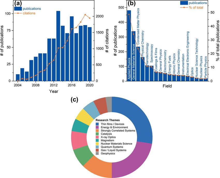
J. Phys.: Condens. Matter 33 (2021) 233001                                                                                    Topical Review

an anode voltage of up to 30 kV and a maximum power of                   limits the exploration of deeper core levels. However, this is
150 W. It can deliver a spot size between 100 and 1000 μm.               partially mitigated by the comparatively good photoelectron
The monochromator is based on a Ge(443) single crystal in                cross sections of many of the lighter elements. The Ag Lα has
a Rowland circle with a diamater of 730 mm. The major-                   found successful application in a number of areas [116–119].
ity of these systems are ambient pressure HAXPES (AP-
HAXPES) setups, including a liquid AP-HAXPES setup at                    2.2.5. Custom systems. A number of custom built systems
the Fritz Haber Institute in Germany and a standard AP-                  are also in operation often combining x-ray sources and anal-
HAXPES system at the University of Manchester in the UK                  ysers from different suppliers.
combining Al and Cr Kα sources. A special case of a labora-                 Kochi University of Technology, Japan, has a double x-ray
tory ambient pressure HAXPES system is beamline 8A at the                source laboratory HAXPES system, which combines
Pohang Accelerator Laboratory, South Korea’s synchrotron.                monochromated Cr Kα and Al Kα x-ray sources from
While the end station uses an undulator-based beamline for               ULVAC-PHI with a Scienta Omicron EW4000 analyser. It
measurement in the soft x-ray regime, it uses an offline Cr              also houses a dual beam charge neutralisation setup using
Kα source installed on the same end station to enable bulk               a low energy electron beam and ion beam (ULVAC-PHI)
measurements in parallel to surface sensitive ambient pressure           and a connected magnetron sputtering deposition system
experiments [114].                                                       (Chemitronics) for in situ characterisation [120–122].
2.2.4. Kratos systems. Kratos started to build systems with                 One of the most recent additions to the family of
hard x-ray sources including Ag and Zr in the 1980s. In the              Cr Kα-based HAXPES systems has been constructed and
newest Kratos spectrometer generation a dual monochromated               commissioned at Temple University (USA). The heart of the
Al Kα/Ag Lα x-ray source is implemented in their AXIS                    system is a custom-built high-flux monochromated 5.4 keV
Supra+ system. The same monochromator crystal is used for                hard x-ray microfocus source, consisting of a 30 kV focused
both, as the photon energy of Ag Lα (hν = 2.984 keV) is                  electron gun (Kimball Physics), a water-cooled thin-film Cr
approximately double that of Al Kα and the second order                  anode, and a large bent-crystal Ge(411) monochromator in a
diffraction of the monochromator crystal used for Al Kα can              730 mm Rowland circle geometry. The x-ray spot size is vari-
be used to also deliver monochromated Ag Lα x-rays. This                 able from 150 to 500 μm. The standard working energy reso-
is not only a very efficient way of delivering two monochro-             lution of the source is
J. Phys.: Condens. Matter 33 (2021) 233001                                                                                         Topical Review

                          Table 2. Selected recent pico- and femtosecond time-resolved photoemission spectroscopy
                          experiments at storage rings and free-electron lasers. The listed experimental parameters are
                          the probe photon energy at which experiments were conducted (hν), the effective pulse
                          repetition rate ( frep ), the effective energy (ΔE), and the time (Δt) resolution. For storage
                          ring-based (FEL-based) experiments, frep is determined by the repetition rate of the pump
                          laser (FEL probe) pulses.
                          Type                   Beamline      Facility    hν/keV f rep /kHz ΔE/eV      Δt/ps Reference
                          Storage ring-based    TEMPO          SOLEIL          0.12    141     0.012    50       [125]
                                                BL07LSU        SPring-8        0.385   208     0.7      50       [126]
                                                11.0.2         ALS             0.95    127              60       [127]
                                                BL19LXU        SPring-8        7.94    1
J. Phys.: Condens. Matter 33 (2021) 233001 Topical Review
3.1. Detection schemes experiments in liquids and at solid/liquid interfaces (as will be
discussed in detail in section 3.3.2) [156, 157].
3.1.1. Energy-dependent HAXPES. Many HAXPES experi-
For both cases with and without elastic scattering being
ment start with the measurement of spectra at a single hard
taken into account, the depth distribution function (DDF) is
x-ray excitation energy in fixed geometry. This can deliver use-
defined as the probability that a photoelectron leaving the sur-
ful information on the bulk of a material, as discussed in the
face originated from a given depth measured normally from the
context of laboratory-based HAXPES (see section 2.2). The
surface, and a number of approaches exist to calculate it [158].
energy-dependence of both photoionisation cross sections and
DDFs can be particularly illustrative when trying to visu-
probing depth can been exploited further by tuning the x-ray
alise the information depth and distribution in multilayered
energy and collecting spectra at a number of energies. In the
samples [89]. Finally, a useful definition in this context is that
case of core level spectra the main interest is to follow changes
of information depth (ID) itself. It is generally defined as the
in chemical states with probing depth, while for valence spec-
depth normal to the sample surface from which useful infor-
tra the motivation is often to distinguish contributions with
mation can be obtained. IDs are given as the percentage of
varying orbital character. Although multi-source laboratory
detected signal across the total depth and common IDs such
systems exist, as discussed in section 2.2, the tunability of
as ID90 , ID95 , and ID99 represent the depths from which 90,
synchrotron sources results in most energy-dependent experi-
95, and 99% of the total signal originate from, respectively.
ments being conducted at synchrotron beamlines. A multitude
of studies has been conducted in this fashion over the years Many HAXPES studies involve the tuning of the excita-
and it is impossible to cover them all, however, a selection of tion energy to collect core level spectra carrying varying depth
specific, exemplary studies is discussed here to illustrate the information to create non-destructive depth profiles. In addi-
applicability of the energy-dependent HAXPES approach. tion, such studies can be used to experimentally determine
Before moving on to individual examples, it is useful to IMFPs for specific systems. Ouardi et al combined these two
recall the concept of information depth in HAXPES at this motivations and performed a systematic exploration of prob-
point, including available models and specific considerations ing depth in HAXPES on the Heusler compound Co2 MnSi,
when using hard x-rays. A number of comprehensive discus- using hard x-ray energies between 1 and 8 keV combined with
sions of the concepts covered here are available in the litera- specifically designed test samples with varying layer thick-
ture, and the following is only a brief summary [47, 143, 144]. nesses [159]. To clearly demonstrate the depth dependence in
In HAXPES experiments the x-ray absorption length is much HAXPES they employed a partially oxidised Ta layer under-
greater than the inelastic mean free path (IMFP) of the excited neath a 1 nm thick Pt layer. With higher excitation energy the
photoelectrons, and therefore the information depth is deter- oxide peak in the Ta 4d core level reduces, reaching equal
mined only by the IMFP. Several approaches for the estimation intensity with the metal peak at 4 keV, and completely van-
of IMFPs have been developed with the most popular being ishing at 7 keV. From these experiments and knowledge of the
those of Seah and Dench, and Tanuma, Powell and Penn (the IMFPs the authors could conclude that the top 3 nm of the
TPP-2M method) [145–148]. Inelastic background modeling Ta layer were oxidised. Such experiments are very useful, to
has been explored to detect deeply buried layers beyond the determine varying oxide thicknesses on the surface of materi-
elastic limit [113, 149]. als. Furthermore, they find application when trying to design
While the IMFP finds wide application to estimate the prob- capping layers to protect oxygen or moisture-sensitive layers
ing depth in HAXPES, and generally in PES, it does not from air exposure, a practice common in PES. By tuning the
include effects from elastic scattering, which can play a signif- energy to probe through capping layers of different thicknesses
icant role in determining the information depth, depending on the oxidation of the underlying layer of interest can be studied.
excitation energy and atomic number. When elastic scattering Beyond the demonstration of a non-destructive depth profile of
effects are important, the effective attenuation length (EAL) the Ta/Pt system, Ouardi et al went on to experimentally deter-
is used instead of the IMFP, which gives smaller information mine the IMFP of Co2 MnSi, by using Ta (40 nm)/Co2MnSi
depths due to losses from elastic scattering [34, 150–153]. It (x)/Pt (1 nm) heterostructures with varying thickness x of the
is important to note that theoretical models generally over- Co2 MnSi interlayer. By recording the intensities of the Pt and
estimate the probing depth of HAXPES [154, 155]. Solokha Ta core level spectra and comparing area ratios they extracted
et al studied the difference between theoretical and experi- experimental IMFP values between 2 and 7 keV, which as
mental EALs in silicon across kinetic energies from 1.5 to expected were lower than those calculated from theory.
8 keV at beamline I09 at Diamond Light Source [154]. The Energy-dependent HAXPES can be approached from two
experimentally determined EALs are significantly smaller than different perspective. One can either vary the excitation energy,
the predicted EALs and IMFPs. One particular observation is as in the previous example, or one can vary the core level
that at higher energies above 15Z 2 eV, where Z is the atomic measured for a specific element to build up non-destructive
number of the element, a better description of the elastic scat- depth profiles. The latter is enabled by the increased number
tering cross section is necessary. Some additional consider- of deeper core levels accessible with HAXPES compared to
ations have to be taken into account when determining the SXPS, as discussed in section 2.2, coupled with the difference
information depth in liquids rather than solids. This is becom- in depth information depending on the kinetic energy of the
ing important in HAXPES as ambient pressure setups enable photoelectrons coming from different core levels. Instead of
11J. Phys.: Condens. Matter 33 (2021) 233001 Topical Review
collecting only one core level for an element at many differ- particularly for deep inner shells and hard x-ray photon ener-
ent excitation energies, multiple core levels of the same ele- gies, Drube et al showed that this approximation in fact breaks
ment with significantly different energies are collected at the down due to the presence of electron correlations and inter-
one (or more) excitation energies. Such an approach has been channel coupling [164]. This process becomes relevant in the
applied successfully in Si-containing systems, including sili- interpretation and analysis of core level spectra, when core
con nanoparticle anodes in lithium ion batteries and SiC/SiO2 level intensities are evaluated quantitatively and the photon
structures used in power electronic devices [89, 160]. The sil- excitation energy is ‘near’ a deep core level threshold. Then,
icon 1s core level together with either the 2s and 2p core the absolute and relative intensities of shallower core levels
levels, which have elemental reference binding energies of may change, potentially leading to misinterpretation. Depend-
1839, 150 and 100 eV, respectively, can be effectively com- ing on the material studied and the photon energy range across
bined to provide a detailed depth profile and aid tuning of the which experiments are performed, this effect can significantly
depth information to layers or interfaces of specific interest. alter the observed core levels, as was shown on the examples
One point of note is that while most energy-dependent stud- of the Ag and In by Drube et al, where interchannel cou-
ies are performed at synchrotron sources due to the tunability pling of the 3d photoionisation channels with the 2p channels
of such sources, this strategy can also be implemented using occurs.
laboratory sources. An in practice tremendously useful aspect of being able
HAXPES is often used to solve arguments about whether to tune the excitation energy is that Auger lines occur at
certain spectral features may be assigned to surface states of the same kinetic energy independent of the excitation energy
some description or whether they are intrinsic to the photoe- and can therefore be moved relative to core levels of inter-
mission in a material. An early study by Payne et al used est to avoid overlap. This is of particular interest in com-
HAXPES at 6 and 7.7 keV in comparison with Al Kα SXPS to plex multi-element materials, such as high entropy alloys and
understand the appearance of two components in the Pb 4f core complex ceramics for solid oxide fuel cells. An example of
spectra of β-PbO2 [161]. They could show that the presence using energy-dependent PES at beamline KMC-1 at BESSY II
of a narrower, low binding energy component with predom- applied to a multicomponent alloy (CrMnFeCoNi) is shown in
figure 5 [165]. When this material is studied using standard
inant Gaussian character together with a broader, high bind-
Al Kα laboratory sources, the overlap in the spectra of Auger
ing energy component with predominant Lorentzian character
electrons and metal 2p photoelectrons impairs the determina-
could be explained by the presence of screened and unscreened
tion of chemical shifts and relative atomic concentrations vital
states, respectively, rather than by an ill-defined surface
to gain an understanding of the material. By tuning the exci-
state.
tation energy at the synchrotron to 2 keV, the same binding
Beyond the information of elemental and chemical distri-
energy region lacks said overlaps due to the change in position
butions with depth, energy-dependent core level HAXPES has
of the Auger lines and a complete analysis and interpretation
also been used to quantify changes in electronic and magnetic
of the transition metal core level spectra is possible. For appli-
behavior with depth. Mukherjee et al studied the distribution
cations, such as the study of the composition of high entropy
of charge carriers in prototypical LaAlO3 /SrTiO3 oxide het- alloys, the opportunity to shift away the Auger spectra removes
erostructures [162]. This system famously exhibits a highly a significant obstacle to a detailed spectroscopic analysis of the
mobile, two-dimensional electron gas (2DEG) at the interface multicomponent alloys of transition metals.
of two insulating, diamagnetic oxides. Using excitation ener- Hard x-rays also open the realm of 1s resonances for the
gies between 2 and 8 keV, La 3d and Sr 3d core level spectra second row elements, starting with P 1s, which has a binding
were recorded to tune the excitation energy to the interface. energy of 2.145 keV. Using tunable hard x-rays the electron
Then, Ti 2p spectra could be used to determine the charge car- emission from a decaying core-excited state can be studied
rier distributions in the system. The authors found that two in kinetic energy regions, where the normal Auger spectrum
distinct distributions could be observed with one distributed manifests, if the photon energy is (far) above the core ioni-
homogeneously throughout the bulk SrTiO3 and another spe- sation threshold. As an example, resonant core excitation of
cific to the interface with a with of ≈1 nm. Pincelli et al S 1s into unoccupied bound states of sulfur atoms in vari-
used excitation energies from 0.8 in the soft x-ray regime to ous environments and the decays of those core excited states
5.94 keV in the hard x-ray regime to determine the critical observed in the S KLL Auger kinetic energy region will be
thickness of electron hybridisation in the spintronic materials discussed here. The electron spectrum in the Auger region is
La1−x Srx MnO3 and (Ga, Mn)As [163]. They used the evolu- considered static for excitations above the ionisation threshold.
tion of the ratio between the peak areas of the main photoion- If the photon energy is stepped over e.g. the K-edge absorp-
isation peak and a well screened peak as a function of the tion resonance of sulfur, a significant variation of intensity
respective IMFPs to show that (Ga, Mn)As and La1−x Srx MnO3 in the x-ray absorption spectrum is noticable. If the electron
have intrinsic limits of 3 and 10 unit cells from the surface kinetic energy spectrum in the S KLL region is recorded at
before bulk properties are restored. the same time, deviations from the normal Auger spectrum
An important fundamental process of photoionisation, that will be readily observed. Parts of the spectrum will remain
is often overlooked in energy-dependent HAXPES, is inter- static in kinetic energy and eventually transition to building
channel coupling. While is broadly assumed that photoionisa- up the normal Auger spectrum, other parts are static in bind-
tion cross sections are dominated by single-particle behavior, ing energy—hence dispersing on the kinetic energy scale. The
12J. Phys.: Condens. Matter 33 (2021) 233001 Topical Review
Figure 5. Transition metal 2p core level and Auger spectra of a CrMnFeCoNi multicomponent alloy collected using a laboratory-based Al
Kα source and 2 keV x-rays at beamline KMC-1 at BESSY II. The Auger region was shifted by 516 eV to aid comparison and illustrate
overlap.
latter parts are intense close to resonance and vanish above the for the crystal and nanoparticle cases, owing to the local char-
ionisation threshold. By recording an electron spectrum for acter of the probe, the composite exhibits a bi-modal charge
photon energy steps over the resonance a map of the decay transfer time distribution as a function of excitation energy.
channels present in the system can be obtained. This forms the The switch between to regimes of charge transfer times is
basis for resonant Auger spectroscopy. attributed to the core excited electron having sufficient energy
Core hole clock spectroscopy (CHCS) utilises the life time to overcome the Schottky barrier created in the MoS2 /graphene
of the core hole (τ ct ) as an internal time-reference and uses the interface.
relative intensity between the channels static in kinetic energy, CHCS has also been combined with polarisation-dependent
denoted Auger channels since the final state is Auger-like even HAXPES, which will be discussed in detail in section 3.2.2.
below the ionisation threshold owing to that the core-excited Using linearly polarised hard x-rays the charge transfer
electron have tunneled away, and those of dispersing states, anisotropy upon resonant core-excitation has been recorded
denoted Raman channels (see Karis et al for details) [166]. If to study a layered SnS2 single crystal [171]. This delivers not
a resonant Auger spectrum is collected at a particular photon only chemical specificity, but also orbital specificity through
energy, the ratio between the intensities of the Raman (I R ) and the difference in alignment of the px , py and pz orbitals rela-
Auger (I A ) channels, I R /I A , multiplied with the core hole life- tive to the photoemission geometry. The resonant Auger spec-
troscopy maps recorded in normal and grazing emission can
time, e.g. that of S 1s (τ 1s ), reveals the time at which the excited
be directly compared to calculations of the band structure with
electrons have moved away from the site of core-excitation.
orbital projections. CHCS can thus be used as a probe of the
Using the core-hole clock method electron dynamics in the
unoccupied band structure of a system using. Another example
fs to tens of as regimes can be studied with the chemical
of combining linearly polarised light with CHCS is the case
specificity that core excitations provide [167, 168].
of black phosphorous explored by Johansson et al [172].
Johansson et al, have used the CHCS methodology to study Black phosphorous is known to have different macroscopic
fs and as electron-transfer dynamics in an organic heterojunc- conductivities in the armchair and zig-zag directions. This
tion [169]. For core excitations close to the K-edge a sig- kind of interlayer charge transfer anisotropy can be studied
nificant variation between charge transfer times is observed with CHCS by aligning the crystal directions with the lin-
depending on the PCPDTBT/PCBM weight ratio in the active early polarised x-rays. Comparing charge transfer times in
layer, with the time reduced by 86% in the 1:2 blend com- the two directions reveals that the armchair direction has a
pared to pristine (1:0) polymer. Excitations close to resonance faster delocalisation of the core excited electron for excita-
facilitate inter-molecular charge transfer between the polymer tion energies close to the P K-edge resonance. At about 2 eV
and PCBM. Farther above resonance the times converge to a excess energy, the electron transfer time is equal for the two
value of 170 as for all blends including pristine polymer, as in directions, but switches over to the zig-zag direction. Together
this regime inter-polymer charge transfer dominates. In MoS2 with out-of-plane results a detailed picture of the electron
charge transfer dynamics have been shown to vary depending dynamics using the orbital projected band structure can be
on the morphology of the sample, including a single crys- achieved [168, 172].
tal, nanoparticles, and a composite of MoS2 sandwiching a Beyond the use of energy-dependent HAXPES for core
graphene backbone, using CHCS [170]. While they are similar level and Auger spectra it is a powerful tool to untangle the
13J. Phys.: Condens. Matter 33 (2021) 233001 Topical Review
Figure 6. Energy-dependent HAXPES experiments of valence states. (a) Valence states of the ruthenium complex Ru(cbpy)2 (NCS)2
collected at 2.8 and 0.1 keV. The two Ru 4d states from varying ligand coordinations can be observed using hard x-rays. (b) and (c)
Experimental valence spectra and LDA + U theory results for CdO. Spectra are shown across a number of excitation energies, including a
soft x-ray measurement at 0.6 keV and four hard x-ray energies (2.5, 4.5, 6.054, and 7.935 keV). Reproduced from [176]. CC BY 3.0.
orbital composition of the valence states of molecules and The detailed knowledge of the electronic structure of mate-
materials [173]. The element-dependent changes in photoion- rials used in electronic and optoelectronic applications is cru-
isation cross section with excitation energy, allow the discrim- cial for the design of efficient heterojunctions in such devices.
ination or enhancement of certain contributions to the valence A clear example of the use of energy-dependent HAXPES in
electronic structure aiding interpretation and comparison with conjunction with DFT to gain an in-depth understanding of the
projected densities of states from DFT calculations. Examples electronic structure of a material is the study on n-type CdO,
of exploited variations in cross sections are relative decreases a transparent conducting oxide (TCO) with great potential in
for the valence orbitals of light elements such as carbon, nitro- photovoltaic applications, by Mudd et al [176]. Experimental
gen, oxygen, and sulfur at higher energies, or the presence results are used to verify different theoretical approaches for
of Cooper minima for some transition metal valence orbitals. the applicability in the case of CdO. Theoretical calculations of
Thus comparing valence photoelectron spectra measured using CdO, like for many metal oxides, suffer from underestimation
varying excitation energies allows the characterisation of the of the binding energy of shallow core states in close proximity
frontier electronic structure. to the valence band, often distorting the level of hybridisa-
An example for an experiment conducted on a molecu- tion present in such systems. Exact binding energy values for
lar sample is shown in figure 6, where the valence states shallow core states and features in the valence band can be
extracted from HAXPES and energy-dependent experiments
of the ruthenium complex Ru(cbpy)2(NCS)2 is measured at
enable the identification of the orbital character of specific
two different photon energies [174]. The valence energy
features in the valence region. Figures 6(b) and (c) show the
levels of this dye molecule are responsible for the light
energy-dependent experimental spectra and photoionisation
absorption properties and for the electron transfer reactions
cross section corrected densities of states, respectively. Feature
occurring during and after light absorption in photoelectro-
I at −3.9 eV shows a strong photon energy dependence, while
chemical processes. Only at higher photon energies can the feature III remains stable. Feature II also shows a response
Ru 4d peaks, so important for the electronic structure of the to changes in photon energy, which appears as a broaden-
molecule, be observed. Ru 4d states show two peaks, as the ing of feature II. From these observed relative changes in the
complex is asymmetric with two different ligand coordinating intensities of valence band features, and in conjunction with
the Ru atoms giving an orbital mix of 1:2. In contrast, in a projected, photoionisation cross section corrected densities of
symmetrical complex such as Ru(bpy)32+ only one Ru 4d fea- states, Mudd et al are able to identify and discriminate between
ture is observed. Complementary studies determining contri- the varying contributions to the valence states. Using energy-
butions to the electronic structures from different ligands were dependent HAXPES it is also possible to observe and identify
further investigated using resonant photoelectron spectroscopy low energy plasmon features that fall within the valence states
(RPES) [175]. This confirmed that the HOMO level is mainly [177]. Another important aspect is that in valence band stud-
of Ru 4d for the Ru(bpy)32+ complex. At the photon energy ies tuning of the photon energy allows precise definition of the
2.841 keV a clear resonance corresponding to excitation from momentum component kz perpendicular to the sample surface.
Ru 2p to unoccupied energy levels, resulting in enhanced emis- An important aspect to take into account when perform-
sion of electrons from the Ru 4d contribution in the valence ing energy-dependent HAXPES is the occurrence of energy-
electronic structure. dependent, recoil-induced binding energy shifts that may
14You can also read