Hepatocellular Carcinoma Case Report from the Phase 3 HOPE-B Gene Therapy Trial in Adults with Hemophilia B
←
→
Page content transcription
If your browser does not render page correctly, please read the page content below
Hepatocellular Carcinoma Case Report from the Phase 3
  HOPE-B Gene Therapy Trial in Adults with Hemophilia B

Manfred Schmidt1, Graham R. Foster2, Michiel Coppens3, Hauke Thomsen1, David Cooper4, Ricardo Dolmetsch4,
                           Eileen K. Sawyer4, Liesbeth Heijink5, Steven W. Pipe6
  1GeneWerk   GmbH, Heidelberg, Germany; 2Barts Liver Centre, Queen Mary University of London, London, UK; 3Amsterdam
University Medical Centers, University of Amsterdam, Amsterdam, Netherlands; 4uniQure Inc. Lexington, MA, USA; 5uniQure BV,
                          Amsterdam, the Netherlands; 6University of Michigan, Ann Arbor, MI, USA

                                                                                                                       1
Disclosures for Steven W. Pipe

In compliance with COI policy, ISTH requires the following disclosures to the
session audience:

     Research Support/P.I.           No relevant conflicts of interest to declare
           Employee                  No relevant conflicts of interest to declare
          Consultant                 No relevant conflicts of interest to declare
      Major Stockholder              No relevant conflicts of interest to declare
       Speakers Bureau               No relevant conflicts of interest to declare
           Honoraria                 No relevant conflicts of interest to declare
   Scientific Advisory Board         No relevant conflicts of interest to declare

                                                                                2
HOPE-B: Phase 3 AAV5-based Gene Therapy Trial for Hemophilia B
• Open label phase 3 study with follow-up of 54 subjects with hemophilia B
receiving a single dose of 2×10¹³ gc/kg of etranacogene dezaparvovec
• The largest AAV gene therapy trial cohort in hemophilia B
reported to date Etranacogene dezaparvovec:
• Mean FIX activity significantly increased to near-normal levels at Hyperactive FIX Padua variant
6 months post-etranacogene dezaparvovec1
• Most common safety findings at 6 months were transaminase
elevations requiring steroid treatment (9 subjects) and infusion- AAV5
related reactions (7 subjects), supporting a safety profile consistent
with early phase studies1,2,3
• Here we present an SAE of hepatocellular carcinoma (HCC) in a trial
subject with multiple pre-existing risk factors for HCC, including the
findings of an independent, expert molecular evaluation that LP-1 Human wild type FIX (codon optimized)
determined this case was unlikely to be related to treatment with (Liver-specific with 2 nucleotide adaptation
etranacogene dezaparvovec promoter)
Hyperactive Padua variant
• Details about the study and interim safety and efficacy data at 52-
weeks after dosing are reported in PB06534
1. Pipe et al; ASH 2020 https://ashpublications.org/blood/article/136/Supplement_2/LBA-6/474189/First-Data-from-the-Phase-3-HOPE-B-Gene-Therapy
2. Leebeek FWG, et al, ASH 2020; Poster #33724
3. Von Drygalski A, et al, ASH 2020; Oral presentation #672.
4. Pipe et al ISTH 2021; 52 Week Efficacy and Safety of Etranacogene Dezaparvovec in Adults with Severe or Moderate-Severe Hemophilia B: Data from the Phase 3 HOPE-B Gene Therapy Trial
AAV, adeno-associated virus; HCC, hepatocellular carcinoma, FIX; factor IX. 3HCC epidemiology and risk factors
• Primary liver cancer is the sixth most common cancer worldwide. 1
• Risk factors for development of HCC includes, but not limited to, Hepatitis C Virus (HCV) and/or
Hepatitis B Virus (HBV), advanced age, gender and cirrhosis. 2
• HCC development has been strongly linked to HBV and HCV infections and is associated with
approximately 80% of HCC cases.3
• Most cases of HCV-related and HBV-related HCC occur among patients with advanced fibrosis or
cirrhosis. However, up to 20% of patients that develop HCC have a non-cirrhotic liver.2
• Other risk factors include high alcohol consumption, obesity, exposure to environmental toxins, and
metabolic disorders such as NAFLD/NASH.2,4
• Although the incidence of HCC is higher in the hemophilic population, it has been correlated with higher
incidence of HCV infection and is not due to the hemophilia phenotype. 5
• Despite clearance of HCV, HCC risk is not eliminated but has been estimated to be reduced by 71%. 6
1. Sung H et al.,CA Cancer J Clin 2021;71:209–249. 2. Desai A, et al. World J Hepatol. 2019;11(1):1-18. 3. El-Serag HB. Gastroenterology. 2012;142(6):1264-1273. 4. Marrero JA,
et al. Hepatology. 2018 Aug;68(2):723-750. doi: 10.1002/hep.29913. 5. Shetty S, et al. Critical Reviews in Oncology/Hematology. 2016;99:129-133. 6. Ioannou GN, et al. J Hepatol,
5 Sep 2017; doi:10.1016/j.jhep.2017.08.030
4Relevant Medical History
• 69-year-old, white, non-Hispanic male with moderately severe Hemophilia B
• 1980 – Diagnosed with HBV (+ve for anti-HBs, anti-HBc and anti-HBe antibodies)
• 1983 – Diagnosed with non-A/non-B hepatitis
• 2003 – Confirmed positive for HCV when testing available
• 2015 – Evaluated for HCV eradication therapy, genotype 1a, no significant fibrosis (Fibroscan 5.7 kPa)
• 2015 – Treated with paritaprevir/ombitasvir/ritonavir, dasabuvir, and ribavirin; achieving a sustained
virologic response
• Social history notable for prior smoking, alcohol consumption of 0-2 units/week
• Familial history notable for cancer
5Timeline of adverse event evaluation/management
July 2019 October 2019 October 2020 October 2020 November 2020 March 2021
Pre-dosing AMT-061 52-week visit Adverse Event Serious AE HCC SAE
Fibroscan Administration ultrasound Liver lesion Likely HCC Unlikely related
Mean FIX activity ~44%
2019 2020 2021
Jul Aug Sep Oct Nov Dec Jan Feb Mar Apr May Jun Jul Aug Sep Oct Nov Dec Jan Feb Mar
Molecular Analysis
November 2019 January 2020 November 2020 March 2021
Per-operative discovery of a 2nd
Chest CT Abdominal Abd CT – 4 phase contrast Post-TACE MRI
Lung nodule eval Ultrasound lesion in liver segment 2/3, S2/3
Needle biopsy
lesion was resected to downstage.
Case conference
Afterwards, he underwent a TACE
procedure for the S8 lesion.
January 2021
Surgical resection
December 2020 Post-op case conference
Surgery scheduled Transplant referral
COVID-19 test positive February 2021
Surgery postponed Transarterial Chemo-
embolization (TACE)
6HCC Analysis - Expectations
Expected findings if Expected findings if
AAV integration drove HCC HCC independent of AAV treatment
Integration • Frequent integrations in HCC • Very infrequent integrations
Site Analysis • Dominant AAV integration site • No dominant integration site
Whole • Integration in/near known • Common HCC oncogene mutations (eg.
Genome oncogenes (eg. TP53, TP53, NFE2L2)
Sequencing NFE2L2) • No AAV integration sites near oncogenes
7Molecular Analysis: Vector Copy Number and Integration Rate
• Molecular analysis for copy number quantification was conducted via qPCR
• Vector copy number (VCN) was calculated by normalizing vector copies to the number of housekeeping-gene copies
(diploid genomes)
Tissue VCN
(copies/diploid genome)
HCC 3.21
HCC-adjacent 4.11
• S-EPTS/LM-PCR*, was used to determine the number of integration sites per cell
• Etranacogene dezaparvovec integration rate into hepatocytes is very low as previously reported for AAV
• Less than 60 cells out of 250,000 (0.027%) had an integration event in the HCC tumor sample
Tissue Integration rate
HCC 0.027%
HCC-adjacent 0.018%
*Shearing Extension Primer Tag Selection Ligation-Mediated PCR 8Molecular Analysis II: Site of Vector Integration
• Integration site (IS) analysis was conducted via whole genome sequencing (WGS)
• No integration event was observed in more than 1 sequence read out of 150 reads
• The low number of sequence reads for each IS indicate that IS are rare in both the HCC and HCC-adjacent samples.
• There is no dominant IS in the HCC sample.
Chromosome Integration site Total sequence count Gene Name
HCC-adjacent Chr19 44924479 1 APOC1P1
Chr5 126472870 1 GRAMD3
Chromosome Integration site Total sequence count Gene Name
Chr19 44924545 1 APOC1P1
HCC
Chr5 54714322 1 EML6
Chr4 91418750 1 CCSER1
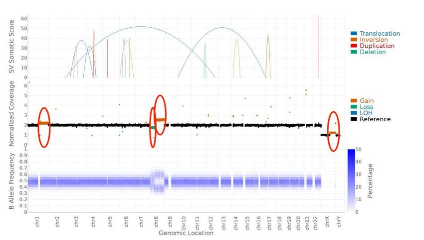
9Molecular Analysis III: genome-wide chromosomal rearrangements
Full Report UNI04GW – v1 Page 22 of 56
Whole Genome Sequence analysis
• Multiple structural variants were
observed in HCC sample,
including mutations in TP53 and
NFE2L2 - known to be common
drivers of HCC - independent of
etranacogene dezaparvovec
• Large chromosomal
rearrangements in Chr 1,8 and X
characteristic of HCC -
independent of etranacogene
dezaparvovec
Figure 5: Coverage plot for HCC versus HCC-adjacent sample WGS. Among the potential differences, large
sized gains on chromosome 1, 8 and X and a loss on chromosome 8 were obvious (marked by red circles).
10HCC Analysis: Summary of Results
Expected findings if Expected findings if
AAV integration drove HCC independent of AAV Actual findings of GeneWerk analysis
HCC treatment
• Etranacogene dezaparvovec integration rate into
hepatocytes is very low as previously reported for AAV
• Frequent integrations in
Integration • No dominant integration event or integration site in the
HCC • Very infrequent integrations
Site HCC sample
• Dominant AAV integration • No dominant integration site
Analysis • Less than 60 cells out of 250K (0.027%) had an
site
integration event in the tumor sample
• Very low rate of vector integration in genes not known to
be associated with HCC
• Common HCC oncogene • Large chromosomal rearrangements in Chr 1,8 and X
Whole • Integration in/near known
mutations characteristic of HCC - independent of etranacogene
Genome oncogenes (eg. TP53,
• No AAV integration sites near dezaparvovec
Sequencing NFE2L2)
oncogenes • Mutations in TP53 and NFE2L2 - known to be common
drivers of HCC - independent of etranacogene
dezaparvovec
11Conclusions and Future Recommendations
• Asymptomatic HCC was identified in an older subject with HBV, prior HCV post SVR on a
routine safety ultrasound 1 year after dosing; the subject has been treated with TACE and
is under evaluation for liver transplant
• HCC development in this case is now considered unlikely related to treatment with
etranacogene dezaparvovec based upon the results of genetic analysis and pre-existing
risk factors
• Short-term and long-term follow-up is important after gene therapy
• Many patients with hemophilia have pre-existing risk factors for HCC
• The risk of HCC after HCV-SVR is still being investigated
• Aging patients may develop risk factors over time unrelated to treatment (age >50, NAFLD/NASH, obesity,
alcohol use)
• Ultrasound monitoring of all participants enrolled in etranacogene dezaparvovec clinical
trials was increased to twice yearly regardless of pre-existing risk factors for HCC as a
conservative approach.
12You can also read