HHS Public Access Author manuscript Sci Total Environ. Author manuscript; available in PMC 2022 January 20.
←
→
Page content transcription
If your browser does not render page correctly, please read the page content below
HHS Public Access
                                 Author manuscript
                                 Sci Total Environ. Author manuscript; available in PMC 2022 January 20.
Author Manuscript

                    Published in final edited form as:
                     Sci Total Environ. 2021 January 20; 753: 142351. doi:10.1016/j.scitotenv.2020.142351.

                    Evaluation of a portable XRF device for in vivo quantification of
                    lead in bone among a US population
                    Xinxin Zhanga, Aaron J. Spechtb, Ellen Wellsa, Marc G. Weisskopfb,1, Jennifer Weuvec,1,
                    Linda H. Niea,*,1
                    aSchool    of Health Sciences, Purdue University, West Lafayette, IN, USA
                    bDepartment       of Environmental Health, Harvard T.H. Chan School of Public Health, Boston, MA,
Author Manuscript

                    USA
                    cDepartment       of Epidemiology, Boston University School of Public Health, Boston, MA, USA

                    Abstract
                         Background: Lead (Pb) concentration in bone is a reliable biomarker for cumulative Pb
                         exposure and studying associated health outcomes. However, the standard K-shell fluorescence
                         (KXRF) bone Pb measurement technology has limitations in large-scale population studies.

                         Objective: We compared measurements from a portable XRF device and a KXRF device.

                         Methods: We measured bone Pb concentrations in vivo using portable XRF and KXRF, each
                         measured at the midtibia bone in 71 people, 38–95 years of age (mean ± SD = 63 ± 11 years)
Author Manuscript

                         living in or near three Indiana communities, US; 10 participants were occupationally exposed. We
                         estimated the correlation between bone Pb concentrations measured by both devices. We also
                         examined the extent to which the detection limit (DL) of the portable XRF was influenced by scan
                         time and overlying soft tissue thickness. Finally, we quantified the associations of estimated bone
                         Pb concentration with age and age with soft tissue thickness.

                         Results: The mean bone Pb concentration measured via portable XRF was 12.3 ± 16.7 mg Pb/kg
                         dry bone. The uncertainty of a 3-minute (N = 60) in vivo portable XRF measurement ranged from
                         1.8 to 6.3 mg/kg, in the context of soft tissue thickness ranging from 2 to 6 mm. This uncertainty
                         was reduced by a factor of 1.4 with 5-minute measurements (N = 11). Bone Pb measurements via
                         portable XRF and KXRF were significantly correlated: r = 0.48 for all participants, and r = 0.73
Author Manuscript

                    *
                     Corresponding author at: School of Health Sciences, Purdue University, 550 Stadium Mall Drive, West Lafayette, IN 47906, USA.
                    hnie@purdue.edu (L.H. Nie).
                    1These authors made equal contributions to this project.
                    CRediT authorship contribution statement
                    Xinxin Zhang: Conceptualization, Methodology, Validation, Data Analysis, Investigation, Data Curation, Project Administration,
                    Writing– Original Draft, Review and Editing. Aaron J. Specht: Methodology, Validation, Data Analysis, Writing – Review and
                    Editing. Ellen Wells: Data Analysis, Writing – Review and Editing. Marc G. Weisskopf, Jennifer Weuve, and Linda H. Nie:
                    Conceptualization, Supervision, Resources, Project Administration, Methodology, Validation, Investigation, Writing – Review and
                    Editing, Funding Acquisition.
                    Declaration of competing interest
                    The authors declare that they have no known competing financial interests or personal relationships that could have appeared to
                    influence the work reported in this paper.
                    Appendix A. Supplementary data
                    Supplementary data to this article can be found online at https://doi.org/10.1016/j.scitotenv.2020.142351.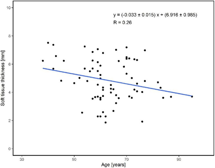
Zhang et al.                                                                                               Page 2

                             among participants with soft tissue thickness < 6 mm (72% of the sample). Bone Pb
Author Manuscript

                             concentrations were higher among participants who were older or were occupationally exposed to
                             Pb. Soft tissue thickness decreased with age.

                             Conclusion: With its ease of use, portability, and comparable sensitivity with conventional
                             KXRF systems, the portable XRF could be a valuable tool for non-invasive quantification of bone
                             Pb in vivo, especially for people with thinner soft tissue.
                       GRAPHICAL ABSTRACT
Author Manuscript

                       Keywords
                             X-ray fluorescence; Biomonitoring; Lead; Bone; In vivo quantification

                       1.     Introduction
                                       Lead (Pb) exposure has declined dramatically over the last several decades due to the
Author Manuscript

                                       regulatory actions prompted by numerous scientific findings that Pb is related to many
                                       adverse health outcomes (Grosse et al., 2002; Hwang et al., 2019; Pirkle et al., 1994).
                                       However, even at the lower exposure levels typical today, there are well-documented health
                                       effects (Bellinger and Needleman, 2003; Canfield et al., 2005; Lanphear et al., 2005;
                                       Lanphear et al., 2018). Pb remains one of the most common chemical toxicants in the
                                       environment and is second, following arsenic, on the substance priority list of the Agency
                                       for Toxic Substances and Disease Registry (ATSDR, 2019). Pb exposure is still a significant
                                       public concern for many populations, especially some vulnerable populations, including
                                       older adults, children, and occupational workers (Centers for Disease and Prevention, 2013;
                                       Lanphear et al., 2005; Lanphear et al., 2018). Blood Pb is a commonly used biomarker of Pb
                                       exposure. However, the half-life of Pb in the blood is relatively short, approximately 35 days
                                       for adults and as low as 7 days for children (Rabinowitz et al., 1976; Specht et al., 2019a).
Author Manuscript

                                       By contrast, Pb in bone has a longer half-life of years to decades (Rabinowitz, 1991),
                                       rendering it a more appropriate biomarker of cumulative Pb exposure, especially for
                                       populations with long-term Pb exposure or exposure that occurred mostly in the past.

                                       K-shell x-ray fluorescence (KXRF2) technology has been used to measure bone Pb in vivo
                                       and to investigate the adverse health effects of cumulative exposure (Navas-Acien et al.,

                       2XRF X-Ray Fluoresences.

                                          Sci Total Environ. Author manuscript; available in PMC 2022 January 20.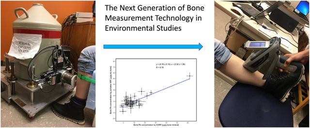
Zhang et al.                                                                                                     Page 3

                                         2007; Nie et al., 2006; Shih et al., 2007; Weisskopf et al., 2015; Weuve et al., 2013).
Author Manuscript

                                         However, the system size, long measurement time, and other operational requirements limit
                                         such studies to only a few research groups who possess the technology. Furthermore, study
                                         participants must visit a central research facility to undergo KXRF measurements, making it
                                         impossible to use this technology in field studies. Our group has developed a portable XRF
                                         approach that employs an easy-to-use device that can noninvasively quantify metal
                                         concentrations in vivo in a few minutes (Specht et al., 2014; Specht et al., 2018; Zhang et al.,
                                         2018). Thus, this device is more practical for use in large-scale epidemiologic studies and
                                         field settings. The portable device may be especially useful among vulnerable and
                                         underrepresented populations, whose participation may be limited by physical mobility or
                                         proximity to a KXRF system. This study compares the use of portable XRF for bone Pb
                                         measurements in vivo in adults against the standard KXRF bone Pb measurement.
Author Manuscript

                       2.     Methods
                       2.1.        Study population
                                         Seventy-six adults were recruited from a community-dwelling population in northwestern
                                         Indiana to participate in this study. Data from five participants were excluded from our
                                         analysis. Three of those participants had a very thick layer of soft tissue overlying their tibia
                                         bone, resulting in unacceptable high uncertainty and unrealistically negative bone Pb
                                         concentrations. External Pb contamination on the portable XRF device interfered with the
                                         measurements of the other two excluded participants. This contamination was eliminated in
                                         subsequent scans by cleaning the surface of the portable XRF. Forty-one participants (22
                                         women and 19 men; ages 43–83 years) were recruited from the vicinity around West
                                         Lafayette, Indiana (USA), among whom one was previously occupationally exposed to Pb.
                                         Nineteen participants (10 women and 9 men; ages 38–95 years) were recruited from East
Author Manuscript

                                         Chicago, Indiana (USA). East Chicago is the site of intense industrial activity over the past
                                         century, including refining Pb ore and recovering Pb from scrap metal and batteries. The
                                         East Chicago-based U.S. Smelter and Pb Refinery Superfund site was added to the National
                                         Priorities List in 2009 (EPA). All East Chicago participants were residents in the operable
                                         unit 1 site, a 322-acre residential area in the Pb superfund site. To cover a broader range of
                                         bone Pb concentrations, we recruited eleven participants (2 women and 9 men, ages 45–87
                                         years) with potential moderate to high historical Pb exposure from the vicinity around
                                         Muncie, Indiana (USA), among whom nine were occupationally exposed to Pb at some time
                                         point of their life.

                       2.2.        Bone Pb exposure assessment
                                         2.2.1. Portable XRF system—The portable XRF used in this study was a customized
Author Manuscript

                                         device from Thermo Fisher Scientific (Thermo Niton XL3t GOLDD+, Billerica, MA). The
                                         x-ray tube has an energy span of up to 50 kV and uses a thermoelectric-cooled silicon drift
                                         detector with a 25-mm2 area and a 1-mm thickness. The x-ray tube settings were optimized
                                         for Pb measurements with a voltage of 50 kV, a current of 40 μA, and a silver (Ag) and iron
                                         (Fe) combination filter. These settings provided the best detection limit (DL)3 for the

                       3DL Detection Limit.

                                              Sci Total Environ. Author manuscript; available in PMC 2022 January 20.
Zhang et al. Page 4
measurements of bone Pb (Specht et al., 2014). In response to irradiation, Pb atoms in the
Author Manuscript
bone generate Pb L-shell-characteristic x-rays with energies of 10.55 keV (Lα) and 12.61
keV (Lβ). However, only the Lβ peak was used for estimating bone Pb concentration
because of the lower background level under the Pb peak region. The peak fitting with the
least-squares algorithm was used to extract the net counts of Pb Lβ. A Gaussian distribution
was fit to the Pb Lβ peak and an exponential function was used to fit the background. The
bone Pb concentration was measured in mg/kg dry bone by the portable XRF (Specht et al.,
2018).
A 3-minute measurement was performed with the portable XRF for participants from West
Lafayette and East Chicago. To determine the extent to which a longer scan time would
reduce the DL of the device, the measurement time was extended to 5 min for Muncie
participants. The induced whole-body effective radiation dose from the 3-minute scan was
about 3.6 μSv (Specht et al., 2016; Specht et al., 2019b), which increased to approximately
Author Manuscript
6.0 μSv with the 5-minute scan. By contrast, the dose for a standard anteroposterior chest x-
ray is 100 μSv, and one day of natural cosmic radiation is equivalent to 10 μSv.
2.2.2. KXRF system—The setup of the 109Cd based KXRF system followed that used in
previous studies (Nie et al., 2004; Nie et al., 2006; Specht et al., 2014; Specht et al., 2016).
A 109Cd source was mounted in front of the detector, emitting gamma-ray with 88.035 keV
energy to irradiate the bone (or bone-equivalent phantoms in calibration procedures). In
response, Pb atoms in the bone emit Pb K-shell-characteristic x-rays with energies of 74.97
keV (Kα1), 72.80 keV (Kα2), and 84.94 keV (Kβ1). The system consists (Specht et al.,
2019a) of four high-purified germanium (HpGe) detectors with a dimension of 16 mm in
diameter and 10 mm in thickness, four feedback resistance pre-amplifiers, four digital signal
analyzers (DSA-1000). This four-detector system is known as a cloverleaf system and has
Author Manuscript
sensitivity about 2–3 times better than the conventional one-unit KXRF systems that are
used in most of the labs with bone Pb measurement capability (Nie et al., 2006). An in-
house peak fitting program was used to calculate bone Pb concentration and its uncertainty
separately with each detector (Nie et al., 2005; Somervaille et al., 1989). The Pb Kα and Kβ
signals were analyzed independently and normalized to the generated coherent peak signal,
and the bone Pb concentration obtained from an individual measurement has a unit of mg
Pb/kg bone mineral. The bone Pb concentration was computed as an average of the Kα and
Kβ signals from all four detectors, with the concentrations weighted by uncertainty
according to the inverse variance weighting method (Todd et al., 2002). The whole-body
effective dose from a 30-minute measurement was approximately 0.26 μSv with a 135
mCi109Cd source for this advanced cloverleaf system (Nie et al., 2007a, 2007b).
Author Manuscript
2.2.3. Calibration of the portable XRF and KXRF systems—The portable XRF
system was calibrated using a 100 mg Pb/kg plaster of Paris Pb-doped bone-equivalent
phantom. A consequence of the low energy of the Pb L x-ray is that the soft tissue over the
bone can attenuate the signal. As the soft tissue thickness increases, the system detects fewer
Pb L x-rays and more Compton scattering counts from the soft tissue. Thus, to simulate the
soft tissue over bone, Lucite plates of 0 to 5 mm were placed over the 100 mg/kg bone
phantom and the phantoms were measured for 15 min to calibrate the portable XRF for bone
Sci Total Environ. Author manuscript; available in PMC 2022 January 20.Zhang et al. Page 5
Pb. The calibration line was established as the Pb Lβ net counts of the 100 mg/kg bone
Author Manuscript
phantom change in relation to the Compton scattering peak counts. The in vivo bone Pb
concentration was calculated by relating an in vivo Pb Lβ net count to a known count per
concentration from the 100 mg/kg bone phantom.
In a previous study (Nie et al., 2011; Specht et al., 2014), a 0 mg/kg bone phantom was used
to estimate the background under the Pb Lβ peak, and the Pb net counts were calculated by
subtracting the estimated background counts from the gross counts under the peak of
interest. However, for in vivo measurements, this background subtraction method
overestimated the background and resulted in an underestimated Pb signal and higher
uncertainty. Instead, the Pb Lβ net count was directly extracted through the peak fitting and
the count from the 100 mg/kg bone phantom at the corresponding thickness was used to
determine concentration.
Author Manuscript
Because Pb K x-rays have much higher energy than the L x-rays, the KXRF signals were not
attenuated as much by the soft tissue over the bone as the portable XRF signals. Hence, the
standard calibration for the KXRF calibration uses nine Pb-doped phantoms with
concentrations ranging from 0 to 100 mg/kg. The phantoms were positioned in front of a
109Cd source, and a 30-minute measurement was performed. The Pb K
α,β peaks were
normalized to the coherent peak for the correction of the measurement geometry, bone
mineral normalization, and soft tissue attenuation. The calibrations lines were plotted as the
ratios of Pb Kα and Kβ peaks over coherent peak versus the bone Pb concentrations.
From the portable XRF calibration data, the relation between Lucite thickness and Compton
scattering counts was also determined. Using this relation, the overlying skin thickness for
an individual participant was estimated directly from the XRF spectrum of their portable
Author Manuscript
XRF bone Pb measurement. This then allowed us to use the Compton scattering counts from
their measurement to correct for the attenuation of the Pb signal caused by overlying tissue.
2.2.4. In vivo measurement of bone Pb—For portable XRF measurement, the
participant sat on a chair with one leg straightened and resting on a chair in front. Before the
measurement, the participant’s mid-tibia area was cleaned with alcohol swabs to eliminate
any extraneous contamination. The portable XRF device was placed in contact with the
participant’s skin right above the tibia on the medial surface of the anterior crest. Three-
minute measurements were taken for West Lafayette and East Chicago participants, and 5-
minute measurements for Muncie participants.
The portable XRF measurement was followed by the KXRF measurement, and the same
mid-tibial spot was cleaned again. The sitting participant’s target leg was immobilized by
Author Manuscript
securing it to the chair leg with Velcro straps and then positioned it about 1–2 mm away
from the 109Cd source. The bone was measured with the KXRF system for 30 min. The
Muncie participants were measured with a new 135 mCi 109Cd source, which had higher
activity than the one used for the West Lafayette and East Chicago participants. This resulted
in a lower uncertainty of the bone Pb concentration measured by the KXRF in the Muncie
participants.
Sci Total Environ. Author manuscript; available in PMC 2022 January 20.Zhang et al. Page 6
The Institutional Review Board of Purdue University, Boston University, and Harvard
Author Manuscript
University approved this study. All participants provided their informed consent forms
before undergoing the study procedures.
2.3. Data analyses
2.3.1. Detection limit of the XRF systems—The DL of the KXRF system was
defined as twice the uncertainty of the measurement of the 0 mg/kg bone phantom. Soft
tissue overlying the tibia bone increases the uncertainty and hence the DL of the portable
XRF measurement. Thus, the DL of portable XRF was calculated using 3-minute
measurements of 0 and 100 mg/kg Pb-doped bone-equivalent phantoms covered with Lucite
of thicknesses ranging from 1 to 8 mm. The DL was calculated as
1
DLportable XRF = 2 × σ0ppm = 2 × ,
1 1
+
Author Manuscript
2
σα, 2
σβ,
0ppm 0ppm
where
BKGα, β, 0ppm /180s
σα, β, 0ppm = 100ppm × ,
Grossα, β, 100ppm − BKGα, β, 0ppm
The DL was estimated from both Pb Lα and Pb Lβ peaks. Grossα, β, 100ppm is the gross
count rate under the Lα and Lβ area measured with a 100 mg/kg Pb bone phantom in 3 min,
and BKGα, β, 0ppm is the total count rate under the Lα and Lβ area measured with a 0 mg/kg
bone phantom.
Author Manuscript
The estimated DL for the phantoms with a given Lucite thickness is the minimum bone Pb
concentration that can be reliably detected with that thickness. However, the discrepancy
between the Lucite plates and overlying soft tissue in vivo can result in slightly lower
estimates of the DL for the phantom with Lucite. Hence, the DL for in vivo measurement is
expected to be slightly higher than that from the phantom. In this study, twice the
uncertainty of in vivo measurement was used to approximately estimate the DL for an in
vivo situation.
2.3.2. Statistical analyses—Because the bone Pb concentration estimated by the
instruments oscillate around the actual value, negative estimates may occur when a
participant’s actual in vivo bone Pb concentration is close to zero. Hence these negative data
points were retained to provide unbiased estimates of the comparison between bone Pb
Author Manuscript
measurements (Kim et al., 1995; Whitcomb and Schisterman, 2008).
The uncertainty of the different measurement times with the portable XRF were compared.
The average uncertainty of in vivo measurement within 3 min and 5 min, at different soft
tissue thickness, were calculated, and the uncertainty reduction factor was determined by
simply calculating their ratio.
Sci Total Environ. Author manuscript; available in PMC 2022 January 20.Zhang et al. Page 7
Estimates of bone Pb concentration generated by the two measurement systems were
Author Manuscript
compared using linear regression models. From these analyses, Pearson correlation
coefficients R was computed and the 95% CI based on Fisher R to Z transformation was
calculated. Overlying soft tissue tends to increase the uncertainty in the portable XRF
estimates. Hence two approaches were taken to evaluate the influence of this uncertainty on
the estimated linear regression and Pearson correlation coefficients. First, we repeated our
analyses that were progressively restricted to participants with thinner overlying soft tissue.
Second, we conducted analyses in which we weighted observations in inverse proportion to
their portable XRF measurement uncertainty (Behinaein et al., 2017).
Associations of the portable XRF bone Pb concentrations with two known predictors of
cumulative Pb exposure, age and occupational exposure to Pb, were evaluated. Older adults
have had more time to accumulate Pb in bone than younger adults, and in the United States,
many older adults who are alive today were exposed to a substantial amount of Pb before the
Author Manuscript
ban of leaded gasoline and paint. Hence, bone Pb concentrations estimated by XRF are
expected to be higher with older participant age. The association between the portable XRF
bone Pb concentrations and the age of the participants were assessed by regressing bone Pb
concentration measured by the portable XRF on age. The bone Pb concentrations of
occupationally and non-occupationally exposed participants were compared. For this
comparison, because the occupationally exposed participants were all male and had a more
restricted age range (N = 10; 63 ± 14 years), the comparison was limited to non-
occupationally exposed males of similar age distribution (N = 24; 68 ± 8 years). Since the
bone Pb concentrations were not normally distributed in these two populations, a two-
sample Kolmogorov-Smirnov (K–S) test was used to evaluate the difference and compute
the p-value. All of these analyses were repeated using KXRF estimates as well.
Author Manuscript
Finally, to further explore the usefulness of the portable XRF in large-scale studies of older
adults, the relation of age to soft tissue thickness was evaluated by regressing soft tissue
thickness on age. An inverse association between the two would indicate that soft tissue
thickness gets thinner with age, and so would be a less dominant contributor to measurement
uncertainty in older adults.
3. Results
3.1. Study population
Table 1 shows selected characteristics of the 71 participants. Ten male participants, ages 45
to 87 years, were occupationally exposed.
Author Manuscript
3.2. Bone Pb exposure assessment
3.2.1. Calibration of the portable XRF and KXRF systems—Compton scattering
counts from the portable XRF increased with thicker Lucite plates (Fig. S1). The Pb Lβ net
counts decreased with progressively higher Compton scattering counts (r2 > 0.99) due to the
signal attenuation from increased Lucite thickness (Fig. S2). Fig. S3a and S3b show the
calibration lines for one of the four detectors of the KXRF system for Kα and Kβ peaks,
Sci Total Environ. Author manuscript; available in PMC 2022 January 20.Zhang et al. Page 8
where Kα/coherent and Kβ/coherent were plotted against the bone phantom Pb
concentrations (r2 = 0.99).
Author Manuscript
3.2.2. Detection limit of the measurement systems—The attenuation of the net
signal caused by soft tissue overlying the bone resulted in a higher DL of bone Pb by the
portable XRF than by the KXRF. These tendencies were borne out in both the measurements
of bone-equivalent phantoms covered with Lucite and the in vivo measurements. With
progressively thicker Lucite covering the bone-equivalent phantom, the DL of the 3-minute
portable XRF measurements was substantially higher, with DLs ranging from 7.6 to
38.63mg/kg for Lucite thicknesses ranging from 4 to 8 mm (Fig. S4). These results extend
those from a previous study, in which DLs calculated with the bone-equivalent phantoms
ranged from 1.2 to 11 mg/kg with Lucite thicknesses from 1 to 5 mm (Specht et al., 2014).
The mean DL of the bone Pb concentration measured by the KXRF using the phantoms was
2.3 mg/kg and varied little by soft tissue thickness.
Author Manuscript
Likewise, the uncertainty of in vivo bone Pb measurements using the portable XRF was
higher with progressively thicker overlying soft tissue, whereas the uncertainty of
corresponding KXRF measurements was not affected by soft tissue thickness. Sixty
participants underwent measurements with the 3-minute portable XRF and the older 109Cd
source (all participants from the West Lafayette and East Chicago sites). Among these
participants, with a maximum soft tissue thickness of 7.5 mm, the mean uncertainty of
portable XRF bone Pb measurement was 10.6 ± 8.2 mg/kg dry bone (Table 2). Among those
with soft tissue thicknessesZhang et al. Page 9
from a gentleman who had been working in a battery factory for many years. If this data was
excluded from the linear regression model, the β coefficient would be 1.05 ± 0.30, the
Author Manuscript
correlation coefficient r would be 0.39, and the p-value would be 0.001 for all the
participants; for participants with soft tissue thickness less than 5 mm, the β coefficient
would be 0.56 ± 0.17, r = 0.48, and p-valueZhang et al. Page 10
the K-x-rays detected by KXRF penetrates the bone more than 1 cm beneath the surface;
Author Manuscript
thus the resulting signal is averaged across that span of bone. If Pb concentrations differ
slightly in these regions, we would not expect the two methods to generate identical
concentration estimates even under ideal conditions.
Because the higher overlying soft tissue thickness leads to higher signal attenuation for the
portable XRF, participants with thicker soft tissue had a higher measurement uncertainty.
Thus, the correlation of bone Pb measured with the two systems for all the participants (N =
71) was less precise than it was for the participants with soft tissue thinner than 6 mm (72%
of the study population). In epidemiologic studies, investigators routinely use bone Pb
measurements less than the DL from the conventional KXRF, as they still provide useful
information in the distribution of the overall measures (Kim et al., 1995). Hence, the DL,
although useful for purposes of determining individual measurement validity, is less useful
in population-level diagnostics. The DL of both XRF systems is approximately twice the
Author Manuscript
mean uncertainty of in vivo measurements. Nonetheless, while the DL of the portable XRF
(~7–10 mg/kg) was higher than that obtained with the cloverleaf KXRF (~4 mg/kg), the
portable XRF DL is roughly the same or less than the DL for conventional KXRF systems
(~8–10 mg/kg) (Nie et al., 2006) that were used in most of the prior research on health
effects of bone Pb, especially if one considers the DL for the portable XRF when tissue
thickness is less than 5 mm (~7 mg/kg).
To help the reader to have a more comprehensive understanding of the characteristics of the
KXRF and portable XRF bone Pb measurement systems, a summary of the comparison of
these two instruments is made in Table S1 (Nie et al., 2006; Nie et al., 2007b; Specht et al.,
2014; Todd et al., 1992). Note that the KXRF system includes both the conventional KXRF
which was used for most of the studies that utilized bone Pb, and the newly developed
Author Manuscript
cloverleaf KXRF which has a better sensitivity than the conventional KXRF instrument.
The correlation of age and soft tissue thickness suggests that the portable XRF may be
especially suitable for studies among older adults. Our results showed that there was a 0.3
mm decrease in soft tissue thickness per 10 years of age for all the participants with age
range from 38 to 95 years. This trend, combined with the minimal logistical barriers of the
portable XRF, makes this instrument especially appealing for Pb exposure research among
older adults and other populations who may have reduced access to study site visits or less
tolerance of the long-measurement times of conventional KXRF.
To further improve the sensitivity of the portable XRF for it to be applied in a large-scale
population study, the measurement time was increased from 3 min to 5 min for the Muncie
participants. The increase of the measurement time reduced the measurement uncertainty by
Author Manuscript
a factor of 1.4 overall. The improvement factor is higher than the theoretically calculated
factor of 1.3. This could be due to the small sample size of population with 5 min
measurements. Even though the whole-body effective dose would be increased from 3.6 μSv
to 6.0 μSv, it is still negligible compared to the dose for a standard AP chest x-ray of 100
μSv or one day of natural radiation from cosmic sources with a dose of 10 μSv. In addition,
as noted above, the DL of the portable XRF is comparable or better than the sensitivity of
Sci Total Environ. Author manuscript; available in PMC 2022 January 20.Zhang et al. Page 11
conventional KXRF, particularly for participants with soft tissue thickness thinner than 5
Author Manuscript
mm, which has been extensively used for past bone Pb epidemiology studies.
It is worth to compare the bone Pb concentrations measured at different time period in some
other studies. As most of the bone Pb measurements for the other studies were made by
KXRF system, the results obtained from KXRF system will be compared. In the current
study, the average bone Pb concentrations in the recruited population were estimated to be
~2 mg/kg, which are significantly lower than the bone Pb concentrations reported by some
of our previous studies. For example, an average tibia Pb concentration of ~20 mg/kg was
reported among an older adult population of over 400 participants (Weisskopf et al., 2004).
Another study reported an average tibia Pb concentration of 10 mg/kg among 500 older
women (Weuve et al., 2009). In a paper published by our collaborative group several years
ago, average tibia Pb was reported to be 7.6 mg/kg for over 100 parkinson’s disease (PD)
patients and controls (Weuve et al., 2013). The Pb cumulation in bone in general population,
Author Manuscript
especially the older adult population born in or before 1960s, was mainly through the air
pollution caused by leaded gasoline. As the other studies were conducted up to 20 years ago,
the main reason for a lower bone Pb concentration in the current study is likely the release of
Pb from bone during bone resorption process in the absence of external exposures.
Moreover, the average age of the participants in the other studies is larger than that in the
current study, which partially contributed the higher bone Pb concentrations in the other
studies. In addition, the population of the other studies are from east coast while the
population in this study is from middle-west, which can also contribute to the difference due
to different exposure environment.
Although the project tested a valuable tool for bone Pb measurement, there are at least two
limitations associated with the study. First, the sample size of the population is relatively
Author Manuscript
small, which means that the results may not be able to be generized to general population. In
addition, the small sample size for the people who had 5-minute measurements and the fact
that the 3-min and 5-min measurements were not conducted among the same people may
affect the credibility of the improvement made by increasing the measurement time. Second,
the number of participants with bone Pb concentrations greater than 20 mg/kg is small (only
a couple), which could affect the correlation analysis of the KXRF and portable XRF. The
correlation will be more convincing if there were more data points between 20 mg/kg and 60
mg/kg (the highest data point).
In conclusion, in this study of community- and occupationally exposed volunteers, bone Pb
concentrations obtained from the portable XRF system were significantly correlated with
concentrations measured with a KXRF system. The correlation was greater when the
Author Manuscript
participants with thicker soft tissue were excluded or a weighted least square regression
method was used to weigh observations based on soft tissue thickness. However, measuring
with the portable XRF for five minutes rather than just three, improved the sensitivity. The
portable XRF is a valuable tool for population studies on Pb exposure, avoiding many of the
disadvantages of the KXRF measurements, and the method is ready to be used for large-
scale population studies where the KXRF is not accessible or not practical.
Sci Total Environ. Author manuscript; available in PMC 2022 January 20.Zhang et al. Page 12
Supplementary Material
Author Manuscript
Refer to Web version on PubMed Central for supplementary material.
Acknowledgment
This work was supported by the National Institutes of Health [NIH/NIEHS-1R21 ES024700]; the National Institute
for Occupational Safety and Health [1K01 OH011648]; and Purdue Bilsland Fellowship. The authors are grateful to
the Community Action Group of East Chicago and Debbie Chizewer for their critical outreach to prospective
participants in East Chicago.
References
ATSDR, 2019 Agency for toxic substances and disease registry, priority list of hazardous substances.
2019. Available. https://www.atsdr.cdc.gov/spl/index.html#2019spl.
Behinaein S, Chettle DR, Fisher M, Manton WI, Marro L, Fleming DE, et al., 2017 Age and sex
Author Manuscript
influence on bone and blood lead concentrations in a cohort of the general population living in
Toronto. Physiol. Meas 38, 431–451. [PubMed: 28067216]
Bellinger DC, Needleman HL, 2003 Intellectual impairment and blood lead levels. N.Engl. J. Med
349, 500–502 (author reply 500–502).
Canfield RL, Jusko TA, Kordas K, 2005 Environmental lead exposure and children’s cognitive
function. Riv Ital Pediatr 31, 293–300. [PubMed: 26660292]
Centers for Disease C, Prevention, 2013 Very high blood lead levels among adults – United States,
2002–2011. MMWR Morb. Mortal.Wkly Rep 62, 967–971. [PubMed: 24280917]
EPA EPA, d. Uss lead superfund site. Available. https://www.epa.gov/uss-lead-superfund-site.
Grosse SD, Matte TD, Schwartz J, Jackson RJ, 2002 Economic gains resulting from the reduction in
children’s exposure to lead in the United States. Environ. Health Perspect 110, 563–569. [PubMed:
12055046]
Hwang YH, Hsiao CK, Lin PW, 2019 Globally temporal transitions of blood lead levels of preschool
children across countries of different categories of human development index. Sci. Total Environ
659, 1395–1402. [PubMed: 31096350]
Author Manuscript
Kim R, Aro A, Rotnitzky A, Amarasiriwardena C, Hu H, 1995 K x-ray fluorescence measurements of
bone lead concentration: the analysis of low-level data. Phys. Med. Biol 40, 1475–1485. [PubMed:
8532760]
Lanphear BP, Hornung R, Khoury J, Yolton K, Baghurst P, Bellinger DC, et al., 2005 Low-level
environmental lead exposure and children’s intellectual function: an international pooled analysis.
Environ. Health Perspect 113, 894–899. [PubMed: 16002379]
Lanphear BP, Rauch S, Auinger P, Allen RW, Hornung RW, 2018 Low-level lead exposure and
mortality in us adults: a population-based cohort study. Lancet Public Health 3, e177–e184.
[PubMed: 29544878]
Navas-Acien A, Guallar E, Silbergeld EK, Rothenberg SJ, 2007 Lead exposure and cardiovascular
disease–a systematic review. Environ. Health Perspect 115, 472–482. [PubMed: 17431501]
Nie H, Chettle DR, McNeill FE, O’Meara JM, 2004 An investigation of the 109cd gamma-ray induced
k-x-ray fluorescence (xrf) bone-lead measurement calibration procedure. Phys. Med. Biol 49,
N325–N334. [PubMed: 15552425]
Author Manuscript
Nie H, Chettle DR, Webber CE, Brito JA, O’Meara JM, McNeill FE, 2005 The study of age influence
on human bone lead metabolism by using a simplified model and x-ray fluorescence data. J.
Environ. Monit 7, 1069–1073. [PubMed: 16252055]
Nie H, Chettle D, Luo L, O’Meara J, 2006 In vivo investigation of a new 109cd gamma-ray induced k-
xrf bone lead measurement system. Phys. Med. Biol 51, 351–360. [PubMed: 16394343]
Nie H, C D, Luo L, O’Meara J, 2007a Dosimetry study for a new in vivo x-ray fluorescence (xrf) bone
lead measurement system. Nucl. Instrum. Methods Phys. Res., Sect. B 263, 225–230.
Nie H, Chettle DR, Luo L, O’Meara J, 2007b Dosimetry study for a new in vivo x-ray fluorescence
(xrf) bone lead measurement system. Nucl. Inst. Methods Phys. Res. B 263, 225–230.
Sci Total Environ. Author manuscript; available in PMC 2022 January 20.Zhang et al. Page 13
Nie LH, Sanchez S, Newton K, Grodzins L, Cleveland RO, Weisskopf MG, 2011 In vivo quantification
of lead in bone with a portable x-ray fluorescence system– methodology and feasibility. Phys.
Author Manuscript
Med. Biol 56, N39–N51. [PubMed: 21242629]
Pirkle JL, Brody DJ, Gunter EW, Kramer RA, Paschal DC, Flegal KM, et al., 1994 The decline in
blood lead levels in the United States. The national health and nutrition examination surveys
(nhanes). JAMA 272, 284–291. [PubMed: 8028141]
Rabinowitz MB, 1991 Toxicokinetics of bone lead. Environ. Health Perspect 91, 33–37. [PubMed:
2040248]
Rabinowitz MB, Wetherill GW, Kopple JD, 1976 Kinetic analysis of lead metabolism in healthy
humans. J. Clin. Invest 58, 260–270. [PubMed: 783195]
Shih RA, Hu H, Weisskopf MG, Schwartz BS, 2007 Cumulative lead dose and cognitive function in
adults: a review of studies that measured both blood lead and bone lead. Environ. Health Perspect
115, 483–492. [PubMed: 17431502]
Somervaille LJ, Nilsson U, Chettle DR, Tell I, Scott MC, Schutz A, et al., 1989 In vivo measurements
of bone lead–a comparison of two x-ray fluorescence techniques used at three different bone sites.
Phys. Med. Biol 34, 1833–1845. [PubMed: 2616639]
Author Manuscript
Specht AJ, Weisskopf M, Nie LH, 2014 Portable xrf technology to quantify pb in bone in vivo. Journal
of Biomarkers 2014, 398032. [PubMed: 26317033]
Specht AJ, Lin Y, Weisskopf M, Yan C, Hu H, Xu J, et al., 2016 Xrf-measured bone lead (pb) as a
biomarker for pb exposure and toxicity among children diagnosed with pb poisoning. Biomarkers :
Biochemical Indicators of Exposure, Response, and Susceptibility to Chemicals 21, 347–352.
Specht AJ, Parish CN, Wallens EK, Watson RT, Nie LH, Weisskopf MG, 2018 Feasibility of a portable
x-ray fluorescence device for bone lead measurements of condor bones. Sci. Total Environ 615,
398–403. [PubMed: 28988075]
Specht AJ, Weisskopf M, Nie LH, 2019a Childhood lead biokinetics and associations with age among
a group of lead-poisoned children in China. Journal of Exposure Science & Environmental
Epidemiology 29, 416–423. [PubMed: 29706621]
Specht AJ, Zhang X, Goodman BD, Maher E, Weisskopf MG, Nie LH, 2019b A dosimetry study of
portable x-ray fluorescence in vivo metal measurements. Health Phys. 116, 590–598. [PubMed:
30624351]
Author Manuscript
Todd AC, McNeill FE, Palethorpe JE, Peach DE, Chettle DR, Tobin MJ, et al., 1992 In vivo x-ray
fluorescence of lead in bone using k x-ray excitation with 109cd sources: radiation dosimetry
studies. Environ. Res 57, 117–132. [PubMed: 1568436]
Todd AC, Carroll S, Geraghty C, Khan FA, Moshier EL, Tang S, et al., 2002 L-shell xray fluorescence
measurements of lead in bone: accuracy and precision. Phys. Med. Biol 47, 1399–1419. [PubMed:
12030563]
Weisskopf MG, Wright RO, Schwartz J, Spiro A 3rd, Sparrow D, Aro A, et al., 2004 Cumulative lead
exposure and prospective change in cognition among elderly men: the va normative aging study.
Am. J. Epidemiol 160, 1184–1193. [PubMed: 15583371]
Weisskopf MG, Sparrow D, Hu H, Power MC, 2015 Biased exposure-health effect estimates from
selection in cohort studies: are environmental studies at particular risk? Environ. Health Perspect
123, 1113–1122. [PubMed: 25956004]
Weuve J, Korrick SA, Weisskopf MG, Ryan LM, Schwartz J, Nie H, et al., 2009 Cumulative exposure
to lead in relation to cognitive function in older women. Environ. Health Perspect 117, 574–580.
[PubMed: 19440496]
Author Manuscript
Weuve J, Press DZ, Grodstein F, Wright RO, Hu H, Weisskopf MG, 2013 Cumulative exposure to lead
and cognition in persons with parkinson’s disease. Movement Disorders : Official Journal of the
Movement Disorder Society 28, 176–182. [PubMed: 23143985]
Whitcomb BW, Schisterman EF, 2008 Assays with lower detection limits: implications for
epidemiological investigations. Paediatr. Perinat. Epidemiol 22, 597–602. [PubMed: 19000298]
Zhang X, Specht AJ, Weisskopf MG, Weuve J, Nie LH, 2018 Quantification of manganese and
mercury in toenail in vivo using portable x-ray fluorescence (xrf). Bio-markers : Biochemical
Indicators of Exposure, Response, and Susceptibility to Chemicals 23, 154–160.
Sci Total Environ. Author manuscript; available in PMC 2022 January 20.Zhang et al. Page 14
HIGHLIGHTS
Author Manuscript
• Portable x-ray fluorescence (XRF) is a valuable tool for non-invasive
quantification of lead in bone in vivo.
• Lead concentrations in bone measured with the portable XRF were
significantly correlated with that measured via the KXRF, among a US
population.
• Lead concentrations in tibia bone were higher among participants who were
older or were occupationally exposed to lead.
• Soft tissue thickness overlying the midtibia bone decreased with age.
Author Manuscript
Author Manuscript
Author Manuscript
Sci Total Environ. Author manuscript; available in PMC 2022 January 20.Zhang et al. Page 15
Author Manuscript
Author Manuscript
Author Manuscript
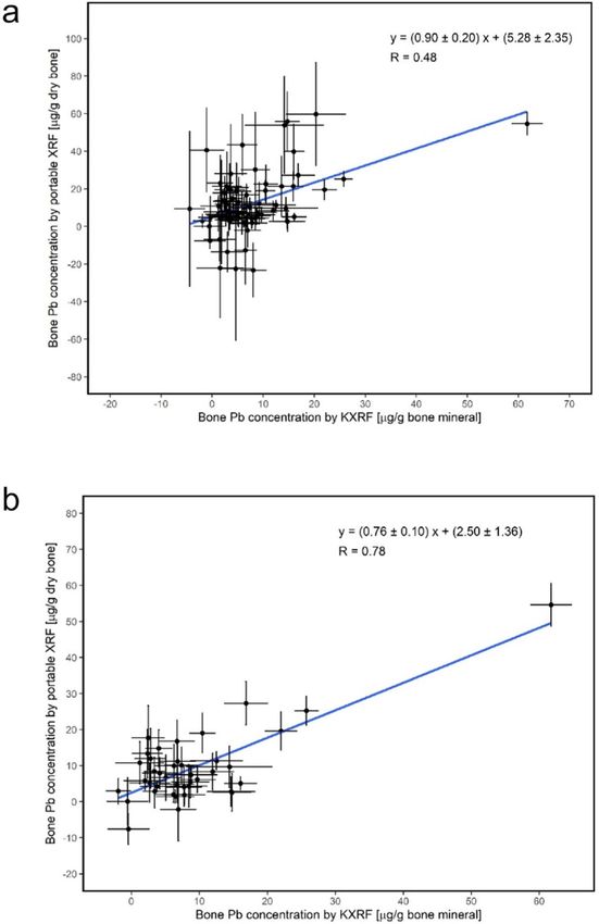
Fig. 1a.
Association of bone Pb measured by the portable XRF and KXRF for all participants. b.
Correlation of bone Pb measured by the portable XRF and KXRF for the participants with
soft tissue thinner than 5 mm.
Author Manuscript
Sci Total Environ. Author manuscript; available in PMC 2022 January 20.Zhang et al. Page 16
Author Manuscript
Author Manuscript
Author Manuscript
Fig. 2.
Bone Pb measured by the portable XRF and KXRF for the participants with soft tissue
thinner than 5 mm and thicker or equal to 5 mm.
Author Manuscript
Sci Total Environ. Author manuscript; available in PMC 2022 January 20.Zhang et al. Page 17
Author Manuscript
Author Manuscript
Author Manuscript
Fig. 3.
Correlation of soft tissue thickness over bone and age of participants.
Author Manuscript
Sci Total Environ. Author manuscript; available in PMC 2022 January 20.Author Manuscript Author Manuscript Author Manuscript Author Manuscript
Table 1
Characteristics of study participants, by site, and previous occupational exposure to Pb.
West Lafayette East Chicago Muncie Occupational exposure to Pb No occupational to Pb Total
Zhang et al.
Participants, N 41 19 11 10 61 71
a 19 (46%) 9 (47%) 9 (82%) 10 (100%) 27 (44%) 37 (52%)
Male, N (%)
Age (Mean ± SD,years) 64 ± 8 62 ± 17 62 ± 12 63 ± 14 63 ± 11 63 ± 11
Soft tissue thickness (Mean ± SD, mm) 4.7 ± 1.4 5.5 ± 1.5 4.2 ± 1.2 4.4 ± 1.0 4.9 ± 1.5 4.8 ± 1.5
Soft tissue thicknessZhang et al. Page 19
Table 2
a
Mean uncertainty , by overlying soft tissue thickness, of 3-minute in vivo bone Pb measurements using the
Author Manuscript
portable XRF and KXRF systems (N = 60).
Soft tissue thicknessZhang et al. Page 20
Table 3
a
Mean uncertainty , by overlying soft tissue thickness, of 5 min in vivo bone Pb measurements using the
Author Manuscript
portable XRF and KXRF systems (N = 11).
Soft tissue thicknessZhang et al. Page 21
Table 4
Correlation between in vivo bone Pb measurements via portable XRF and KXRF (N = 71).
Author Manuscript
Soft tissue thicknessAuthor Manuscript Author Manuscript Author Manuscript Author Manuscript
Table 5
Mean difference in bone Pb concentration per 5 years in age, as estimated with bone Pb measured via the portable XRF and KXRF, by soft tissue
thickness.
Zhang et al.
Soft tissue thicknessYou can also read