Histopathological Spectrum of Neoplastic and Non Neoplastic Lesions of Urinary Bladder- A Retrospective Study - NJLM
←
→
Page content transcription
If your browser does not render page correctly, please read the page content below

Original Article                                                                                                       DOI: 10.7860/NJLM/2021/46894:2537

     Histopathological Spectrum of Neoplastic

                                                                                                                                                          Pathology Section
     and Non Neoplastic Lesions of Urinary
     Bladder- A Retrospective Study
     Priyamvada Singhal1, Mitali Singhal2, Mamta Gupta3, Rani Bansal4

     ABSTRACT                                                                 Results: Total 252 cases were evaluated. A total of 200 (79%)
     Introduction: Urinary bladder encompasses a wide variety                 cases were neoplastic and 52 (21%) cases were diagnosed as
     of lesions, both neoplastic and non neoplastic responsible for           non neoplastic with majority of cases being cystitis. Male to
     significant morbidity and mortality throughout the world. All            female ratio was 7:1. The most common age group was 4th to 7th
     bladder lesions require biopsy because of their lack of distinctive      decade. In neoplastic category, urothelial tumours constituted
     features. Urinary bladder cancer is the 9th most common cancer           194 (97%) cases with Infiltrating Urothelial Carcinoma (IUC)
     worldwide accounting for 6% and 2% of the cancer incidence in            being 118 (60.8%) cases. In non invasive lesions majority
     men and women, respectively.                                             27 (35.6%) cases were Papillary Urothelial Neoplasm of Low
                                                                              Malignant Potential (PUNLMP) followed by urothelial carcinoma
     Aim: To analyse the histopathological spectrum of bladder
                                                                              in situ and papillary urothelial neoplasm of low grade. Entities
     specimens with neoplastic and non neoplastic lesions, and
                                                                              other than urothelial tumours encountered in the present study
     categorising them according to recent 2016 World Health
                                                                              were primary adenocarcinoma, small cell carcinoma, melanosis
     Organisation (WHO) classification.
                                                                              and metastasis from prostate. TNM staging showed lamina
     Materials and Methods: The present study was conducted                   propria invasion (pT1) in 59 (37%) cases, followed by pT2
     from January to June 2020 at Subharti Medical College,                   tumours invading muscle in 56 (35%) cases.
     Meerut, Uttar Pradesh, India. Retrospective data was retrieved
                                                                              Conclusion: Proper knowledge of histologic characteristics of
     from a period of 10 years from January 2010-December 2019.
                                                                              various bladder lesions is of utmost importance as few benign
     Histopathological analysis of all the urinary bladder biopsies and
                                                                              conditions mimic neoplastic and few serve as preneoplastic
     radical cystectomy/cystoprostectomy specimens received during
                                                                              conditions, misdiagnosis may cause further any unnecessary
     this period was done on basis of light microscopic examination
                                                                              treatment procedure. With a multidisciplinary approach, early
     of Haematoxylin and Eosin (H&E) stained slides. Lesions were
                                                                              diagnosis and immediate intervention can have a better survival
     categorised into non neoplastic and neoplastic. The neoplastic
                                                                              and provide a more comfortable life to the patient.
     lesions were classified based on WHO classification 2016 and
     staging as per 8th edition of American Joint Committee on Cancer
     (AJCC). Descriptive data analysis was done.

                                                                  Keywords: Cystitis, Cystectomy, Transurethral biopsies, Urothelial carcinoma

     INTRODUCTION                                                             was therefore conducted with an aim to study the spectrum of
     Urinary bladder lesions cause significant morbidity and mortality        non neoplastic and neoplastic lesions in urinary bladder. Also,
     [1]. Non neoplastic lesions like cystitis are barely lethal but they     due to paucity of studies in this region as per the recent updates
     deteriorate the quality of life. On the contrary, malignant neoplasms    in categorisation of the bladder neoplasms which has included
     of the bladder have therapeutic consequences.The bladder                 new morphologic variants and better reproducible grading
     responds to chronic irritation through several reactive/metaplastic      systems. The authors reviewed these lesions to classify and
     to hyperplastic/proliferative lesions which should be distinguished      stage them according to the WHO classification 2016 and AJCC
     from malignant processes. Clinical, macroscopic, and radiologic          staging 8th edition [7,8].
     findings for these entities may overlap; mandating a histologic
     evaluation [2].                                                          MATERIALS AND METHODS
     Differentiating these lesions is also important because of differences   The present retrospective study was conducted from January to
     in patient management and clinical outcome [3]. Cystoscopy is the        June 2020 at Subharti Medical College, Meerut, Uttar Pradesh, India.
     primary diagnostic tool and useful in localising bladder tumours and     Retrospective data was archived for a period of 10 years (January
     biopsies of the suspected lesions [4]. Urinary bladder cancer is the     2010-December 2019). During this period, specimens submitted
     9th most common cancer worldwide accounting for 6% and 2% of             either in the form of transurethral biopsies or radical cystectomy/
     the cancer incidence in the men and women respectively [5].              cystoprostectomy, were retrieved from the surgical Pathology
                                                                              department. The study was approved by Institutional Ethical
     The 2016 WHO classification emphasises the ability of
                                                                              Committee (reference number SMC/UECM/2021/231/146).
     urothelial neoplasms to exhibit divergent differentiation, multiple
     morphologic variants and diverse genome, which may be utilised           Inclusion criteria: All cystoscopic biopsies/cystectomy/
     for selection of therapy [6,7]. Biopsy is the first line investigation   cystoprostectomy specimen, received in Pathology department,
     for all cases with clinico-radiological features of bladder mass or      were considered for the study.
     diffuse bladder wall thickening. Many inflammatory and infectious        Exclusion criteria: Inadequate/Inconclusive bladder biopsies were
     diseases may mimic neoplastic conditions. The present study              excluded.
20                                                                                     National Journal of Laboratory Medicine. 2021 Oct, Vol-10(4): PO20-PO24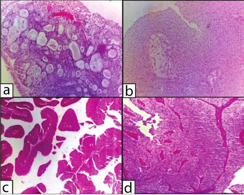
www.njlm.net                                                                                              Priyamvada Singhal et al., Spectrum of Urinary Bladder Lesions

Histopathological analysis was carried out on formalin fixed, paraffin               Type of lesion                                         Number of cases            %
embedded tissue sections of urinary bladder lesions stained with
                                                                                     Cystitis                                                        43               82.6
haematoxylin and eosin. The lesions were classified into non
                                                                                     • Chronic nonspecific cystitis                                  16               30.7
neoplastic and neoplastic lesions based on microscopic examination
of H&E stained slides. The neoplastic lesions were further categorised               • Acute on chronic cystitis                                     14               26.9
based on the 2016 WHO classification of urinary bladder [6]. TNM                     • Eosinophilic cystitis                                         6                11.5
staging was according to the 8th edition of AJCC [8].                                • Foreign body giant cell reaction                              2                 3.8
                                                                                     • Chronic granulomatous cystitis                                1                 1.9
STATISTICAL ANALYSIS                                                                 • Acute cystitis                                                1                 1.9
Data was entered in Excel sheet and descriptive data analysis was
                                                                                     • Polypoidal cystitis                                           1                 1.9
performed.
                                                                                     • Follicular cystitis                                           1                 1.9
RESULTS                                                                              • Interstitial cystitis                                         1                 1.9
In this retrospective study 271 patients with urinary bladder biopsy/                Urinary bladder diverticulum                                    2                 3.8
Transurethral Resection of Bladder Tumour (TURBT)/ cystectomy/                       Inflamed urachal cyst                                           1                 1.9
cystoprostectomy procedures, were evaluated. Out of 271, 18 biopsies
                                                                                     Urethral caruncle                                               1                 1.9
were inadequate/inconclusive, one biopsy showed no significant
                                                                                     Malakoplakia                                                    1                 1.9
pathology, and in 252 cases pathologic diagnosis was made. Non
neoplastic lesions were diagnosed in 52 cases (21%) and neoplastic                   Cystitis glandularis                                            3                 5.8

lesions were detected in 200 cases (79%).                                            Cystitis cystica                                                1                 1.9

Most common age group was 4 to 7 decade with maximum
                                           th     th                                 Total                                                           52               100
13 (25%) cases in 51-60 years age group for non neoplastic lesions                   [Table/Fig-2]: Histopathological spectrum of non neoplastic lesions.
and 65 cases (33%) in 61-70 years age group for neoplastic lesions.
The male to female ratio was 7:1. In the present study, 39 (75%)
cases and 183 (91.5%) cases were males in non neoplastic and
neoplastic lesions, respectively [Table/Fig-1].

                  Non neoplastic lesions               Neoplastic lesions
 Age group
 (years)       Male     Female    Total   (%)   Male     Female      Total   (%)
 0-10            1         0        1      2      0         0          0      0
 11-20           2         2        4      7      0         0          0      0
 21-30           1         1        2      4      4         4          8      4
 31-40           2         1        3      6      14        2         16      8
 41-50           9         1       10      19     33        3         36     17.6
 51-60          10         3       13      25     53        2         55     27.7
 61-70           9         3       12      23     60        5         65     32.7
 71-80           3         2        5      10     14        0         14      7
 81-90           2         0        2      4      5         1          6      3
 Total          39        13       52     100    183        17       200     100
 [Table/Fig-1]: Age wise and gender wise distribution of both Non neoplastic and     [Table/Fig-3]: Non neoplastic lesions: a) Interstitial cystitis (H&E, 4X), b) Cystitis
 neoplastic lesions.                                                                 glandularis (H&E, 10X); c) Follicular cystitis (H&E, 10X); d) Eosinophilic cystitis
                                                                                     (H&E, 40x).
The spectrum of pathologic lesions revealed cystitis being most
common among non neoplastic lesions constituting 43 cases                           Out of 252 cases, there were 10 specimens of cystectomy/
(82.6%), majority were chronic non specific cystitis, followed by                   cystoprostectomy. A 9/10 cases were diagnosed as invasive urothelial
other variants like acute or chronic, eosinophilic and foreign body                 carcinoma while one case reported as urothelial carcinoma on biopsy
giant cell reaction. There were three cases of cystitis glandularis and             from outside was reported as follicular cystitis with fibrosis after
two cases of urinary bladder diverticulum with one case each of                     extensive sampling.
inflamed urachal cyst, urethral caruncle, malakoplakia and cystitis                 Of the total 200 neoplastic lesions, staging was rendered in 161
cystica [Table/Fig-2,3]. Out of all neoplastic lesions of various                   cases which had been diagnosed as invasive urothelial carcinoma,
histomorphological categories, urothelial tumours were most                         carcinoma-in situ, non invasive papillary urothelial carcinoma low and
common with 194 cases (97%). Of 118 cases of IUC, conventional                      high grade, mucinous adenocarcinoma and small cell neuroendocrine
IUC constituted 88 (74.5%) cases while 30 (25.5%) cases were                        carcinoma. Non invasive papillary carcinoma (pTa) was found in
constituted by IUC with divergent differentiation. The various                      24 (15%) cases while carcinoma in situ (pTis) was seen in 16 (10%)
histologic entities with divergent differentiation were squamous                    cases. Lamina propria invasion (pT1) was observed in 59 (37%)
(24 cases), glandular (two cases) poorly differentiated (two cases),                cases, followed by pT2 with tumours invading muscle 56 (35%)
micropapillary and mixed glandular and microcystic (one case each)                  cases. pT3 and pT4 tumours were found in 2 (1.3%) and 4 (2.7%)
variety. Two cases each of primary adenocarcinoma and metastasis                    cases respectively. In remaining 39 neoplastic cases AJCC, TNM
from prostate, one case of small cell neuroendocrine carcinoma and                  staging was not done. These included five benign lesions comprising
melanosis were also seen. In non invasive lesions out of 76 cases,                  of urothelial papilloma in 1 (1.3%), inverted urothelial papilloma in
Papillary Urothelial Neoplasm of Low Malignant Potential (PUNLMP)                   3 (4%), and melanosis 1 (0.5%). Tumours with unspecified, borderline
constituted 27 (35.6%) cases followed by urothelial carcinoma in situ               or uncertain behaviour in 27 (35.6%) cases of PUNLMP, urothelial
16 (21%) cases, papillary urothelial neoplasm- low grade 16 (21%)                   proliferation of uncertain malignant potential in 2 (2.6%) and urothelial
cases and papillary urothelial neoplasm- high grade 8 (10.5%) cases                 dysplasia/atypia in 3 (4%) cases. 2 (1%) cases were metastasis
[Table/Fig-4-6].                                                                    from prostate.
National Journal of Laboratory Medicine. 2021 Oct, Vol-10(4): PO20-PO24                                                                                                       21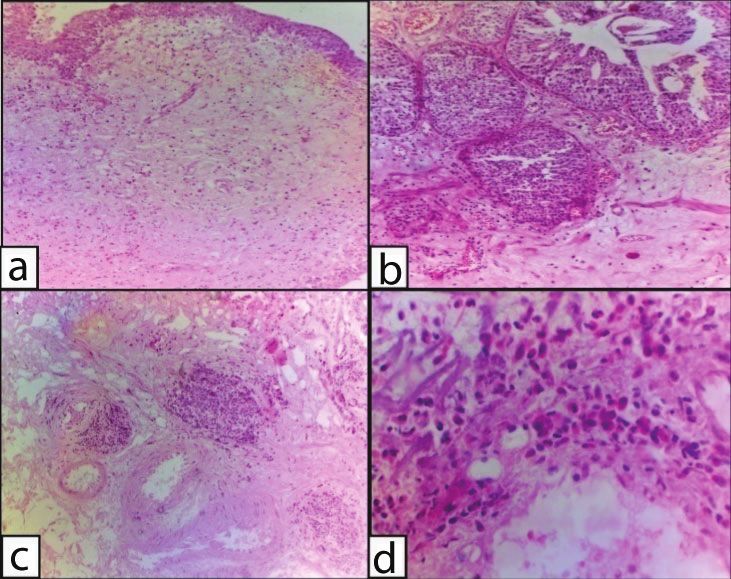
Priyamvada Singhal et al., Spectrum of Urinary Bladder Lesions www.njlm.net
Category No. of cases %
1. Urothelial tumours 194 97
• Invasive/Infiltrating urothelial carcinoma 118 60.8
a. Infiltrating urothelial carcinoma 88 74.5
b. Infiltrating urothelial carcinoma with divergent
30 25.5
differentiation
• Non invasive urothelial neoplasia 76 39.2
Urothelial carcinoma in-situ 16 21
Papillary urothelial carcinomalow grade 16 21
Papillary urothelial carcinomahigh grade 08 10.5
Papillary urothelial neoplasm of low malignant potential 27 35.6
Urothelial proliferation of uncertain malignant potential 02 2.6
Urothelial papilloma 01 1.3
Inverted urothelial papilloma 03 4
Urothelial dysplasia/atypia 03 4
2. Glandular neoplasm 04 2
• Mucinous adenocarcinoma (Primary) 02 1
• Metastasis from prostate (Secondary) 02 1
3. Neuroendocrine tumour
• Small cell neuroendocrine carcinoma 01 0.5
4. Melanocytic tumour
• Melanosis 01 0.5
Total 200 100
[Table/Fig-4]: Histopathological spectrum of neoplastic lesions.
[Table/Fig-6]: Urothelial carcinoma with invasion of lamina propria (H&E, 40x);
b Muscle infiltrating IUC (H&E, 400x); c) IUC with poor differentiation and necrosis
(H&E, 100x); d) IUC with micropapillary differentiation; e) IUC with glandular
and microcystic differentiation (H&E, 100x); f) IUC with squamous differentiation
showing keratin pearls (H&E, 100x); g) Mucin secreting adenocarcinoma showing
muscle invasive pool of mucin with presence of atypical cells (H&E, 4X); h) Small
cell neuroendocrine carcinoma with sheets of hyperchromatic cells lying below
unremarkable urothelium (H&E, 10x).
S et al., Goyal VK et al., and Shah PY et al., however the difference
in ratio in the present study as compared to others could be due to
the low access of females to healthcare in this area or exposure to
environmental factors [11-14].
Majority of the bladder lesions noted were neoplastic lesions
accounting for 79.3% of the cases. This finding was well correlated
[Table/Fig-5]: Non invasive urothelial lesions: a) Urothelial papilloma with both
endophytic and exophytic proliferation (H&E,10X); b) PUNLMP showing orderly with other studies done by Vaidya S et al., (77.57%), Goyal VK et al.,
arrangement of cells within papillae with minimum abnormality in architecture and (96.87%), and Dravid NV et al., (62.58%) [12,13,15]. Amongst the wide
atypia (H&E, 10X); c) Papillary urothelial carcinoma low grade showing isolated
papillae (H&E, 4x); d) Papillary urothelial carcinoma high grade showing fused spectrum of non neoplastic lesions (20.6%) most common lesions
papillae, nuclear atypia (H&E, 40x). observed was cystitis (82.6%) with histological variants like chronic
non specific cystitis, acute on chronic, eosinophilic, granulomatous,
DISCUSSION follicular, polypoidal and interstitial cystitis. Similar distribution of
In bladder cancer histomorphology is the most powerful tool to predict
non neoplastic lesions was documented by various other authors
the risk of recurrence, progression and therapeutic response [9]. In
[1,14,16,17]. However, it is to be worth mentioning herein that one
most of the lesions, diagnosis is fairly easy, occasionally, it can pose
case reported as invasive papillary urothelial carcinoma on TURBT
diagnostic challenges. Therefore, pathologist play an important role in
from outside, was diagnosed as follicular cystitis with fibrosis after
not just labelling the diagnosis but also to give additional information
extensive sampling of the cystectomy specimen. Follicular cystitis
that can have an impact on the treatment [10]. The present study
is a benign proliferative lesion usually secondary to bladder outlet
aimed to present the histopathological spectrum of bladder lesions.
obstruction or dysfunction. It can also be found adjacent to invasive
The authors archived 252 cases of bladder pathology comprising or in situ bladder carcinomas representing a host response. Also,
of 242 bladder biopsies/TURBT and 10 cases of cystectomy/ patients receiving intravesical chemotherapy or Bacillus-Calmette-
cystoprostectomy. Guerin therapy may develop follicular cystitis [18]. In the present
In present study, the male to female ratio was 7:1, which was higher case, entire tumour was possibly resected following TURBT with no
as compared to other studies conducted by Ploeg M et al., Vaidya residual tumour in cystectomy specimen.
22 National Journal of Laboratory Medicine. 2021 Oct, Vol-10(4): PO20-PO24www.njlm.net Priyamvada Singhal et al., Spectrum of Urinary Bladder Lesions
Cystitis cystica and cystitis glandularis are reactive process in carcinoma 30 cases (25.5%) exhibited features of divergent differentiation
response to chronic irritation, infection, calculi, outlet obstruction comprising 24 cases of squamous differentiation, 2 cases of glandular
and catheterisation. These are extremely common and seen in differentiation, 2 cases of poorly differentiated carcinoma and one case
60% of normal bladders at autopsy but majority of these cases are each of micropapillary and mixed glandular and microcystic variant.
asymptomatic incidental findings therefore frequency in bladder Goyal VK et al., and Sushmitha S et al., also found majority cases
biopsies is quite low [19,20]. However, these conditions are benign of conventional urothelial carcinoma with 7.27% and 23% cases of
mimickers of invasive carcinoma so it is of utmost importance to divergent differentiation respectively [13,27]. The study conducted
diagnose these lesions correctly on histomorphology [15,21]. In
by Black PC et al., also stated that approximately 60% of tumours
the present study, the authors found 3 (5.8%) cases of cystitis
exhibit squamous differentiation and 10% of urothelial carcinomas
glandularis and 1 (1.9%) case of cystitis cystica.
contain foci of glandular differentiation [28].
Bladder cancer has a lower incidence in women that reflects an
In the present study, Glandular neoplasms comprised two cases
approximate 3:1 male-to-female (M:F) ratio globally [22]. However,
each of primary mucinous adenocarcinoma and secondary
in present study this male:female ratio in neoplastic lesions was
adenocarcinoma (metastases from prostate). Studies have shown
found to be 7:1, which was quite high as compared to various other
that Primary adenocarcinoma of bladder is uncommon and accounts
studies. In studies by Srikoustubha et al., the male:female ratio was
for 0.5% -2% of all bladder tumours. Metastatic adenocarcinoma is
5.25:1 and Shah PY et al., it was found to be 2.29:1 [1,14].
commoner than primary tumours [29]. One case each of small cell
In the neoplastic category, most common tumour observed in the neuroendocrine carcinoma and melanosis were also seen, although
present study was urothelial tumour (97%). Among all the urothelial both are rare neoplasms with very few cases of both these entities
tumours, IUC (60.8%) was the most common subtype followed by documented in literature [30,31]. Most of the studies from India
PUNLMP and non invasive papillary urothelial neoplasm. These showed concurrence in prevalence of various carcinomas of bladder
findings are comparable with other studies [13,17,23]. with the present study, the study done in West African region by Darre
Non invasive tumours can be papillary or flat. Grading of urothelial T et al., showed high prevalence of squamous cell carcinoma and
tumours is important in Non invasive papillary neoplasms [6]. The adenocarcinoma as compared to urothelial carcinoma [32]. A plausible
PUNLMP is a low-grade, small, solitary neoplasm that neither cause for this high prevalence could be association with urinary
invades nor metastasizes. In the present study out of total 76 cases schistosomiasis and mechanical and chemical vesical irritant factors
of non invasive neoplasias, 35.5% were PUNLMP and 31.5% cases [33]. Comparison of prevalence of various carcinomas of bladder in
were papillary urothelial carcinoma, of which low grade cases were different studies is shown in [Table/ Fig-7] [13,15,23,32,34-36].
twice (21%) to high grade (10.5%). Distinction of PUNLMP from Pathologic staging of bladder cancer is important to patient prognosis
low-grade carcinoma may be difficult because approximately 35% and treatment decision [37]. In the present study, invasive urothelial
of PUNLMPs recur and 11% progress in grade [7]. carcinomas, showed laminal invasion (pT1) in 37.3% cases while
Conventional urothelial carcinoma constitutes about 75% of all cases muscle invasion (pT2) was seen in 33.5% cases. Comparison
[24]. The authors observed 88 (74.5%) cases of conventional IUC out of TNM staging in various other studies is shown in [Table/Fig-8]
of 118 IUC cases. [12,15,38,39].
“Invasive urothelial carcinoma with divergent differentiation” was A new entity “urothelial proliferation of uncertain malignant potential”
introduced in the recent WHO classification (2016) of bladder tumours. was introduced replacing the older term of urothelial hyperplasia
These tumours exihibit component of “usual type” urothelial carcinoma and better categorisation of entity “urothelial dysplasia” was done
combined with other morphologies [7]. The divergent morphology in spectrum of flat and non invasive lesions of urinary bladder in
include squamoid, glandular, small cell and trophoblastic differentiation the new WHO classification [7]. On re-assessment of biopsies
[24,25]. Many of these variants have important prognostic or therapeutic according to the 2016 WHO classification [7] the authors found three
implications worth knowing by the urologist and oncologist. Awareness cases of urothelial proliferation of uncertain malignant potential and
of these unusual patterns is critical to avoid diagnostic misinterpretations two cases of urothelial dysplasia among 76 cases of non invasive
[26]. In the present study, out of 118 cases of invasive urothelial urothelial neoplasia.
Mylsamy S and
Agrawal S
Mahesh Kumar Goyal VK Dravid NV et Altaf J et al., Kanakasabapathi
et al., [36]
U et al., [23] Darre T et al., [13] al., [15] [34] D [35] Present study,
Moradabad,
Bijapur, India et al., [32] Bikaner, Dhule, India Pakistan Coimbatore, India Meerut, India
India (2019)
Neoplastic lesion (2012) Africa (2014) India (2015) (2016) (2017) (2017) (2021)
Urothelial carcinoma 28 (46.6%) 25 (26.04%) 93 (93%) 77 (53.39%) 86 (90.5%) 38 (74.5%) 38 (84%) 118 (46.8%)
Squamous cell
2 (3.33%) 36 (37.5%) 2 (2%) 7 (5.03%) 6 (6.3%) 5 (9.8%) 2 (4%) Nil
carcinoma
Adenocarcinoma 2 (3.33%) 32 (33.3%) 1 (1%) 3 (2.15%) 3 (3.2%) 2 (3.9%) 1 (2%) 4 (1.6%)
Small cell carcinoma Nil Nil Nil Nil Nil Nil Nil 1 (0.4%)
[Table/Fig-7]: Comparison of prevalence of carcinomas of bladder in different studies.
Kong CH et al., [38] Vaidya S et al., [12] Dravid NV et al., Benhayoune K et
TNM stage 2010 2013 [15] 2016 al., [39] 2018 Present study 2021
pTa: Non invasive papillary carcinoma 23 (33.3%) 39 (48.14%) 20 (21.73%) 37 (23.7%) 24 (15%)
pTis: Carcinoma in situ - - - - 16 (10%)
pT1:Invades lamina propria 15 (20.0%) 18 (22.22%) 49 (53.26%) 35 (22.4%) 59 (37%)
pT2:Invades muscularispropria 8 (10.7%) 24 (29.63%) 18 (19.56%) 56 (36.2%) 56 (35%)
pT3:Invades perivesical tissue 9 (12.0%) - 3 (3.26%) 15 (9.9%) 02 (1.3%)
pT4:Directly invades prostatic stroma, seminal vesicles,
14 (18.7%) - 2 (2.17%) 11 (7.2%) 04 (2.7%)
uterus or vagina, pelvic wall or abdominal wall
[Table/Fig-8]: Comparison of TNM stage of bladder neoplasms in various studies.
National Journal of Laboratory Medicine. 2021 Oct, Vol-10(4): PO20-PO24 23Priyamvada Singhal et al., Spectrum of Urinary Bladder Lesions www.njlm.net
Limitation(s) [16] Manjula K, Kalyani R, Kumar H. Spectrum of lesions in urinary bladder biopsies: A
histopathological study. Int J of Clinical and Diagnostic Pathology. 2020;3(1):302-04.
The limitation of present study is that clinicopathological correlation [17] Aparna C, Thumma RR, Devi CP, Jyothi Vanapalli SVRL, Mounika TDN. Histological
could not be, due to retrospective nature of study clinical details and spectrum of urothelial lesions–experience of a single tertiary care institute.
follow-up was not available in all cases. International Journal of Contemporary Medical Research. 2016;3(6):1731-33.
[18] Trombetta M, Packard M, Ferrara D, Werts ED. The use of radiotherapy in
the management of follicular cystitis refractory to conservative and surgical
CONCLUSION(S) management. Rare Tumours. 2012;4(2):e25.
Proper knowledge of histologic characteristics of the bladder [19] Grignon DJ, Sakr W. Inflammatory and other conditions that can mimic carcinoma
lesions is utmost important as few benign conditions mimic in the urinary bladder. Pathol Annu. 1995;30(Pt 1):95-122.
[20] Wiener DP, Koss LG, Sablay B, Freed SZ. The prevalence and significance of
neoplastic and few serve as pre-neoplastic conditions, tumour Brunn’s nests, cystitis cystica and squamous metaplasia in normal bladders. J
grading is a significant predictor for all patient outcome variables. Urol. 1979;122(3):317-21.
The present study has potential areas of further research with the [21] Amin MB, Young RH. Intraepithelial lesions of the urinary bladder with a discussion
of the histogenesis of urothelial neoplasia. Semin Diagn Pathol. 1997;14(2):84-97.
use of immunohistochemical stains which can aid to establish [22] Hansel DE, Amin MB, Comperat E, Cote RJ, Knüchel R, Montironi R, et al. A
urothelial origin in bladder tumour with unusual histology and also to contemporary update on pathology standards for bladder cancer: Transurethral
distinguish between reactive atypia and carcinoma in situ in difficult resection and radical cystectomy specimens. Eur Urol. 2013;63(2):321-32.
[23] Mahesh Kumar U, Yelikar BR. Spectrum of lesions in cystoscopic bladder
cases. This distinction is critical because of the therapeutic and
biopsies-A histopathological Study. Al Ameen J Med Sci. 2012;5(2):132-36.
prognostic implications. [24] Processali T, Diminutto A, Cerruto MA, Antonelli A. The impact of histological
variants on bladder cancer outcomes. AME Med J. 2020;5:4.
REFERENCES [25] Daniel AA, Trpkov K. What is new in Genitourinary Pathology? Recent
[1] Srikousthubha, Sukesh, Raghuveer CV, Hingle S. Profile of lesions in cystoscopic developments and highlights of the new 2016 World Health Organization
bladder biopsies: A histopathological study. J Clin Diagn Res. 2013;7(8):1609-12. classification of tumours of the urinary system and male genital organs. Applied
[2] Harik LR, O’Toole KM. Nonneoplastic lesions of the prostate and bladder. Arch Cancer Res. 2016;36:1.
Pathol Lab Med. 2012;136(7):721-34. [26] Amin MB. Histological variants of urothelial carcinoma: Diagnostic, therapeutic
[3] Wong-You-Cheong JJ, Woodward PJ, Manning MA, Sesterhenn IA. Neoplasms and prognostic implications. Mod Pathol. 2009;22:96-118.
of the urinary bladder: Radiologic-pathologic correlation. Radiographics. [27] Sushmitha S, Patil GS, Patil SB. A histological analysis of urinary bladder
2006;26(2):553-80. specimens with elaboration of various neoplastic lesions. Indian J Pathol Oncol.
[4] Felix AS, Soliman AS, Khalad H, Zaqhloul MS, Banerjee M, El-Baradie M, et al. 2019;6(4):688-94.
The changing patterns of bladder cancer in Egypt over the past 26 years. Cancer [28] Black PC, Brown GA, Dinney CP. The impact of variant histology on the outcome
Causes Control. 2008;19(4):421-29. of bladder cancer treated with curative intent. Urol Oncol. 2009;27:03-07.
[5] Pudasaini S, Subedi N, Prasad KBR, Rauniyar SK, Josi BR, Bhomi KK. [29] Suba G, Gayatri J, Jayprakash HT. Histopathological overview of cystoscopic
Cystoscopic bladder biopsies: A histopathological study. Nepal Med Coll J. bladder biopsies- A retrospective analysis. Tropical Journal of Pathology and
2014;6(1):09-12. Microbiology. 2017;3(2):229-34.
[6] Humphrey PA, Moch H, Cubilla AL, Ulbright TM, Reuter VE. The 2016 WHO [30] Çamtosun A, Çelik H, Altıntaş R, Akpolat N. Primary small cell carcinoma in
Classification of tumours of the urinary system and male genital organs- Part B: urinary bladder: a rare case. Case Reports in Urology. 2015;2015:789806.
Prostate and Bladder Tumours. Eur Urol. 2016;70(1):110-15. [31] Yau SE, Singer EJ, Sun Y, Johnson MH. Bladder melanosis with concurrent
[7] Moch H, Humphrey PA, Ulbright TM, Reuter VE. (Eds): WHO classification of urothelial carcinoma. Urol Case Rep. 2017;15:30-32.
tumours of the urinary system and male genital organs. 4th Ed.IARC: Lyon 2016. [32] Darré T, Amégbor K, Kpatcha M, Tengue K, Sonhaye L, Doh K et al. Urologic cancers
[8] Bochner BH, Hansel DE, Efstathion JA, Konety B, Lee CT, Mckiernan JM, et in Togo: Histo-epidemiological profile of 678 cases. J Afr Cancer. 2014;6:27-31.
al. Urinary Bladder. In: Amin M.B. (eds). AJCC cancer staging manual. 8th Ed. [33] Desgrippes A, Meria P, Cortesse A, Cochand-Priollet B, Cariou G. Carcinome
Springer nature; USA. 2018; Pp.765-74. épidermoïde de la vessie.Epidermoid carcinoma of the bladder. Prog Urol.
[9] Reuter VE. Bladder. Risk and prognostic factors-a pathologist’s perspective. Urol 1998;8(3):321-29.
Clin North Am. 1999;26(3):481-92. [34] Altaf J, Mahesar MA, Jatoi T. Clinicopathological features of bladder tumours in
[10] Young RH, Eble JN. Non-neoplastic disorders of the urinary bladder. Urologic a single institution in Hyderabad, Sindh, Pakistan. Int J Clinical & Case Studies.
Surgical Pathology. St Loius: Mosby; 1997;166-212. 2017;1(1):22-29.
[11] Ploeg M, Aben KK, Kiemeney LA. The present and future burden of urinary [35] Mylsamy S, Kanakasabapathi D. Histopathological study TURBT biopsies of
bladder cancer in the world. World J Urol. 2009;27(3):289-93. urinary bladder cancer. Trends in Medical Research. 2017;12:51-54.
[12] Vaidya S, Lakhey M, Sabira KC, Hirachand S. Urothelial tumours of the urinary [36] Agarwal S, Dutta S, Awasthi S, Ashutosh KA, Arora D. Histopathological
bladder: A histopathological study of cystoscopic biopsies. J Nepal Med Assoc. spectrum of urinary bladder biopsies. Int J Med Res Prof. 2019;5(2):94-97.
2013;52:475-78. [37] Cheng L, Montironi R, Davidson DD, Lopez-Beltran A. Staging and reporting of
[13] Goyal VK, Vyas SP, Kothari DC. Spectrum of lesions in urinary bladder biopsies: urothelial carcinoma of the urinary bladder. Mod Pathol. 2009;22(Suppl 2):70-95.
Histopathological study. Int J Dent Med Res. 2015;1(6):42-46. [38] Kong CH, Singam P, Hong GE, Cheok LB, Azrif M, Tamil AM, et al.
[14] Shah PY, Nanavati M, Patel RG, Goswami HM. Spectrum of lesions in urinary Clinicopathological features of bladder tumours in a single institution in Malaysia.
bladder- A histopathological study. Int J Cur Res Rev. 2016;8(4):19-24. Asian Pac J Cancer Prev. 2010;11(1):14.
[15] Dravid NV, Rajeshwari K, Karibasappa GN, Patil A. Histomorphological profile [39] Benhayoune K, Tahiri L, Mellas S, Tazi F, Khallouk A, El-Fassi J, et al.
of lesions in cystoscopic bladder biopsies- A prospective study in North Histoprognostic factors in bladder cancer: A case series of 156 patients. Arch
Maharashtra. Int Clin Pathol J. 2016;3(1):161-66. Can Res. 2018;6(3):13.
PARTICULARS OF CONTRIBUTORS:
1. Assistant Professor, Department of Pathology, Subharti Medical College, Meerut, Uttar Pradesh, India.
2. Assistant Professor, Department of Pathology, Subharti Medical College, Meerut, Uttar Pradesh, India.
3. Associate Professor, Department of Pathology, Subharti Medical College, Meerut, Uttar Pradesh, India.
4. Professor and Head, Department of Pathology, Subharti Medical College, Meerut, Uttar Pradesh, India.
NAME, ADDRESS, E-MAIL ID OF THE CORRESPONDING AUTHOR: PLAGIARISM CHECKING METHODS: [Jain H et al.] Etymology: Author Origin
Dr. Mitali Singhal, • Plagiarism X-checker: Sep 24, 2020
Department of Pathology, Subharti Medical College, Swami Vivekanand Subharti • Manual Googling: Feb 20, 2021
University, NH58, Meerut, Uttar Pradesh, India. • iThenticate Software: May 28, 2021 (20%)
E-mail: drsinghal2020@gmail.com
Author declaration:
• Financial or Other Competing Interests: None Date of Submission: Sep 23, 2020
• Was Ethics Committee Approval obtained for this study? Yes Date of Peer Review: Dec 02, 2020
• Was informed consent obtained from the subjects involved in the study? Yes Date of Acceptance: Mar 17, 2021
• For any images presented appropriate consent has been obtained from the subjects. Yes Date of Publishing: Oct 01, 2021
24 National Journal of Laboratory Medicine. 2021 Oct, Vol-10(4): PO20-PO24You can also read