Hughston Health Alert - COVID-19 response
←
→
Page content transcription
If your browser does not render page correctly, please read the page content below
Hughston Health Alert
                                6262 Veterans Parkway, PO Box 9517, Columbus, GA 31908-9517 • www.hughston.com/hha

                                                                                VOLUME 32, NUMBER 1 - WINTER 2020

                                                      Top view of right shoulder
                                                                                   Clavicle
  Inside...                                               Acromioclavicular
                                                          ligament                 (collarbone)
                                                                                                   First rib
  • Stress Fractures in Adolescent
    Athletes                                       Acromion
                                                   of scapula
  • Hallux Rigidus                                 (shoulder                                                      Sternum
  • Fallen Arches                                  blade)                              Coracoclavicular           (breast-
                                                                                       ligaments                  bone)
   • COVID-19 response

                                                                                        Coracoid

                                                                              Humerus

AC Joint Injuries                                                                    The Hughston Foundation, Inc. ©2020

   Injury to the acromioclavicular (AC)
joint is common; in fact, it makes up              Fig. 1. Above: Anatomy of normal acromioclavicular (AC)
approximately 9% of shoulder injuries.             joint. Below: An athlete experiencing a direct hit to the
Injury of the AC joint is often referred to        shoulder, which can cause AC joint injuries.
as an AC sprain or an AC separation with
treatment ranging from nonoperative to
surgical. Orthopaedists define this injury
as stretching or tearing of the ligaments
(tissues that connect bones) surrounding the
acromioclavicular joint and the ligaments
between the coracoid and the clavicle
(Fig. 1). As part of the diagnosis, doctors
label the injury as a type I through a type
VI, depending on the severity of the injury.
   The AC joint is the articulation in the
shoulder between the clavicle (collarbone)
and the acromion of the scapula (shoulder
blade). The clavicle is the only bony
connection between the arm and the
axial skeleton (central bones of the chest).
The AC joint is a small joint stabilized
by the AC capsule, (multiple ligaments
that encapsulate the joint) as well as the                                                               The Hughston
ligaments between the clavicle and the                                                                 Foundation, Inc.
coracoid process (small bony projection of                                                                     ©2020
the scapula).

FOR A HEALTHIER LIFESTYLE
Risk factors                                                      tendons, and ligaments) of the shoulder if other injuries are
  AC joint injuries are usually the result of a direct blow       suspected. Injuries can range from a sprain (stretching of
to the shoulder. The injury can occur after severe trauma,        the ligaments) to complete tears of both the AC capsule and
such as motor vehicle collision; but it more often happens        coracoclavicular ligaments. In the case of complete tears, the
after less severe activity such as sports participation. The      clavicle is usually significantly displaced (dislocated) from the
injuries are prominent in contact sports, such as football,       acromion.
wrestling, rugby, or hockey, which may account for why
male athletes are more prone to the injury than females.          Types of AC joint injuries
However, you can also experience AC joint pain from                  Your orthopaedist will label your injury as a type I, II, III,
chronic irritation that results from repetitive overhead          IV, V, or VI, depending upon the extent of injury and number
sports such as throwing a baseball, or work-related pain          of ligaments involved. The type of injury can usually be
from doing repetitive movements on your job.                      determined with a physical examination and x-rays.
                                                                     Type I injuries involve a partial tear of the
Symptoms                                                          acromioclavicular ligament with no injury to the
  Patients often describe pain on top of the shoulder             coracoclavicular ligament. This causes a tender AC joint
and can sometimes touch a specific tender area at the             that often has mild swelling and usually heals within a
AC joint. You may experience increased pain while lying           few weeks. Type II injuries involve a complete tear of
on your side or pain that increases with lifting or during        the acromioclavicular ligament but the coracoclavicular
overhead or across body movements. Upon examination,              ligaments are stretched, but remain intact (Fig. 2). This
your doctor may find swelling and bruising along the              causes a tender AC joint, often with significant swelling that
shoulder, and a visible bump on the top of the shoulder.          heals within a few weeks. Type III injuries involve a complete
You may notice a decrease in strength and range of                tear of both the acromioclavicular and coracoclavicular
motion. During movement you may hear a popping                    ligaments. The AC joint will appear abnormal, although
sound and feel a catching sensation.                              swelling may obscure the degree of injury. Type III injuries
                                                                  often take several months to heal and there is debate among
Screening and diagnosis                                           surgeons on whether or not these injuries need to be fixed.
  If you have had a shoulder injury and have either                  Type IV, V, VI injuries are described as the most severe
continued pain or pain with movement, you should see              cases because with these injuries both the acromioclavicular
an orthopaedist for evaluation. Your doctor will take a           and coracoclavicular ligaments are completely torn and the
thorough history, perform a physical exam, and order              clavicle is displaced (Fig. 3). During these traumatic injuries
radiographs (x-rays) to evaluate the degree of separation.        the force applied to the shoulder creates a separation
Your physician may also obtain magnetic resonance                 between the clavicle and the acromion. Doctors distinguish
imaging (MRI scan that shows the bones, muscles,                  these injuries by the distance and direction of the displaced
                                                                  clavicle. To treat these injuries, surgeons use reduction and
                                            Fig. 2. Type II AC
                                                                  fixation surgical treatment techniques to repair the ligaments
                                        joint injury involves a
                                                                  and hold the clavicle back in its original place.
                                         complete tear of the
                                  acromioclavicular ligament      Nonsurgical treatment
                                     but the coracoclavicular       If you have a type I or II injury, you may start to feel
                                     ligament remains intact.     better within a few days, but it can take 6 weeks before
            Enlarged view                                         the ligaments heal. During that time you should take the
                                              Clavicle
                                              (collarbone)        pressure off the AC joint and protect it from stretching the
     Torn                                                         immature scar tissue. This would include sling immobilization
     acromioclavicular                                            for approximately 1 to 2 weeks. At follow-up, your physician
     ligament                                                     will obtain repeat radiographs (x-rays) and if there are no
                                                                  concerning changes, such as increase in separation, you can
                                                                  begin gentle physical therapy that focuses on shoulder range
                                                                  of motion.
        Injured                                                     For more severe injuries, your doctor will work with you
        coracoclavicular                                          and a physical therapist to develop a plan for your recovery.
        ligaments                                                 The goal of physical therapy is to reduce pain, improve
                                                                  range of motion, and then to improve strength and shoulder
                                                                  function. After 4 to 6 weeks, most patients see improvements
                                                                  and can progress to advance activities
                           The Hughston Foundation, Inc. ©2020
                                                                  as tolerated.
2 FOR A HEALTHIER LIFESTYLE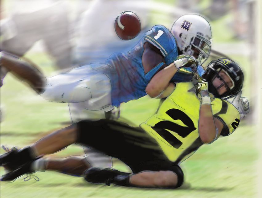
Fig. 3. Type V AC joint injury
involves a complete tear Displaced clavicle
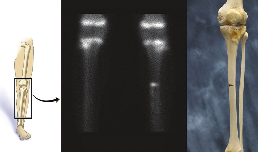
Stress Fractures in
of the acromioclavicular (collarbone) Adolescent Athletes
ligament and
coracoclavicular More and more young athletes are
ligaments as well as participating in a single competitive sport
the clavicle being year-round instead of multiple sports through
displaced. different seasons. This change in athletic
Torn competition has led to an increase in overuse
Torn
acromioclavicular injuries in the pediatric population. Sports
coracoclavicular
ligament medicine physicians often attribute the cause
ligaments
of these overuse injuries to a combination of
an underdeveloped musculoskeletal system,
increased duration and intensity of competition,
The Hughston Foundation, Inc. ©2020 and early sports specialization. One type of
overuse injury that can have a detrimental
Surgical treatment impact on sports performance and the overall
Most AC injuries don’t require surgery; however, more severe health of an adolescent athlete is a stress
injuries that are associated with instability have better outcomes fracture.
when operatively treated. Surgery focuses on reconstruction the Stress fractures occur when abnormal
coracoclavicular ligaments. Surgeons use different techniques; stresses are placed on the trabecular bone
therefore, your surgeon will determine which treatment best fits your (cancellous bone that is highly porous and
needs. These techniques involve suturing or using an allograft tendon located at the end of long bones) resulting in
(a tendon from a donor), or both, to go through or wrap around microfractures. Due to lack of adequate rest
the coracoid and clavicle. Other treatments include using plates and recovery or underlying abnormalities in
to put over the top of the clavicle and acromion. These plates can bone metabolism, these microfractures are
sometimes be prominent and are often removed a few months after unable to heal and remodel and can propagate
the initial surgery. The goal of surgery is to reduce (place back into the over time. Continued repetitive loads exceeding
original position) the clavicle to the acromion so that the ligaments the body’s normal intrinsic bone healing can
can heal, creating scar tissue that holds the clavicle in place. eventually lead to complete fracture through
the affected bone. The most common site for
Complications
a stress fracture is the anterior tibia (shinbone)
Outcomes for both nonsurgical and surgical treatments are
(Fig. 1), but the injury can also occur in the
favorable for AC joint injuries. However, complications can include
femoral shaft (straight part of the thighbone),
chronic instability and pain if you do not seek treatment. Arthritis
femoral neck (below the ball of the hip joint)
of the AC joint can erupt years after the injury causing residual pain
and posterior ribs (back ribs). Stress fractures
between 30 to 50% of the time. Additionally, surgical fixation failure
can occur on the compression side (pushing
can happen, which includes loosening of the graft used to reconstruct
together) or the tension side (pulling apart) of
the ligaments. Furthermore, fracture of the coracoid and clavicle can
bones. Physicians can treat stress fractures on
arise if tunnels are drilled within the bones during surgery.
the compression side without surgery because
Don’t shoulder the pain alone the site is stable and has an adequate blood
AC joint injuries often result from a direct blow to the shoulder supply. However, stress fractures that occur
and can cause significant pain. Because the injury often results after on the tension side of a bone have poor
trauma, there is no foolproof prevention plan; however, you can healing potential and usually require surgical
reduce your risk considerably by wearing protective equipment intervention.
during sporting activities. Simple treatments include a short period
Who is at risk?
of immobilization for mild injuries while surgery may be required
Two major risk factors for the development
for more significant injuries. Chronic symptoms can be alleviated
of stress fractures are rapid increases in high
with therapy and activity modification. For some patients, early
intensity activity and abnormalities in normal
management can prevent late complications, such as arthritis;
bone metabolism. High intensity athletes as
therefore, don’t shoulder the pain alone, seek medical attention.
well as new military recruits are subject to
rapid increase in training intensity especially
Roman Ashmyan, DO, and Ryan Mahoney, DO
during conditioning periods and basic training.
Columbus, Georgia
FOR
FORAAHEALTHIER
HEALTHIERLIFESTYLE
LIFESTYLE 3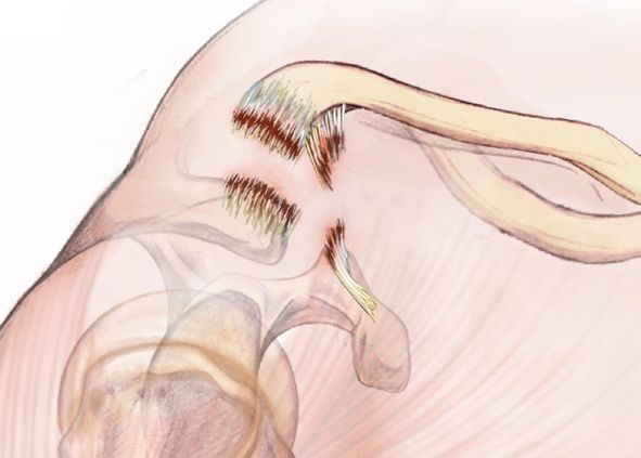
Fig. 1. Mid-tibial stress                        Magnified bone scan
   fracture in an adolescent.

                                                                                               Knee
                                                         Growth
                                                         plates

                                                          Stress                                  Stress
                                                          fracture                                fracture

                                                                 Tibia                                 Tibia
                                                                 (shin-                                (shin-
                                                                 bone)                                 bone)

                                        Right leg                         Left leg           Left leg bones
   The Hughston                         (normal)
   Foundation, Inc. ©2020

Pediatric and adolescent muscles are underdeveloped and           thigh that increases with running or physical activity. Over
subsequently subject to early fatigue causing compensatory        time pain can occur even with simple daily activities like
gait changes with increased stresses applied to the bone.         walking or at rest. Symptoms usually develop insidiously
This loss of the body’s ability to absorb shock from fatigued     and unrelated to a known injury.
muscles combined with poor mechanics and improper                    Evaluation by a physician begins with a detailed history
training regimens can also cause overloading of these             of symptoms and physical exam. The physician often
deconditioned muscles ultimately resulting in overuse             orders radiographs (x-rays) which can demonstrate cortical
injuries.                                                         thickening, or stress fracture. There may be signs on x-ray
   Ninety percent of a child’s bone mass is attained by           of bone healing, such as cloudy immature bone forming
the age of 18. Essential vitamins, such as vitamin D and          within 3 weeks of injury. If x-rays are negative, magnetic
calcium, play a vital role in a child’s developing skeleton.      resonance imaging (MRI, a test that shows the bones,
Athletes that are at risk of suboptimal bone mass and             muscles, tendons, and ligaments) can help to evaluate the
stress fractures include distance runners, gymnasts, and          location of stress fractures as well as to assess the growth
elite dancers. These sports require lean body mass and            plate and surrounding soft tissues. A MRI can demonstrate
are often associated with disordered eating habits and            subtle findings with exquisite detail of bone edema (fluid
irregular menstrual cycles in young female athletes. There        builds up as a response to injury) and fracture to confirm
has been a dramatic increase in the number of healthy             diagnosis without radiation exposure to the child.
children and adolescents who are at risk of vitamin D                First line treatment for compression-sided stress fractures
deficiency. Lifestyle changes that place children at risk for     is protected weight bearing on the affected extremity for 4
vitamin D deficiency include lack of outdoor activities,          to 6 weeks or until the pain resolves. Doctors usually avoid
increased screen time, and poor diets. Inadequate oral            prescribing nonsteroidal anti-inflammatory medications
intake of calcium and vitamin D, lack of outdoor physical         (NSAIDs), which reduce inflammation, such as aspirin,
activity, and abnormal estrogen levels can disrupt normal         Aleve®, and Advil® because these medications interfere
body homeostasis needed for appropriate bone growth and           with the inflammation that your body uses for bone
remodeling.                                                       healing. Tension-sided stress fractures are less likely to heal
                                                                  with conservative management and often require operative
Symptoms, diagnosis, and treatment                                fixation. Treatment also consists of a progressive return-to-
  Patients with lower extremity stress fractures most often       play protocol to avoid recurrence of the stress fracture.
present with complaints of pain in the anterior tibia, hip, or
4 FOR A HEALTHIER LIFESTYLE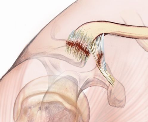
Don’t confuse a stress fracture with shin splints
   Stress fractures are commonly confused with tibial stress
                                                                 Hallux Rigidus
syndrome, also known as shin splints. Shin splints differ          In 1888, British surgeon J.M. Cotterill was first to
from stress fractures in that shin splints are an inflammation   describe hallux rigidus as an osteoarthritic condition of
of the superficial portion of the bone called the periosteum     the metatarsophalangeal joint of the great toe. Today,
(Fig. 2). The underlying periosteum becomes inflamed             orthopaedists recognize this condition as the most common
from the repetitive pull of the muscles originating in the       form of osteoarthritis (a type of joint disease that results
anterior (front) or posterior (back) compartment of the          from the breakdown of joint cartilage and underlying bone)
lower leg. Patients most commonly report vague pain              in the foot. It affects roughly 2.5% of all people over the
that runs along the middle to distal aspect of the tibia         age of 50, is more common in females, and can involve
that increases during the start of exercise but improves         both the left and right great toes. Besides arthritis, the
with prolonged activity. Physicians often order x-rays for       causes of hallux rigidus include previous injury, trauma,
the patients to rule out stress fracture. Treatment for shin     and various deformities of the great toe, including bunion,
splints involves modifying activities until the pain subsides    hypermobility of the metatarsophalangeal joint, and
which can involve decreasing running distance, performing        avascular necrosis.
cross-training exercises, and avoiding running on hills or
hard, uneven surfaces. Physicians sometimes order physical       Anatomy
therapy as an adjunctive treatment to help with stretching          Three bones of the foot, the metatarsal, proximal
and strengthening the ankle and leg muscles, tendons             phalanx, and the distal phalanx, form the great toe. The
(tissues connecting muscle to bones), and ligaments (tissue      space between the metatarsal and the proximal phalanx
connecting two bones).                                           is the first metatarsophalangeal joint, which healthcare
                                                                 professionals often call the MTP joint (Fig.). This joint
Prevention is key                                                consisting of articular cartilage (smooth tissue that covers
  The most important factor for preventing stress fractures      the ends of bones) and synovial fluid (reduces friction
in young athletes and children is to recognize risk factors      between articular cartilage in a synovial joint) allows
early. Young children should eat a healthy diet that consists    significant motion of the great toe. The MTP joint is also
of essential vitamins and protein for normal skeleton            surrounded by ligaments (tissues connecting bones), a
development. Children of all ages should also participate in     joint capsule (a dense fibrous connective tissue), and small
at least 60 minutes of physical activity per day; this regular   muscles, and tendons (tissues connecting muscles to bones)
physical activity also helps to promote the development of       that allow the great toe to move up and down. In patients
bone strength. Additionally, coaches, parents, and athletic      who have hallux rigidus, the articular cartilage of the joint
                                          trainers can help      wears down causing each bone to rub together during
 Fig. 2. Anterior                         identify athletes      movement. Physicians often describe this as “bone on
 compartment                              and children at        bone” osteoarthritis. As the joint space narrows, the joint
 shin splint                              risk for overuse       becomes stiff or rigid. The process also leads to contracture
                                          injuries and stress    (permanent tightening) of the surrounding soft tissues.
                                          fractures as well
                                          as encourage           Evaluation and diagnosis
                              Tibia       adequate                  Patients who have hallux rigidus present with painful
                              (shin-      recovery and rest.     and limited range of motion at the MTP joint of the great
                              bone)       Furthermore, young     toe. On physical examination, the orthopaedist may find
                                          athletes should        the MTP joint swollen and enlarged with bone spurs at
                            Muscle
                                          be encouraged          the metatarsal head. In moderate to severe cases, patients
                                          to seek treatment      may alter gait patterns to compensate for their pain and
                                          to prevent             stiffness, especially since walking or increasing physical
                       Periosteum         complications or       activity can elicit the symptoms. Patients frequently
                                          need of surgical       complain of difficulty wearing high-heeled shoes and joint
                                          intervention from      stiffness.(Fig.)
                                          their overuse             The diagnostic imaging of choice is weight bearing
                                          injuries.              radiographs (x-rays) of the foot. Depending on the
                                                                 severity of disease, the x-rays can demonstrate joint space
                                              Julia Fink, DO     narrowing, subchondral sclerosis (hardening of bone),
                                          Columbus, Georgia      deformity of the metatarsal head, and osteophytes (bony
The Hughston
Foundation, Inc. ©2020                                           projections, or bone spurs) surrounding the joint.

                                                                                               FOR
                                                                                                FORAAHEALTHIER
                                                                                                     HEALTHIERLIFESTYLE
                                                                                                               LIFESTYLE 5
An arthrodesis or fusion
 Fig. Hallux rigidus (highlighted in red) and a
 x-ray of a foot in high-heel shoe (right).                                                           of the MTP joint is the
                                                                                                      most common procedure
                                                                                                      performed for the
                                                                                                      condition and represents
                                                                                                      the current gold standard
                                                                                                      for managing severe hallux
                              Left foot bony                                                          rigidus. Arthrodesis involves
                              anatomy                                                Heel             removing the remaining
                                                                                     elevation        articular cartilage of the
                                                                                     support          MTP joint and fusing
                                                                                     bar              the proximal phalanx to
                                                                                                      the metatarsal to form
                                                                                                      a single bone. Often,
                                                                                                      surgeons use a metal
                             Metatarsal                                                               plate and screws to hold
                                                                                                      the fusion together. The
                              Metatarsophalangeal                                                     current medical literature
                              joint (MTP joint)                                                       reports significantly high
                                Proximal                                                              patient satisfaction scores,
                                phalanx                                           Foot outline        pain relief, and durability
                                                    Shoe outline                                      for the procedure. One
                                Distal                                                                study published in Foot
                                phalanx                                                               and Ankle International
                                                                                                      reports the activity levels of
Treatment                                                                                             patients after undergoing
  Nonsurgical treatment for hallux rigidus is often the first    MTP    arthrodesis.   The  study showed   that preoperative
option offered to patients. The treatment consists of activity   activities were reestablished in 92% of patients who hiked,
modification, shoe modifications, and anti-inflammatory          80% who played golf, 75% who jogged, and 75% who
medicines. Generally, your orthopaedist will recommend           played tennis.
a variety of shoe modifications including a Morton’s                Lastly, researchers and surgeons have developed newer
extension orthotic, rocker bottom stiff soled athletic shoe,     orthopedic     technology regarding joint replacement
or shoes with high and wide toe boxes to take the pressure       surgery   and   synthetic cartilage. Many companies have
off the dorsal aspect of the joint. In conjunction with shoe     advertised    implants  to replace the MTP joint of the great
modifications, your doctor may prescribe nonsteroidal anti-      toe in order to preserve range of motion and provide pain
inflammatory medicines such as ibuprofen or naproxen             relief. Medical device manufacturers make the implants
to help decrease swelling and pain. Additionally, your           from a variety of materials including metal or plastic or a
physician can recommend a corticosteroid injection into          combination of both. Short term studies have reported
the arthritic joint space to help with pain and swelling.        good outcomes with these implants; however, long term
  Unfortunately, 2 out of 3 patients who have moderate to        studies have reported high rates of implant removal and
severe disease will fail conservative treatment. After more      complications. Thus, orthopaedic surgeons offer this
than 6 months of nonsurgical treatment without results,          surgical option only when the patient meets strict criteria.
your orthopaedist may recommend surgical management
                                                                 Seek treatment early
as your next option. Although there are numerous surgical
                                                                    The management of hallux rigidus varies from conservative
treatments published in the medical literature, most
                                                                 treatment to surgical options depending on disease severity.
orthopaedists focus on 3 procedures.
                                                                 The key to a good outcome is to seek treatment early in the
  The first treatment, known as a cheilectomy, is ideal for
                                                                 process. If you begin to experience pain and swelling and
patients who have mild to moderate arthritis and pain
                                                                 find it difficult to bend your big toe, see your orthopaedist.
during the extremes of motion. Cheilectomy involves
                                                                 There are treatment options available to help relieve pain
removing bone spurs from the metatarsal head and
                                                                 and get you back on your feet.
removal of about 30% of the articular surface. Overall, a
review of the medical literature on the procedure shows
                                                                                                               A. Gianni Ricci, DO
satisfaction rates of 88% to 95% with an increase in range
                                                                                                                Columbus, Georgia
of motion by approximately 20 degrees.
6 FOR A HEALTHIER LIFESTYLE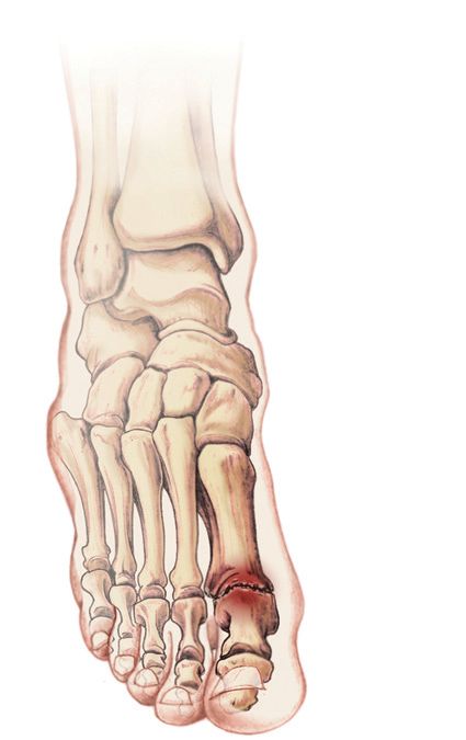
Fallen Arches                                                       evaluation can be
                                                                    completed if you have
                                                                                            Fig. 2. Short foot exercise

                                                                    access to an athletic
   About 20 to 30% of people have pes planus, or fallen                                            Relaxed
                                                                    trainer at a local high        foot core
arches, which we commonly call flatfeet. Physicians divide
                                                                    school or can see
arch types into 3 categories: fallen arch, neutral or medium
                                                                    another professional
arch, and high arch. When someone has a fallen arch, the
                                                                    such as a physician
entire sole of the foot touches the floor. Unfortunately, people
                                                                    or orthopaedist.
with this condition are unable to absorb forces from the
                                                                    These professionals                                   The Hughston
ground properly when walking, running, or landing from a                                                                  Foundation, Inc.
                                                                    use specific tools,
jump, and the joint motions are not as efficient in the ankles,                                    Contract
                                                                                                                          ©2020
                                                                    instruments, and
knees, and hips. Therefore, unnecessary stress is placed                                           foot core
                                                                    techniques to provide          muscles to
on the body’s bones and soft tissues (muscles, tendons,
                                                                    a more detailed                arch the
ligaments) that are not designed to absorb the pressure.                                           foot
                                                                    assessment.
What causes fallen arches?
                                                                    Treatment & exercises
   All infants and toddlers have flatfeet because your foot
                                                                      Whether you have
arch does not develop until the age of 5 or 6. Causes of
                                                                    inherited flatfeet or
flatfeet include genetics, injury to the soft tissue in the arch
                                                                    acquired it from age
of the foot, and other health conditions such as arthritis and
                                                                    or injury, be sure to select the most comfortable shoe that
nerve problems that can lead to the development of fallen
                                                                    provides the support you need to prevent the development
arches. Flatfeet can simply develop over time as we age or
                                                                    of pain and injuries. Flatfeet can lead to painful, debilitating
it can develop after years of walking, running, and standing
                                                                    conditions including plantar fasciitis, Achilles tendonitis,
weakens the posterior tibial tendon (tendons attach muscles
                                                                    medial tibial stress syndrome (shin splints), and pain in
to bone). The posterior tibial tendon starts at the muscle in
                                                                    the knees, hips, and low back due to disruption of proper
the calf, travels down the inside of the lower leg and attaches
                                                                    alignment and mechanics of the body. General treatment
to the bones on the inside of the foot. The main function of
                                                                    of these conditions includes anti-inflammatory medication,
the tendon is to hold up the arch and support your foot while
                                                                    ice, rest, stretches of the calf muscles and Achilles tendon,
you walk. If you tear the posterior tibial tendon or it becomes
                                                                    and physical therapy exercises. Exercises that strengthen
inflamed, the arch can collapse, causing a fallen arch.
                                                                    the arch and foot muscles and the posterior tibialis tendon
   Many people have fallen arches and experience no
                                                                    and muscle include the short foot exercise (Fig. 2), towel
problems, but high levels of activity can lead to pain in the
                                                                    scrunches, and marble pickups.
foot, ankle, lower leg, knees, hips, and even the lower back.
                                                                      If you have flatfeet and increasing pain with activity, you
Individuals with non-neutral foot types are at increased risk
                                                                    may benefit from a different shoe selection or an orthotic
for pain and injury during activity; therefore, you should
                                                                    insert. An athletic shoe with a wide sole, good arch support,
select the appropriate footwear and orthotics for your foot
                                                                    and wide toe box can help alter the way your feet and
type to reduce the risk.
                                                                    ankles move to help reduce unnecessary stress on the area.
How to determine your foot type                                     Arch support orthotics should be considered for people who
  Simple ways to determine your foot type at home are the           have a required or standard type of shoe, such as military
“Wet Test” and “Wear Test.” For the “Wet Test”, wet your            personnel, athletes, and factory workers who are experiencing
foot and step on a piece of paper to make a footprint. (Fig.        discomfort and problems associated with flatfeet.
1). The “Wear Test” includes observing the wear pattern
                                                                    A common problem
on the bottom of your frequently worn tennis shoe. If the
                                                                      Flatfeet are more common than people think. You should
sole of the shoe is worn away on the inside of the foot, you
                                                                    know your foot type and arch height to determine if you are
are more likely to have flatfeet. However, a more accurate
                                                                    at increased risk of injury or development of certain chronic
                                               Fig. 1. "Wet Test"   conditions of the feet and lower legs. Rest, ice, stretching,
                                               sample footprints    and rehabilitation exercises can help ease the symptoms
                                               to help classify     that may develop due to flatfeet. If over the counter inserts
                                               foot type.           do not improve symptoms or you have redness, soreness,
                                                                    or swelling in the foot that lasts more than a few days, seek
                                     The Hughston
                                     Foundation, Inc.               medical advice from your physician.
                                     ©2020

    Flat         High       Normal                                                                        Kathryn Boylan, LAT, ATC
                                                                                                               Columbus, Georgia
                                                                                                   FOR
                                                                                                    FORAAHEALTHIER
                                                                                                         HEALTHIERLIFESTYLE
                                                                                                                   LIFESTYLE 7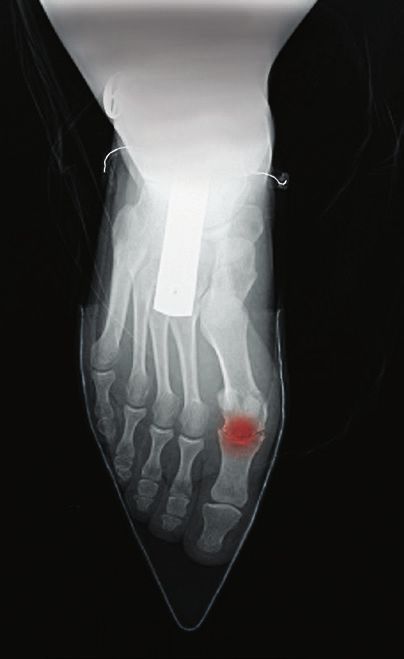
Hughston Health Alert                                                                                                      NONPROFIT ORG
                                                                                                                                       US POSTAGE
                                                                                                                                          PAID
                        The Hughston Foundation, Inc.                                                                                 COLUMBUS GA
                                                                                                                                      PERMIT NO 99
                        6262 Veterans Parkway P.O. Box 9517
                        Columbus, Georgia 31908-9517
                                                                                                                   2002-2019

                                                                                                                   Editor - Garland K. Gudger, Jr., MD
                                                                                                                   Managing Editor - Dennise Brogdon
                                                                                                                   Associate Editor - Roman Ashmyan, DO
                                                                                                                   Art Director - Belinda J. Klein, MA
                                                                                                                   Layout Editor - Tiffany C. Davis, MS
                                                                                                                   Editorial Board
                                                                                                                   Mark A. Baker, PT, CEO
                                                                                                                   Devin Collins, DO
                                                                                                                   William C. Etchison, MS
                                                                                                                   Andy J. Grubbs, Jr., MEd, ATC
                                                                                                                   Rob Hopkins, PT, SCS
                                                                                                                   William Kuerzi, PT; Cert. DN
                                                                                                                   Cholly P. Minton

   The Hughston Health Alert is a quarterly publication of the Hughston Foundation, Inc. The Foundation’s
mission is to help people of all ages attain the highest possible levels of musculoskeletal health, fitness, and
athletic prowess. The content of the Hughston Health Alert, including text, graphics, images, and all other
material considered “content,” is published for educational purposes only. It is not intended to be a
substitute for professional medical advice, diagnosis, or treatment. Always consult your physician or other
qualified healthcare provider about any questions or concerns you may have regarding a medical condition.
You should never delay seeking professional medical advice, disregard medical advice, or change or
discontinue medical treatment based on information found in the Hughston Health Alert or on the Hughston
website. Moreover, the Hughston Health Alert does not recommend or endorse any specific physicians,
products, tests, procedures, or opinions mentioned therein. Reliance on any information published in the
newsletter or appearing on the website is solely at your own risk.
   Special written permission is required to reproduce, by any manner, in whole or in part, the material
herein contained.
   Send inquiries to Medical Writing, The Hughston Foundation, Inc.,
P.O. Box 9517, 6262 Veterans Parkway, Columbus GA 31908-9517 USA.
   Copyright 2020, The Hughston Foundation, Inc.               ISSN# 1070-7778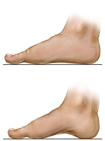
You can also read