INSTITUTE OF HUMAN GENETICS - October 2012
←
→
Page content transcription
If your browser does not render page correctly, please read the page content below

INSTITUTE OF
          HUMAN GENETICS
                                Scientific Report

                                    CNRS UPR 1142 - MONTPELLIER - FRANCE
                                            http://w w w.igh.cnrs.fr

               I G       H

INSTITUT DE GENETIQUE HUMAINE

                                    October 2012
LAYOUT & DESIGN
Catherine Larose

PICTURES
Office de tourisme Montpellier
Cyril Sarrauste de Menthière, IGH Montpellier

COVER ILLUSTRATIONS
Polycomb group proteins are conserved chromatin factors critically required for the regulation of multiple
target genes during cell differentiation and development. The cover picture shows a tangle of ropes on a
boat, metaphorically illustrating the tangled state of chromatin in the nucleus. Green cylinders represent
examples of specific 3D interactions among PcG-bound chromatin elements. Photo and Artwork credit:
Cyril Sarrauste de Menthière. IGH Montpellier

                http://www.igh.cnrs.fr

CONTACT / DIRECTION

Institut de Génétique Humaine

Dr. Giacomo CAVALLI

141 Rue de la Cardonille
34396 MONTPELLIER cedex 5
FRANCE

Phone : +33 (0)4 34 35 99 04 / + 33 (0)4 34 35 99 70
Fax : +33 (0)4 34 35 99 99

Giacomo.Cavalli@igh.cnrs.fr
Contents
Introduction                                   2

GENOME DYNAMICS Department                     8

Giacomo Cavalli                                9
Séverine Chambeyron                            11
Jérôme Déjardin                                13
Bernard de Massy                               15
Nicolas Gilbert                                17
Rosemary Kiernan                               19
Marcel Méchali                                 21

GENETICS & DEVELOPMENT Department              24
Brigitte Boizet                                25
Jean-Maurice Dura                              27
Anne Fernandez & Ned Lamb                      29
Kzrysztof Rogowski                             31
Hervé Seitz                                    33
Martine Simonelig                              35

MOLECULAR BASES OF HUMAN DISEASES Department   38
Monsef Benkirane                               39
Angelos Constantinou                           41
Pierre Corbeau                                 43
Dominique Giorgi & Sylvie Rouquier             45
Marie-Paule Lefranc                            47
Domanico Maiorano                              49
Philippe Pasero                                51

ADMINISTRATION AND OTHER SERVICES              54

SEMINAR SPEAKERS                               64
PUBLICATIONS                                   72
Giacomo Cavalli Director
                                                                                         Deputy Director             Secretariat Assistant
                                                                    Philippe Pasero
                                                                                                                    Anne-Pascale Botonnet
                                                                    Brigitte Mangoni     Secretary General

                                                    SAB
                                                  PI Board                                     Institute Council
                                            Steering Committee

                               RESEARCH LABS                                           Administration & Human
                                                                                                                            Common Services
                                                                                            Ressources
                    « Genome Dynamics » Department
                            Director : B. De Massy
                                                                                                                        Information Technology (IT)
Giacomo Cavalli : Chromatin and Cell Biology                                           Administrative Secretariat
                                                                                                                       Guillaume Gielly - Jacques Faure
Séverine Chambeyron : RNA Silencing and Control of Transposition                           Silke Conquet
                                                                                                                                Alfred Vriese
Jérôme Dejardin : Biology of Repetitive Sequences
Bernard De Massy : Meiosis and Recombination                                             Financial Management                 IT Development for
Nicolas Gilbert : Mobile Elements, Integrity and Plasticity of the Human Genome        Sahondra Rakotondramasy                 Research Support
Rosemary Kiernan : Gene Regulation                                                        Marie-Claire Merriot           Cyril Sarrauste de Menthière
Marcel Méchali : Réplication et Genome Dynamics
                                                                                                                             Cell Imaging Facility
                                                                                            Communication                          Julien Cau
                   « Genetics and Development » Department                                 Training Program                 Julio Mateos-Langerak
                             Director : M. Simonelig                                       Catherine Larose                   Technical servicing
Brigitte Boizet: Development and Pathology of the Gonad                                                                        Daniel Bellenoue
Jean-Maurice Dura : Neurogenetics and Memory
Ned Lamb, Anne Fernandez : Cell Cycle and Myogenesis                                      Health and Safety                          Store
Krzysztof Rogowski : Tubulin Code                                                            Robert Orti                         Faïza Laachir
Hervé Seitz : Systémic impact of small regulatory RNAs                                    Stéphane Bocquet                   Animal housing facility
Martine Simonelig : mRNA Regulation and Development                                        Aymeric Chartier                 Audrey Combe-Sainseau

                     « Molecular Bases of Human Diseases »                                                             Washing / sterilization & media
                      Department - Director : M. Benkirane                                                                  preparation facility
Monsef Benkirane : Molecular Virology                                                                                     Marie-Thérèse Molinier
Angelos Constantinou : DNA Damage Response and Human Diseases                                                             Séverine Arena-Nadaud
Pierre Corbeau : Homing, Immune Activation and Infection
                                                                                                                               Drosophila facility
Dominique Giorgi , Sylvie Rouquier : Microtubules and Cell Cycle
                                                                                                                             Stéphanie Chalmeton
Marie-Paule Lefranc : IMGT, international ImMunoGeneTics information systems
                                                                                                                               Mustapha Hanyn
Domenico Maiorano : Genome Surveillance and Stability
                                                                                                                                Fabienne Mazur
Philippe Pasero : Maintenance of Genome integrity during DNA Replication
GIACOMO CAVALLI
                  Director

                                                      PHILIPPE PASERO
                                                      Deputy Director

    The Institute of Human Genetics (Institut de
Génétique Humaine, IGH) is a CNRS unit located
in the fast growing Arnaud de Villeneuve
biomedical campus of Montpellier that
includes several CNRS and INSERM laboratories
(Centre de Biochimie Structurale (CBS), Institut
de Génomique Fonctionnelle (IGF), etc.), the
future University of Montpellier School of
Medicine (University of Montpellier 1) and
academic hospitals. It is close to the site of the
University of Montpellier 2 and the Center for           The IGH aims at providing a first
Cancer Research (IRCM). The Institute occupies       class scientific environment for the
a surface of 3800 m2.                                development of innovative research
    It hosts more than 200 people, including         projects. The excellence of the research
scientists (36 CNRS, 11 INSERM and 9 University      carried out at the IGH is attested by the
and     Hospital     researchers),  engineers,       quality of the scientific production, the
technical and administrative staff (40), post-       number of awards attributed to scientists
doctoral fellows (37), graduate students (33),       working at the Institute as well as the
undergraduate students and visiting scientists.      prestigious grants that support their
                                                     research, particularly three grants from
                                                     the European Research Council (ERC).
                                                        Currently, the IGH houses 20 research
                                                     groups distributed in the three scientific
                                                     departments      (Genome      Dynamics,
                                                     Genetics & Development and Molecular
                                                     Bases of Human Diseases).

                                                                            OVERVIEW

                                                                                         2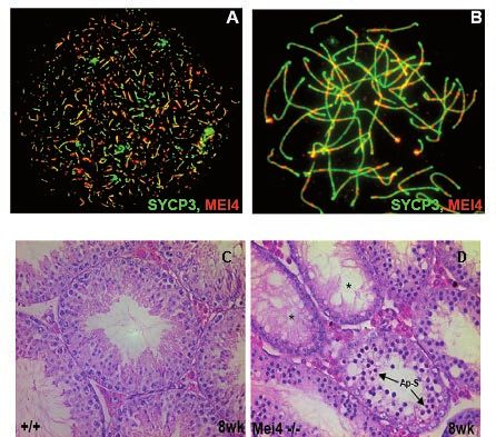
Director’s foreword

        The “Institut de Génétique Humaine” (IGH) is a high-profile institute devoted to basic biomedical
research. Throughout its 13 years of life, it has provided an excellent environment in which it is possible to carry
out innovative, frontier-breaking science and where the quality of the technical facilities, infrastructure and
administrative department matches and supports the high scientific output of the IGH.

IGH scientific life

        The IGH is characterized by a dynamic day-to-day activity that boasts both scientific and extra-scientific
events which contribute to the exciting science and the pleasant daily atmosphere of the Institute. Furthermore,
routine events are complemented by special meetings every year. The main activities that characterize the IGH
community life are:

- weekly «external» seminars given by invited scientists. Most of these seminars are given by internationally-
renowned researchers and all are held in English;

- the annual IGH Seminar Series in which leading are invited by the Institute’s departments to give keynote talks
on their research work;

- weekly «internal» seminars where scientists, post-doctoral fellows and PhD students expose their results and
research projects. The lively informal discussions characterizing these seminars are continued in a friendly
atmosphere during the Pizza time after the seminars;

- scientific retreats (every second year) organized by each Department in order to facilitate scientific interaction
in beautiful places free from the everyday worries of laboratory life;

- the IGH Retreat, a meeting that brings together all the Institute staff every second year, alternating with the
department retreats;

- organization of various high-level meetings like the «IGH 10th Anniversary Meeting», with lectures by outstanding
scientists including the Nobel laureate David Baltimore (see http://congres.igh.cnrs.fr/IGH/IGH10ans.pdf for an
overview of the program); or other prestigious international conferences (such as EMBO conference series and
others). For an exhaustive list, see http://www.igh.cnrs.fr/EN/seminaire.php;

- IGH researchers are frequently involved in the organization of practical courses (Ateliers INSERM and others) to
train scientists in specific approaches on which they have high-profile expertise.

Teaching activities

       The IGH is strongly involved in teaching and has a close relationship with the Universities of (Universities
of Montpellier 1 and 2). Several Professors and Associate Professors carry out their research activities at the IGH.

        The Doctoral School «Biology and Health» (CBS2) of the Universities of Montpellier 1 and 2 is housed
at the IGH and its secretary is a CNRS employee of the Institute. Every year, about thirty graduate students are
pursuing their PhD program at the Institute, and 8-10 of them defend their thesis. In addition, about 20 Master
students do their practical laboratory training at the IGH each year.

                                                                                                   3
Technical facilities

        The IGH offers an excellent technical environment and all the infrastructures needed to carry out cutting-
edge molecular, cellular and developmental biology research. It also possesses two biosafety L3 laboratories.
One of the main strengths of the Institute is its capacity to react rapidly to the need of updating its facilities
in response to the fast technological progress of science. For the last three years we have been running an
«Agence de Biomédecine»-certified laboratory devoted to the study of human embryonic stem cells. In 2009, we
opened a state-of-the-art 100 m2 imaging facility. This facility, called MRI – IGH, has imaging equipment which is
worth more than 3 million Euros, including 3 confocal microscopes and more than 10 top-level epifluorescence
microscopes. We have recently acquired the “OMX” super-resolution fluorescence microscope, which puts our
imaging facility at the absolute forefront in fluorescence imaging acquisition/analysis in France and Europe. The
IGH has also equipped the «Montpellier GenomiX» genomic facility with an Illumina HiSeq instrument, which joins
the already existing Illumina Genome Analyzer IIx and microarray equipment. Together with their bioinformatic
analysis pipeline, these instruments allow high throughput genomic analyses. This facility is installed in the new
building of the Institute of Functional Genomics (IGF) that communicates directly with the IGH. The Institute also
has rodent, Drosophila and Xenopus housing facilities.

        Finally, the IGH is a member of “Biocampus”, the new CNRS-funded servicing unit that provides easy
access to all technical facilities available in the city to the whole Montpellier research community. The facilities
located at the IGH (particularly the animal house and the imaging facility) are thus available to the whole scientific
community of Montpellier.

Institute Governance

         The acting director, Giacomo Cavalli, and the deputy director, Philippe Pasero, took up their functions in
January 2011. They are assisted by a steering committee, composed by the department heads (Martine Simonelig
for Genetics and Development, Bernard de Massy for Genome Dynamics, Monsef Benkirane for Molecular Bases of
Human Diseases and Marcel Méchali, head of the upcoming Genopolys). Scientific issues are discussed within the
group leader board and they are further examined, along with budget and other policy issues, by the 15-member
Institute Council, composed by the directors and a mix of nominated and elected members from all the personnel
bodies: researchers, post-doctoral fellows, PhD students, engineers, technicians and administrative managers.
Starting from 2011 the IGH Scientific Advisory Board (SAB) started its activity. The SAB includes Hervé Chneiweiss,
University Paris Descartes, Paris, France; Denis Duboule, University of Geneva, Switzerland; Edith Heard, Institut
Curie, Paris, France; and Stéphane Noselli, Institute of Developmental Biology and Cancer, Nice, France. A further,
former SAB member, Ron Laskey stepped down this year for personal reasons. We wish to thank him for the great
help he gave while in service, and wish him all the best for the continuation of his life and career. Two new SAB
members, both from the Ecole Polytechnique Fédérale de Lausanne, joined the IGH in 2012; Didier Trono, expert
in virology and Joachim Lingner, expert in telomere biology and replication. We wish to welcome them warmly
and wish them a good collaboration with the IGH governance team, PIs, researchers and personnel. Together with
the four other current SAB members, Hervé Chnieweiss, Denis Duboule, Edith Heard and Stéphane Noselli, they
cover well the research fields of the three IGH departments. They will examine the overall Institute activity every
two years, starting this November when they will participate in the Institute Retreat during which all groups and
scientific facilities present their ongoing and past work. They will also take part in the laboratory evaluations and
will give their advice on new hiring and other scientific policies.

A year of thrilling science

        Last year’s scientific achievements have been particularly striking. With 55 peer reviewed papers published
in peer-reviewed journal under the responsibility of IGH PIs, the average impact factor is above 12, placing IGH
among the very best research units in the country. It would be too long to discuss all the main discoveries published
by the IGH groups but it is remarkable to see how several laboratories have published striking discoveries. We are
particularly delighted to see that two junior laboratories published their research in top journals.

                                                                                                        4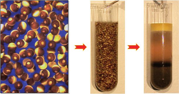
The laboratory of Rosemary Kiernan published in Cell (Wagschal et al, 2012) a regulatory role for
microprocessor in HIV expression. Microprocessor, a complex containing RNase III endonuclease, Drosha, and
a RNA binding subunit Dgcr8, was shown to act in an RNAi-independent manner to regulate transcriptional
pausing at the HIV promoter and, possibly, of many other promoters. Shortly before, the laboratory of Séverine
Chambeyron published in Genome Research (Grentzinger et al. 2012) their discovery that maternal inheritance
of piRNAs can contribute a form of non-chromatin mediated epigenetic inheritance in Drosophila that can help
controlling transposition of retroelements in changing environmental parameters (different temperatures, aging
conditions, …).

        It is particularly rejoicing to see young PIs succeed in their research, and we wish many more of these
discoveries to come in the coming years.

New groups at IGH

        Following the last IGH evaluation by the AERES agency (http://www.aeres-evaluation.fr/index.php/
Etablissements/UNIVERSITE-MONTPELLIER-1, see Institut de Génétique Humaine), we have set up clear policies
concerning laboratory space and the status of junior groups.

        At the end of last year, the junior laboratory of Hervé Seitz “Systemic Impact of small regulatory RNAs”
has joined IGH, strengthening the “Genetics and Development” Department. The junior laboratory of Reina
Fernandez de Luco will join the Genome Dynamics department in 2013. We wish both of them great fun doing
science at IGH, and to become world leaders in their respective fields.

        Finally, the IGH has issued a junior group leader recruitment call in mid 2012. This call received about 80
applicants. 7 of them were short listed for interview and we are finalizing the interview process, hoping to finish
it soon and to welcome a new junior PI in our institute by the end of 2013.

IGH and the initiative “investissements d’avenir” (investments for the future) of the French Ministry of
Research

         To increase French scientific competitiveness, the French Ministry of Research launched two years ago
a large investment campaign in order to fund various research-related components, such as acquisition of large
equipment, large facilities and infrastructures, Centers of excellence and Campuses of excellence.

         The IGH PI Marcel Méchali is coordinating a Center of Excellence (Labex) called EpiGenMed: From Genome
and Epigenome to Molecular Medicine. In total, 49 internationally renowned research laboratories working
in different fields (mathematics, biophysics and biochemistry, molecular, cellular and developmental biology,
cancer biology, infectiology and neurobiology) joined forces to address the following main questions:

- How do genome and epigenome regulations impact on cell proliferation, differentiation and development?

- What are the interactions between host and infectious pathogens, how do they induce diseases and how can
we use this knowledge to cure the world’s most critical infectious diseases?

- What are the molecular bases of the cell signaling processes in the central nervous system and in the sensory
organs and how do signaling dysfunctions induce neurological, neurodegenerative and sensory disorders?

                                                                                                         5
The next years will see these laboratories and others that may join them along the way take an innovative
interdisciplinary research approach in which the knowledge from single molecule research will be followed all
the way up to the development of novel diagnostic and therapeutic approaches. The project will run until 2021
thanks to massive funding that will serve to support PhD and post-doctoral fellowships, group leader hires,
research, teaching and scientific communication activities as well as the clinical exploitation of the results.
EpiGenMed started its activity with a first round of PhD and Postdoc programs that have been heavily subscribed
by excellent applications. IGH researchers are heavily involved in the EpiGenMed research programs and they
coordinate 3 of the 5 programs (biophysics and systems biology; epigenetics and genome dynamics; cell cycle,
cell fate and development; infectious disease and immunology; cell signaling and neurobiology). Thus, IGH will
be a major steering force of this innovative large-scale project.

Enjoy the future!

        In summary, IGH has achieved strong scientific goals and has improved its organization in many ways
during the last year. As always, we are committed to further enhance the quality and impact of our science, while
maintaining a friendly and easy-going atmosphere. It is thus my pleasure to wish a great year to come to all IGH
members.

                                                                                                          6
7
Genome Dynamics
   Department
  Director : Bernard De Massy

             General Statement about the
                     Department
             The research groups of the department of Genome Dynamics focus their
    research on understanding the genome functions by analyzing different aspects of its
    biology in various model systems (Drosophila, Xenopus, mouse, human cells). These
    aspects include DNA replication and recombination, chromatin structure and dynamics,
    mobile elements and gene expression.
             Research on DNA replication aims at identifying origins of replication,
    understanding the molecular mechanisms of origin firing and how these events
    are regulated in order to take place at the right time and only once per cell cycle. A
    special form of the cell cycle is the meiotic division that generates gametes, and our
    department is exploring the processes that ensure the proper transmission of the
    genome by studying the mechanisms of recombination and chromosome segregation
    during meiosis. Specific projects are focused on understanding the mechanism of the
    programmed induction of DNA double strand breaks during meiosis. How genome
    integrity is maintained in the germline, particularly via the control of the activity of
    mobile elements, is also addressed through the analysis of the regulation of a small RNA
    family called piRNAs. Studies directly aimed at identifying the mechanism of insertion
    of mobile elements, such as the human L1 retrotransposons, in the genome provide
    a complementary approach to understand processes that could represent a threat to
    genome stability.
             Several projects also want to determine how the organization of the genome,
    at the level of chromosomes and chromatin, can influence several of its activities.
    Specifically, we aim at understanding how the closed, compact chromatin structure
    called heterochromatin is regulated and its biological relevance for development and
    genome stability in regions of the genome, such as telomeres, pericentromeres and
    rDNA. How local chromatin modifications and the three-dimensional organization of
    chromosomes in the nucleus are integrated and how they impact on gene expression
    is also addressed through the study of the Polycomb and Trithorax protein families. At
    the gene level, factors that are involved in activation or silencing of gene expression,
    through direct or indirect interactions with the transcription machinery, and their links
    with cellular processes of RNA metabolism are investigated.
             Our department has a strong expertise in a variety of approaches, particularly in
    biochemistry, genetics and molecular and cellular biology. State-of-the-art microscopy,
    imaging and bio-informatics for the analysis of next-generation sequencing data
    have also been recently developed by several groups. The department research
    groups are engaged in several collaborations that are fueled by common interests,
    an excellent scientific atmosphere and by formal laboratory interactions, such as the
    department retreats. In addition to the interactions within the department, several of
    our teams collaborate with laboratories in the two other departments of the Institute
    to understand how genome regulation drives development and its relationship with
    human pathologies.

                                                                                      8
GENOME DYNAMICS
Chromatin and Cell Biology
GIACOMO CAVALLI
Giacomo.Cavalli@igh.cnrs.fr
Giacomo Cavalli
Research Director CNRS
Thierry Cheutin,
Research Scientist CNRS
Anne-Marie Martinez, We are more than our DNA! In the last couple of decades it has become
Lecturer, clear that chromosomal components such as histones, regulatory proteins and
University Montpellier 2 noncoding RNAs contribute to regulate all aspects of DNA function and contribute
to heredity. Our lab has mainly focused on the analysis of proteins of the Polycomb
Bernd Schuttengruber, and Trithorax groups: key regulators of the expression of major developmental genes
Research Scientist INSERM that coordinate the processes of cell differentiation and cell proliferation. Polycomb
proteins are able to silence gene expression, while Trithorax proteins counteract
gene silencing in the appropriate cells. At the molecular scale, we have studied
Aubin Thomas,
how Polycomb and Trithorax proteins are recruited to DNA and how they may
Engineer CNRS
interact with other regulatory elements, such as chromatin insulators. Moreover,
we published the first large-scale mapping of the distribution of Polycomb group
Inma Gonzalez, proteins along Drosophila chromosomes at different developmental stages. We also
Post-doctoral Fellow demonstrated that polyhomeotic, a Polycomb group gene, is a tumor suppressor
that controls cell proliferation by regulating Notch signaling.
Nicola Iovino,
Post-doctoral Fellow A distinctive feature of these proteins is their ability to maintain the memory
of gene regulatory states through successive mitotic divisions in the different cell
Thomas Sexton, lineages and research in our laboratory has analyzed this phenomenon. We showed
that the regulation of chromosome architecture by these proteins contributes the
Post-doctoral Fellow
transgenerational epigenetic inheritance of chromatin states by revealing that
the transmission of this mitotic and meiotic cellular memory can bring into play
Filippo Ciabrelli, long-distance chromosomal interactions in the three-dimensional space of the cell
PhD student nucleus.
Anne Delest, We recently extended the analysis of chromosome architecture by
PhD student analyzing at genome-wide scale the contacts made by each locus with all other
chromosome loci in the genome. From this study, we deduced the principles
Philip Yuk Kwong Yung, governing chromosome organization and the functional implications of regulation
PhD student of genome architecture. We will pursue this analysis In the coming years.
Caroline Jacquier-Labroche
Engineer
9GENOME DYNAMICS
- Sexton, T., Yaffe, E., Kenigsberg, E., Bantignies, F., Leblanc, B., Hoichman, M., Parrinello, H., Tanay, A., and Cavalli, G. (2012). Three-dimensional
folding and functional organization principles of the Drosophila genome. Cell, 148, 458-472
-Bantignies, F., Roure, V., Comet, I., Leblanc, B., Schuettengruber, B., Bonnet, J., Tixier, V., Mas, A., and Cavalli, G. (2011). Polycomb-dependent
regulatory contacts between distant Hox loci in Drosophila. Cell , 144, 2, 214-226.
- Martinez, A.M., Schuettengruber, B., Sakr, S., Janic, A., Gonzalez, C., and Cavalli, G. (2009). Polyhomeotic has a tumor suppressor activity
mediated by repression of Notch signaling. Nat Genet., 41, 10, 1076-1082.
- Grimaud, C., Bantignies, F., Pal-Bhadra, M., Ghana, P., Bhadra, U., and Cavalli, G. (2006). RNAi Components Are Required for Nuclear
Clustering of Polycomb Group Response Elements. Cell, 124, 957-971.
- Dejardin, J., Rappailles, A., Cuvier, O., Grimaud, C., Decoville, M., Locker, D., and Cavalli, G. (2005). Recruitment of Drosophila Polycomb
group proteins to chromatin by DSP1. Nature, 434, 533-538.
Figure 2. 3D distribution of PcG proteins (polycomb in
green and polyhomeotic in red) compared to histone
H3K27me3 (blue) inside the cell nucleus. They co-
localize inside sub-nuclear volumes called polycomb
bodies.
Figure 1. Hi-C, a molecular biology method to map interactions between
chromatins sequences in vivo, is used to explore the spatial organization
of the genome in Drosophila embryos. A cluster of strong interactions
between two myoblast-specific genes, hbs and sns, located ~6 Mb
apart on one chromosome arm, is highlighted. This interaction was
also shown to occur at high frequency by DNA FISH (fluorescent in situ
hybridization).
Figure 3. Mutation of the polyhomeotic locus (second panel from the left) induces over-proliferation of the mutant tissue
(in green, compare to control on the left). Most larvae die but around 10% survive and, in that case, the mutant tissue over-
proliferates (the mutant eye in the second panel from the right is larger than wild type eye on the left) and forms tumors.
RESEARCH GROUPS
10GENOME DYNAMICS
JUNIOR LABORATORY
RNA Silencing & Control
of Transposition
SEVERINE CHAMBEYRON
severine.chambeyron@igh.cnrs.fr
We are interested in understanding the mechanisms involved in the
Séverine Chambeyron control of transposable elements (TEs) that are essential for the maintenance of
Research Scientist CNRS genome integrity. They involve a class of small RNAs, the piRNAs (piwi-interacting
RNAs). Since the piRNA silencing pathway is not well known, we propose to
characterize in the Drosophila ovary the biogenesis and the role of this class of
Alain Pelisson, small non coding RNAs.
piRNAs may be considered as key elements of a sort of bipartite immune
Research Director CNRS
system: one genetic component is encoded by heterochromatic loci (named
piRNA clusters) that contain defective copies of TEs producing antisense piRNAs;
Christine Brun, the other component is achieved by sense piRNAs produced by the functional
Technician CNRS copies of TEs located in euchromatin. In the so-called “ping-pong” biogenesis
pathway, primary antisense piRNAs, produced by an unknown mechanism
Bruno Mugat, from piRNA clusters, target the transcripts of functional TEs that are cleaved to
Engineer CNRS produce sense piRNAs. These sense piRNAs then target the transcripts of the
piRNA clusters that are then cut to produce secondary antisense piRNAs.
Abdou Akkouche, Using two TE models, the I element in the germline and the gypsy
Post-doctoral fellow retrotransposon in the somatic ovarian cells, we are studying the biogenesis
of the piRNAs, the mechanism of the piRNA-mediated TE repression, and the
epigenetic mechanisms involved in the maternal inheritance of the silencing.
Thomas Grentzinger,
PhD student Our recent results provide evidence that secondary piRNAs can repress
the I element in the female germline. The I element is an excellent model
because it is one of the rare transposable elements which has not yet invaded
all Drosophila melanogaster strains. This invasion can therefore be reproduced
at will in the lab and the establishment of the silencing followed in real time. A
cryptic production of secondary piRNAs by the piRNA clusters was discovered
which explains how flies submitted to various environmental treatments (aging,
temperature,…) are better prepared to resist the invasion (Fig. 1). The “ping-
pong” amplification loop in the ovary seems to be boosted if the egg already
contained such maternally deposited cryptic piRNAs. So, secondary piRNAs are
the molecular basis of the non-chromatin-mediated epigenetic memory of the
environmental treatment (Fig. 2).
The “ping-pong” amplification loop does not occur in somatic ovarian
cells, where TE silencing is only achieved by primary piRNAs (Fig. 3). In this tissue,
we are studying the relationships between piRNA- and both the micro- and the
siRNA pathways in the regulation of somatic TEs. We are also assessing to what
extent the piRNA-loaded Piwi protein can affect the expression of TEs and their
flanking genes by changing the chromatin landscape in and around TEs. In the
germ cells, another layer of TE silencing occurs post-transcriptionally through
the sequestration of TE transcripts inside the nucleus (Fig. 4).
.
11GENOME DYNAMICS
- Grentzinger, T., Armenise, C., Brun, C., Mugat, B., Serrano, V., Pelisson, A., Chambeyron, S. (2012)piRNA-mediated transgenerational
inheritance of an acquired trait. Genome Res., 22, 10, 1877-1888. PMID : 22555593.
- Chambeyron, S., Popkova, A., Payen-Groschêne, G., Brun, C., Laouini, D., Pelisson, A., Bucheton, A. (2008) piRNA-mediated nuclear
accumulation of retrotransposon transcripts in the Drosophila female germline. Proc. Natl. Acad. Sci. U S A. 105, 39, 14964-14969.
- Mevel-Ninio, MT., Pelisson, A., Kinder, J., Campos, AR., Bucheton, A.(2007) The flamenco locus controls the gypsy and ZAM retroviruses
and is required for Drosophila oogenesis. Genetics, 175, 4, 1615-1624.
- Pelisson, A., Payen-Groschene, G., Terzian, C., Bucheton, A. (2007) Restrictive Flamenco Alleles Are Maintained in Drosophila
melanogaster Population Cages, Despite the Absence of Their Endogenous Gypsy Retroviral Targets. Mol. Biol. Evol., 24, 2, 498-504.
- Pelisson, A., Sarot, E., Payen-Groschene, G., Bucheton, A. (2007) A novel rasiRNA-mediated silencing pathway downregulates sense
gypsy transcripts in the somatic cells of the drosophila ovary. J. Virol., 81, 4, 1951-1960.
Figure 2 : Secondary piRNAs are produced
Figure 1 : Female aging rescues the sterility due in the ovary of the daughter, in response to
to I element derepression in the ovaries lacking the maternal deposition of cognate piRNAs
cognate piRNAs. in the eggs laid by the aged mother .
Figure 3 : Piwi-dependent repression of a gypsy-
LacZ reporter in the somatic cells of the female Figure 4 : Nuclear sequestration of I element
gonad (X-Gal staining). transcripts detected by FISH
RESEARCH GROUPS
12GENOME DYNAMICS
JUNIOR LABORATORY
Biology of Repetitive Sequences
JEROME DEJARDIN
Jerome.Dejardin@igh.cnrs.fr
Chromatin can be viewed as a highly complex mixture of proteins and
nucleic acids that orchestrate DNA-based processes in the eukaryotic genome. Most
Jérôme Déjardin of the mammalian genome is assembled into heterochromatin, a ‘closed’ structure
Research Scientist INSERM imposed by several enzymatic activities. Such activities act on histones and the DNA
itself to impinge on transcription, replication or repair.
Satoru Ide, Most of the heterochromatic fraction of the genome can be found at critical
Post-doctoral Fellow loci. These include telomeres, repetitive sequences around centromeres and a portion
(about half ) of the gene units encoding ribosomal RNAs. Defects in the regulation
Agnieszka Nowak, of these loci have therefore disastrous consequences on cell identity and can lead
Post-doctoral Fellow to developmental problems, cancer, premature aging or immune deficiencies. How
precisely heterochromatic enzymes affect the composition of target loci has remained
Nehme Saksouk, elusive and research in our laboratory primarily focuses on this question.
To understand how heterochromatin acts at the molecular level, we are
Post-doctoral Fellow
looking at the effect of abrogating important heterochromatic activities, such
as histone and/or DNA methyl-transferases, on the overall composition of key
Alexandra Lawera, heterochromatic loci (telomeres, pericentromeres and rDNA).
PhD student In particular, we are interested in:
(i) How telomere compositional changes upon loss of heterochromatin function can
Paulina Marzec, explain the appearance of the ALT (Alternative Lengthening of Telomeres) pathway
PhD student observed in certain cancers.
(ii) How the situation at ALT telomeres can be compared to the changes observed
Claudia Armenise, at human satellite 2 sequences upon loss of DNA methylation in ICF cells. Indeed,
Engineer satellite 2 regions recombine aberrantly and localize to PML bodies in ICF cells, a
‘behavior’ also observed in the case of ALT telomeres.
(iii) How pericentric heterochromatin is regulated by such enzymatic activities during
Elodie Rey-Sahinovic, development, differentiation and why such regulation matters for genome stability.
Engineer (iv) Characterizing the new SMCHD1 chromatin protein which possibly links DNA
methylation and non-coding RNAs.
(v) How is rDNA expression regulated?
We have initiated these studies using a quantitative version of the
PICh technology, qPICh, which couples SILAC with PICh. This approach allows the
unbiased characterization of proteins bound to a specific locus in vivo (see figure).
By correlating compositional and phenotypic changes at distinct loci, we hope our
research will uncover important determinants of gene expression and genome
stability. Importantly, because PICh has been adapted to quantitative approaches, we
are now able to precisely monitor the dynamics of heterochromatin in vivo.
For more information, please, see:
Déjardin J and Kingston R (2009). Locus specific chromatin proteomics. Cell 136(1):175-
86.
13GENOME DYNAMICS
- Déjardin, J., Kingston, R.E. (2009) Purification of proteins associated with specific genomic Loci. Cell, 139, 1, 175-186
- Dejardin, J. (2012) How chromatin prevents genomic rearrangements: Locus colocalization induced by transcription factor binding.
Bioessays, 34, 2, 90-93. doi: 10.1002/bies.201100122
Input
JSat
Scr
Tel
WT
WT
WT
WT
KO
KO
KO
KO
191
97
64 38 126
51
362
39
28
19 Telomeres
14
B
A
Purification of major satellites (Sat) and telomeres from mouses embryonic stem cells in WT or
Suv39h1+h2 K.O. backgrounds. Composition of both loci is established in the two backgrounds,
allowing to determine:
- the signature of constitutive heterochromatin in mammals, i.e. proteins found enriched in
common at both loci (e.g. HP1 isoforms, etc ...)
- the role of Suv39h in the biology of these targets : specific proteins are lost or gained at telomeres
or pericentric chromatin in the absence of this important heterochromatin enzyme.
RESEARCH GROUPS
14GENOME DYNAMICS
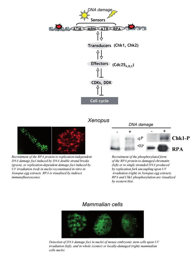
Meiosis and Recombination
BERNARD DE MASSY
Bernard.de-Massy@igh.cnrs.fr
In sexually reproducing species, meiosis allows the formation of
haploid gametes from diploid cells. The halving of the DNA content results from
Bernard de Massy a specialized cell cycle, where a single phase of DNA replication is followed by
Research Director CNRS two divisions. In most species, the proper segregation of chromosomes at the
first meiotic division requires connections between homologous chromosomes
Frédéric Baudat, that result from reciprocal homologous recombination events or crossovers.
Research Scientist CNRS Crossovers also generate new allele combinations and thus increase genetic
diversity. The absence of crossover leads to segregation defects and sterility,
Jérôme Buard, and alteration of the meiotic recombination pathway can lead to genome
Research Scientist CNRS rearrangements and aneuploidy.
Our group is investigating several aspects of the mechanism and
Corinne Grey,
regulation of meiotic recombination using the mouse as a model system. Meiotic
Research Scientist CNRS recombination events are initiated by the formation of DNA double-strand
breaks (DSBs), the repair of which leads to both crossovers and non-crossovers
Thomas Robert, (gene conversion without crossover) (Fig. 1). Several hundreds DSBs, catalyzed
Research Scientist CNRS by the SPO11 protein, are formed at the beginning of the first meiotic prophase
in mouse meiotic cells. SPO11 is homologous to the catalytic subunit of the Topo
Rajeev Kumar, VI family of type II DNA topoisomerases, and is conserved among eukaryotes.
Post-doctoral Fellow We are interested in understanding how the frequency and distribution
of these DSBs are regulated, and how DSB formation and repair are coordinated.
Boubou Diagouraga, We have recently discovered a major component that determines the sites
where DSBs are formed in mammals: the Prdm9 gene. This gene encodes a
PhD student
protein with a methyl-transferase activity and a tandem array of C2H2 zinc
fingers. PRDM9 recognizes specific DNA motifs in the genome and is thought to
Denis Dunoyer de Segonzac promote trimethylation of lysine 4 of Histone H3 at these sites (Fig. 2). How does
PhD student this protein actually function in vivo and how its activity allows the recruitment
of the recombination machinery remains to be determined. In addition, a
Yukiko Imai, remarkable property of PRDM9 is its rapid evolution and diversity. We are
PhD student currently investigating both its molecular and evolutionary features.
DSB formation is expected to be a highly coordinated process given
the potential threat to genome integrity, and studies in yeast have shown that,
in addition to SPO11, several other proteins are necessary for DSB formation.
We have recently identified two mouse proteins that are orthologs of the yeast
Rec114 and Mei4 proteins and shown that Mei4 is required for DSB formation in
mice (Fig. 3). We are currently investigating the activities and functions of these
proteins using biochemical, molecular, cytological and genetic approaches.
15GENOME DYNAMICS
- Grey C, Barthès P, Chauveau-Le Friec G, Langa F, Baudat F and de Massy B. (2011) Mouse PRDM9 DNA-Binding Specificity Determines Sites
of Histone H3 Lysine 4 Trimethylation for Initiation of Meiotic Recombination. PLoS Biol., 9, 10, e1001176.
- Lichten, M., de Massy, B. (2011) The impressionistic landscape of meiotic recombination. Cell, 147, 267-270.
- Baudat, F. *, Buard, J. *, Grey, C. *, Fledel-Alon, A., Ober, C., Przeworski, M., Coop, G., and de Massy, B. (2010) PRDM9 is a Major Determinant
of Meiotic Recombination Hotspots in humans and mice. Science, 327, 836-840.
- Kumar, R., Bourbon, HM., and de Massy, B. (2010) Functional conservation of Mei4 for meiotic DNA double-strand break formation from
yeasts to mice. Genes Dev., 24, 1266-1280.
- Grey, C., Baudat, F. and de Massy, B. (2009) Genome wide regulation of meiotic recombination, PLoS Biol., 7, 2, e35.
- Fig.1. DNA and cytological events during meiotic
prophase.
Meiotic recombination is initiated by DSBs, which
are catalyzed by SPO11 and visualized by the
appearance of H2AX (the phosphorylated form of
H2AX). DSB repair, with the strand exchange activity
of RAD51 and DMC1, leads to crossover (CO) and
non- crossover (NCO) events. CO sites are visualized
by the presence of MLH1 on chromosome axes
(SYCP3) at the pachytene stage.
- Fig.3. Mei4 is essential for male and female fertility.
MEI4 (red) localizes as discrete foci along unsynapsed
- Fig.2. Model of PRDM9 specification of meiotic recombination initiation sites chromosome axes (labeled with SYCP3, green) at leptotene
in mammals. (A) and zygotene-like stages (B) in Spo11-/- and wild type
PRDM9 binds to a DNA motif through its zinc finger domain and induces (not shown) spermatocytes. Spermatogenesis in wild type
H3K4Me3 on adjacent nucleosomes (beige cylinder and histone post- (C) and Mei4 -/- (D) mice: meiotic arrest and apotosis are
translational modifications as red balls). Additional chromatin modifications observed in Mei4 -/- mice. *, empty tubules; Ap-S, Apoptotic
and/or remodeling may take place and other proteins may be recruited. spermatocytes.
SPO11 is then recruited, binds to DNA and promotes DSB formation.
RESEARCH GROUPS
16GENOME DYNAMICS
JUNIOR LABORATORY
Mobile elements, Integrity and
Plasticity of the Human Genome
NICOLAS GILBERT
Nicolas.Gilbert@igh.cnrs.fr
Nicolas Gilbert
Research Scientist INSERM Interspersed repeat sequences are present in almost all eukaryotic
genomes. The LINE-1 (Long Interspersed Element-1, or L1) retrotransposon is
Oliver Siol, the most abundant mobile element of the human genome.
Post-doctoral Fellow
Approximately 500,000 copies of L1 are present in the human genome
and represent ~17% of human DNA. The vast majority of these copies are
considered as molecular fossils. However, ~100 elements remain potentially
active (RC-L1). Because of its activity, L1 can induce genetic diseases by
insertional mutation in either coding or regulatory regions. Moreover, due to
its high representation in the genome, L1 can generate deleterious genomic
rearrangements induced by non-allelic homologous recombination.
Although L1 mobility can induce genetic instability, the mechanism
of L1 retrotransposition is still poorly understood. Our group focuses on
understanding the molecular mechanisms of L1 transposition and its impact
on the genome. We are particularly interested in the L1 ribonucleoprotein
complex formation, an intermediate of retrotransposition.
We also would like to understand the interplay between DNA repair
mechanisms and the resolution of L1 insertion. We use two complementary
approaches. First, we utilize a cell culture assay that allows us to control
L1 retrotransposition. It will help us to decorticate the different steps of L1
retrotransposition. Second, we perform in silico analyses to support our
molecular approach and to determine L1 implication in genomic variability
and evolution of mammalian genomes.
.
17GENOME DYNAMICS
- Doucet, A., Hulme A., Sahinovic, E., Kulpa, D., Moldovan, J., Kopera, H., Athanikar, J., Hasnaoui, M., Bucheton, A., Moran, J., Gilbert, N. (2010)
Characterization of LINE-1 Ribonucleoprotein Particles, PLoS Genetics : 6, 10, pii: e1001150.
- Hasnaoui, M., Doucet, a.J., Meziane, O., Gilbert, N. (2009) Ancient repeat sequence derived from U6 snRNA in primate genomes
GENE, 448, 2, 139-144.
- Garcia-Perez, JL., Doucet, AJ., Bucheton, A., Moran, JV., Gilbert, N. (2007) Distinct mechanisms for trans-mediated mobilization of cellular
RNAs by the LINE-1 reverse transcriptase. Genome Res., 17, 5, 602-611.
- Gasior, SL., Preston, G., Hedges, DJ., Gilbert, N., Moran, JV., Deininger, PL. (2007) Characterization of pre-insertion loci of de novo L1
insertions. Gene, 390, 1-2, 190-198.
Figure 1: Structure of an L1 and model of
retrotransposition.
ORF2 encodes enzymatic activities
essential for L1 mobility, EN for
endonuclease and RT for reverse
transcriptase. ORF2 presents also a
cysteine-rich domain important for L1
retrotransposition in its carboxyl end, but
of unknown function (C). The essential
steps (a to h) of the mechanism are shown.
TPRT stands for Target-site Primed Reverse
Transcription, i.e. the endonuclease
domain of ORF2p cleaves the DNA target
site (step f ) and reverse transcription is
initiated at this site by the RT domain (step
g).
Figure 2: Cell localization of L1-
encoded proteins and RNA.
Immunofluorescence/RNA FISH
was carried out using pAD3TE1-
transfected U-2 OS cells 48 hours
post-transfection. T7-tagged
ORF1p (green), TAP-tagged ORF2p
(blue), L1 RNA (red) and DAPI
(turquoise) staining are shown in
the four micrographs on the left.
A merged image is shown in the
rightmost panel. The schematic of
pAD3TE1, our engineered active
L1 element, is shown above the
micrographs.
RESEARCH GROUPS
18GENOME DYNAMICS
JUNIOR LABORATORY
Gene Regulation
ROSEMARY KIERNAN
Rosemary.Kiernan@igh.cnrs.fr
Rosemary Kiernan
Research Scientist CNRS
All organisms must regulate gene expression to achieve the
Xavier Contreras, silencing of certain genes and the activation of others during development
Research Scientist INSERM and homeostasis.
Poornima Basavarajaiah, Deregulation of gene expression frequently has dire
consequences, and can lead to pathologies such as cancer. The regulation
Post-doctoral Fellow
of gene expression occurs at different levels, all of which depend on a
multitude of factors.
Daniel Latreille,
PhD student Chromatin is a primary regulator of gene expression. Physical
compaction of the genome into chromatin controls accessibility to the
Lisa Bluy, transcription machinery.
Engineer
Studies performed over recent years have revealed the enormous
complexity involved in modifying chromatin to regulate gene expression.
Once the genome becomes accessible, the engagement of
the transcription machinery is a highly orchestrated process involving
the recruitment of hundreds of factors that co-operate to achieve gene
expression.
Finally, transcription of a gene is linked to cellular processes
required for the maturation and export of the mRNA in order to achieve
gene expression.
The Gene Regulation Laboratory is interested in understanding
the mechanisms that contribute to the silencing or activation of
mammalian genes. We use the promoter of the human immunodeficiency
virus (HIV-1) as a model to study gene regulation in mammalian cells.
19GENOME DYNAMICS
Using this model, we have shown that the ubiquitin-proteasome system (UPS) strongly regulates HIV-1
transcription through recruitment of the 19S subunit to HIV-1 chromatin. We determined that a proteasome-associated
protein, PAAF1, is a potent co-activator of transcription from the HIV-1 promoter. Ongoing studies are aimed at further
characterizing the role of 19S and PAAF1 in transcription from HIV-1 and cellular promoters.
We have also recently shown that HIV-1 transcription is controlled by premature termination induced by the co-
operative activities of microprocessor, Setx, Xrn2 and Rrp6. A subset of cellular genes and an endogenous retrovirus were
also found to be regulated by this pathway.
- Wagschal, A., Rousset, E., Basavarajaiah, P., Contreras, X. , Harwig, A., Laurent-Chabalier, S. , Nakamura, M., Chen, X., Zhang, K., Meziane,
O., Boyer, F., Parrinello, H., Berkhout, B., Terzian, C., Benkirane, M., Kiernan, R. (2012) Microprocessor, Setx, Xrn2 and Rrp6 Co-Operate to
Induce Premature Termination of Transcription by RNAPII. 2012. Cell, 150, 6, 1147-1157. doi: 10.1016/j.cell.2012.08.004. PMID: 22980978
- Nakamura, M., Basavarajaiah, P., Rousset, E., Beraud, C., Latreille, D., Henaoui, I.S., Lassot, I., Mari, B., Kiernan, R. (2012) Spt6 levels are
modulated by PAAF1 and proteasome to regulate the HIV-1 LTR. Retrovirology, 9, 13.
- Sobhian, B., Laguette, N., Yatim, A., Nakamura, M., Levy, Y., Kiernan, R., Benkirane, M. (2010) HIV-1 Tat assembles a multifunctional
transcription elongation complex and stably associates with the 7SK snRNP. Molecular Cell, 38, 439-451.
- Lassot, L., Latreille, D., Rousset, E., Sourisseau, M., Linares, L.K., Chable-Bessia, C., Coux, O., Benkirane, M., Kiernan, R. (2007) The Proteasome
Regulates HIV-1 Transcription by Both Proteolytic and Non-Proteolytic Mechanisms. Molecular Cell, 25, 369-383.
RESEARCH GROUPS
20GENOME DYNAMICS
Replication & Genome Dynamics
MARCEL MECHALI - Marcel.Mechali@igh.cnrs.fr
Paradoxically, a major cell function such as the faithful duplication of the genome remains
poorly understood in metazoans. During embryonic development chromosomes should
Marcel Méchali be duplicated while maintaining memory of the specific on-going transcription programs,
Research Director CNRS because, in multicellular organisms, cell proliferation must not only deal with cell growth,
but also with cell differentiation. In mammals, DNA replication starts at around 30 000-
Christelle Cayrou, 50 000 sites along chromosomes. These sites are called DNA replication origins. As they
do not share any detectable consensus sequence, unveiling their common features
Research Scientist CNRS
remains an ambitious challenge. We wish to decipher the code of DNA replication origins
in metazoans and unravel its involvement in cell identity. We also aim at dissecting the
James Hutchins, molecular mechanisms used to build a chromosomal DNA replication origin and wish to
Research Scientist CNRS analyze how epigenetic mechanisms control the organization of chromatin domains for
replication.
Magali Kitzmann, We have used different approaches to identify replication origins (Figure 1) including a
Research Scientist CNRS genome-wide analysis of mouse pluripotent stem cells and differentiating cells as well as
of Drosophila cells. To this aim, we purified nascent DNA strands synthesized at replication
Malik Lutzmann, origins and identified their distribution along chromosomes by micro-array analysis and
Research Scientist CNRS high-throughput sequencing. We could characterize several new features of replication
origins and we found that they are conserved, including a new genetic element that we
called Origin G-rich Repeated Element (OGRE) and can forms G-quadruplexes. We also
Stéphane Bocquet, analyzed the global organization of origins by DNA combing (Figure 1). Bioinformatic
Research Assistant CNRS simulations using the data obtained suggest a flexible replicon model in which origins
Isabelle Peiffer, are organized in groups of adjacent potential origins that define a replicon. Moreover, a
Engineer CNRS single origin is activated in each replicon and the chosen one can vary from cell to cell.
Other studies mimicking the nuclear transfer experiments used for animal cloning allowed
Post-doctoral Fellows : us to observe a dramatic reorganization of chromosomes and replication origins when
Hanane Agherbi, differentiated nuclei are exposed to a mitotic embryonic context. We further showed that
Philippe Coulombe, Xenopus egg extracts can efficiently reprogram differentiated mouse cells to become
Michail Fragkos, pluripotent cells, in a reaction that also requires mitotic events (Figure 2).
In the second axis of our project, we exploit in vitro systems derived from Xenopus eggs
Olivier Ganier,
(Figure 3) as well as mammalian cells to identify and characterize new replication proteins.
Sabine Traver During the past decade, we have characterized several replication factors, including Cdt1,
MCM8, MCM9 and MCM-BP. We found that Cdt1 and geminin form a complex that acts
PhD students : as an ON/OFF switch at replication origins. We also reported two new members of the
Marta Rodriguez-Martinez, MCM helicase family, MCM8 and MCM9, and found that they play distinct roles during
Fabien Velilla, DNA replication. We also discovered that MCM8 and MCM9 form a new complex involved
in the control of recombination, DNA repair and animal fertility.
Emmanuelle Beyne, The dissociation of replication complexes at the end of S phase is crucial to avoid mitotic
Engineer defects. We found that Topoisomerase II couples termination of DNA replication with the
clearing of the replication complexes at the end of S phase. The ORC complex, in addition
to its known role in the assembly of the replication initiation complex in G1, is also required
Silke Conquet,
for its disassembly at mitotic entry. Specifically, MCM-BP, a protein that interacts with the
Secrétary MCM2-7 helicase, contributes to MCM complex dissociation from DNA at the end of DNA
synthesis. Further information is available at: http://www.igh.cnrs.fr/equip/mechali/
21GENOME DYNAMICS
- Lutzmann, M., Grey, C., Traver, S., Ganier, O., Maya-Mendoza, A., Ranisavljevic, N., Bernex, F., Nishiyama, A., Montel, N., Gavois, E., Forichon,
L., de Massy, B., and Méchali, M. (2012) MCM8- and MCM9-Deficient Mice Reveal Gametogenesis Defects and Genome Instability Due to
Impaired Homologous Recombination. Mol Cell. 47, 523-534
- Cayrou C., Coulombe P, Vigneron A., Stanojcic S., Ganier O., Peiffer I., Rivals E., Puy A., Laurent-Chabalier S., Desprat R., and Méchali M.
(2011) Genome-scale analysis of metazoan replication origins reveals their organization in specific but flexible sites defined by conserved
features. Genome Res., 9, 1438-1449.
- Ganier O., Bocquet, S., Peiffer, I., Brochard, V., Arnaud, P., Puy, A., Jouneau, A., Feil, R., Renard, J.P., and Méchali M. (2011) Synergic
reprogramming of mammalian cells by combined exposure to mitotic Xenopus egg extracts and transcription factors. Proc Natl Acad
Sci U S A., 108, 17331-17336.
- Méchali, M, (2010), DNA replication origins: many choices for appropriate answers. Nature Rev Mol Cell Biol, 11, 728-738.
- Lutzmann, M., and Mechali, M. (2008) MCM9 binds Cdt1 and is required for the assembly of prereplication complexes. Mol Cell, 31, 190-
200.
- Lemaitre, JM., Danis, E., Y Vassetzky, Y., Pasero. P., and Méchali., M. (2005) Mitotic remodeling of the replicon and chromatin structure,
Cell, 123, 787-801.
- Maiorano, D., Cuvier, O., Danis, E., and Méchali, M. (2005) MCM8 is an MCM2-7 related protein that functions as a DNA helicase during
replication elongation and not initiation. Cell, 120, 315-28.
Fig.2. Mouse embryonic fibroblasts reprogrammed by
Xenopus egg extracts express OCT4, a marker of pluripotency.
Left, phase-contrast image. Right, fluorescence image
showing cell clones expressing GFP under the control of the
Oct4 promoter.
Fig.1. From replication foci to the replication
origin code.
A) A nucleus, in which replication foci are
labeled with BrdUTP, followed by fluorescence
imaging; (B) when two consecutive pulses of
labeling (red and then green) are performed
and the DNA combed on silanized glass,
replication origins can be visualized, with
the red labeling the origin and the green
highlighting the progressing replication forks;
(C) nascent strand isolation and microarrray
analysis allow genome-wide identification of
replication origin sequences, the positions of
which (D) in the chromosomes can then be
visualized (shown for two cell lines: E5 and P19).
Fig.3. From Xenopus eggs to DNA replication extracts
RESEARCH GROUPS
2223
Genetics and
Development Department
  Director : Martine Simonelig

            General Statement about the
                    Department
               Developmental Genetics aims at understanding how the genetic
      information is translated into the production of many different cell types that
      are coherently organized in a complete organism. Groups in the Department of
      Genetics and Development are interested in various aspects of developmental
      genetics, from the establishment of cell polarity in the egg, to muscle differentiation,
      or the formation of an extremely complex structure such as the adult brain.
      Research topics in the Department include the identification of the molecular and
      signaling pathways that control the cell cycle as well as those involved in stem
      cell biology, in the development of the gonads and of the germ line and in muscle
      differentiation. Another topic concerns the ligand/receptor interactions in axonal
      guidance during the development and function of the central nervous system.
      Several groups are interested in deciphering specific molecular regulations that
      control developmental processes, such as RNA silencing by small non-coding RNAs
      (microRNAs and piRNAs) and post-translational regulations.
               These fundamental biological questions are addressed using model
      organisms (Drosophila and the mouse) and a variety of approaches. Groups in
      the Department have strong expertise in classical and cutting-edge genetic
      techniques, biochemistry, molecular and cell biology, advanced light microscopy
      and bioinformatics.
               All the groups in the Department of Genetics and Development
      work towards understanding the molecular mechanisms of human diseases.
      Tumorigenesis is an important question addressed in the Department, through
      the utilization of cell and mouse models. Several groups have also developed
      Drosophila models of human diseases (e.g. muscular dystrophy, motoneural
      dysfunction, sterility), in which sophisticated genetic approaches can be applied to
      gain insights into the molecular pathways involved in these diseases. The analysis
      of multipotent stem cells showing regenerative potential is another important
      topic of research in the Department.
               The Department of Genetics and Development has strong transversal
      interactions with other groups at the IGH and groups located in the close-
      by Institute of Functional Genomics that are also interested in some aspects of
      embryonic and germ line development, neurogenesis or muscle differentiation.
      The Department organizes each year the IGH Seminar Series on Genetics and
      Development.

                                                                                     24
You can also read