Intercostal Muscle Cavernous Haemangioma: A Chest Wall Pandora's Box - European ...
←
→
Page content transcription
If your browser does not render page correctly, please read the page content below
European Journal
                                                                        of Case Reports in
                                                                    Internal Medicine

  Intercostal Muscle Cavernous Haemangioma: A Chest Wall Pandora’s Box
                                       Klein Dantis1, Yashwant Kashyap1, Aghosh Raju2, Swastik Bhardwaj2
                                                    1
                                                        All India Institute of Medical Sciences, Raipur, India
                                                    2
                                                     All India Institute of Medical Sciences, Bhopal, India

                                 Doi: 10.12890/2021_002248 - European Journal of Case Reports in Internal Medicine - © EFIM 2021

Received: 26/12/2020
Accepted: 04/01/2021
Published: 27/01/2021

How to cite this article: Dantis K, Kashyap Y, Raju A, Bhardwaj S. Intercostal muscle cavernous haemangioma: a chest wall pandora's box. EJCRIM 2021;8:
doi:10.12890/2021_002248.

Conflicts of Interests: The Authors declare that there are no competing interests.
Acknowledgements: We are grateful to Dr. Melisha R Pinto for proof reading and editing the article.

This article is licensed under a Commons Attribution Non-Commercial 4.0 License

ABSTRACT
Background: Haemangiomas are uncommon chest wall tumours arising outside the rib cage. Their occurrence in intercostal muscle is
extremely rare.
Aim: We describe a case of intercostal muscle cavernous haemangioma as a differential diagnosis for chest wall swelling.
Case description: We describe an 18-year-old male patient with an asymptomatic left-sided chest wall swelling. Contrast-enhanced computed
tomography revealed a well-defined homogenously non-enhancing mass lesion arising from the seventh intercostal muscle with differential
diagnoses of various chest wall tumours. Clinical presentation and imaging findings were inconclusive, but histopathological examination
following excision biopsy revealed a cavernous haemangioma. The present case emphasizes the importance of histopathological diagnosis
when clinical and radiological examination is inconclusive. Hence, it is necessary to consider intercostal muscle haemangiomas as a
differential diagnosis for chest wall tumours in the absence of a feeding vessel.
Conclusion: Despite its rare occurrence, intercostal muscle haemangioma must be considered as a differential diagnosis in chest wall
tumours even in the absence of a feeding vessel. We believe that histopathology can provide a definitive diagnosis when most investigative
procedures are inconclusive.

LEARNING POINTS
• Haemangiomas are rare chest wall tumours and even rarer when they originate from intercostal muscle.
• Intercostal muscle haemangiomas should be included in the differential diagnosis of chest wall tumours even in the absence of a feeding
   vessel.
• The present case emphasizes the importance of histopathological diagnosis when clinical and radiological examinations are inconclusive.

KEYWORDS
Cavernous haemangioma, chest wall tumour, histopathology, surgery

INTRODUCTION
Haemangiomas are neoplastic entities arising from blood vessels. They can occur in many parts of the body, with those arising from
intercostal muscle being very rare with an incidence of 0.01% of all benign haemangiomas [1]. They usually occur at birth or in the third
decade of life, with no sex difference.

DOI: 10.12890/2021_002248                                                                  European Journal of Case Reports in Internal Medicine © EFIM 2021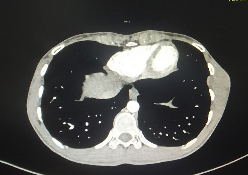
European Journal
of Case Reports in
Internal Medicine
CASE DESCRIPTION
An 18-year-old man presented with a 4-month history of an asymptomatic swelling on the left hemithorax. Needle aspiration in the second
month was inconclusive but aggravated the size of the swelling and pain developed. Physical examination disclosed a swelling measuring 4×2
cm confined to the left posterolateral aspect of the hemithorax. The plane of the swelling was intramuscular with no obvious thrill palpable
or bruit heard on auscultation. A posterior-anterior (PA) chest x-ray showed soft tissue opacity in the left lateral chest wall (Fig. 1). Contrast-
enhanced computed tomography (CECT) revealed a well-defined 4.1×1.5×4.2 cm homogeneously non-enhancing lesion in the left lateral
chest wall arising from the seventh intercostal space with no feeding vessel, bony erosion or sclerosis (Fig. 2). Thus, the differential diagnoses
included glomus tumour, neurofibroma and schwannoma. We did not perform magnetic resonance imaging (MRI) as it was expensive and
the lesion was well localized and delineated on CECT.
.
Figure 1. Chest X Ray Figure 2. Contrast-enhanced computed tomography of the thorax
Under general anaesthesia with endotracheal intubation, the patient underwent wide local excision of the swelling with tumour-free margins
followed by reconstruction of the resected portion with an overlay of latissimus dorsi muscle. The postoperative course was uneventful.
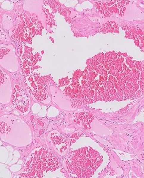
The gross specimen was a grey-brown mass of tissue measuring 6×4.5×4 cm with fatty tissue material with a homogenous yellowish-white
appearance on the cut section (Fig. 3). Histopathological examination revealed dilated and congested vascular vessels with fibrous stroma
suggesting cavernous haemangioma (Fig. 4). Regular follow-up for a 1 year did not show any recurrence.
.
Figure 3. Gross specimen
DOI: 10.12890/2021_002248 European Journal of Case Reports in Internal Medicine © EFIM 2021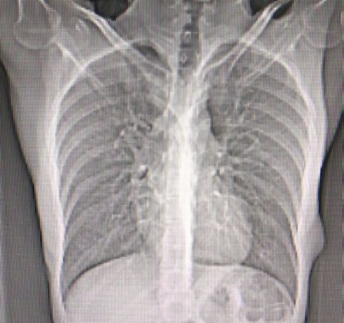
European Journal
                                                                of Case Reports in
                                                            Internal Medicine

.

                                                                         Figure 4. Microscopy demonstrating arteriovenous malformations

DISCUSSION
Chest wall tumours are rare and account for less than 1% of all tumours [2]. Haemangiomas are uncommon and arise outside the rib cage,
seldom originating from intercostal muscle. Watson and McCarthy suggested that they are congenital and traumatic in origin [2]. Although
chronic liver disease and hormonal imbalance have definite roles in the development of the lesion, a congenital origin is the most common
aetiology. Haemangiomas are asymptomatic, but pain occurs as a result of compression of the adjacent intercostal nerve or intervening
neurovascular bundle. Histologically, they are categorized into five subtypes: capillary, cavernous, venous, arteriovenous or mixed [3].
Preoperative evaluation with a detailed medical history, clinical examination and radiological investigations is necessary. Imaging, especially
CECT and MRI, plays an important role in the diagnosis. CECT for haemangiomas shows a heterogeneous mass with a low level of attenuation
due to fatty, fibrous and vascular tissue elements with phleboliths in 30% of cases [4]. The presence of a feeding vessel on CT angiography
paves the way for the diagnosis of a vascular lesion. Elbawab et al. [5] in their report demonstrated a vascular mass with a feeding vessel
supplied by a branch of the internal mammary artery. Our case presentation was similar to theirs, but the lack of a vascular element in the
CT images directed us towards a differential diagnosis of lesions of non-vascular origin. CT of glomus tumours reveals a soft tissue mass with
erosion of the adjacent bone. Heterogenous enhancement has been seen with neurofibromas, while with schwannomas, attenuation has
been slightly equal to or greater than the muscle [4]. MRI is the preferred modality for the evaluation of chest wall tumours. It can accurately
delineate the tumour and enable characterization of tumour tissue. For haemangiomas, MRI demonstrates a homogenous mass with signal
intensity similar to that of skeletal muscle on T1-weighted and high signal intensity on T2-weighted images. [2]. For glomus tumours, MRI
shows a tumour displacing a major vessel, encircled by tortuous vessels arborizing from a vascular pedicle [4]. The characteristic target
sign on T2-weighted images is commonly seen in neurofibromas, and has been reported in up to 50% of schwannomas [4]. Since there is a
paradox in the diagnosis of these lesions, histopathological confirmation along with preoperative radiological investigations is of paramount
importance. However, when clinical and radiological investigations remain inconclusive, histopathological examination following excision
biopsy determines the exact aetiology.
Wide local excision with a tumour-free margin is an accepted treatment of choice [2]. Reconstruction of the chest wall should be considered
in lesions involving a major portion of the chest wall (three or more ribs) or an anterolateral defect of more than 5 cm [3]. Various traditional
techniques including muscle flap, meshes (biologic/titanium) and methyl methacrylate have been used for chest wall reconstruction, but
more recently titanium plates have been used as a prosthetic device as they are biologically inert, less corrosive, resistant to traction and

DOI: 10.12890/2021_002248                                                        European Journal of Case Reports in Internal Medicine © EFIM 2021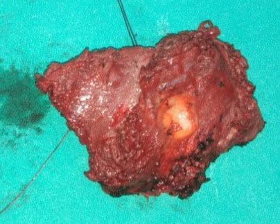
European Journal
                                                                                of Case Reports in
                                                                           Internal Medicine

compatible with MRI [6]. Preoperative embolization is palliative and may lead to tumour debulking or reduced bleeding, but surgical excision
is mandatory in order to prevent the recruitment of a collateral blood supply [2]. Transformation to malignancy or recurrence is rarely seen.

CONCLUSION
Despite their rarity, intercostal muscle haemangiomas must be considered as a differential diagnosis in chest wall tumours even in the absence
of a feeding vessel. We believe that histopathology provides a definitive diagnosis when most investigative procedures are inconclusive.

REFERENCES

1.   Scott JE. Haemangioma in skeletal muscle. Br J Surg 1957;44:496–501.
2.   Ulku R, Onat S, Avci A, Ozmen CA. Resection of intercostal hemangioma with involved chest wall and ribs: in an 11-year-old girl. Tex Heart Inst J 2010;37(4):486–489.
3.   Dzian A, Hamzík J. Intercostal hemangioma of the chest wall. Kardiochir Torakochirurgia Pol 2016;13(1):58–60.
4.   Nam SJ, Kim S, Lim BJ, Yoon CS, Kim TH, Suh JS, et al. Imaging of primary chest wall tumors with radiologic-pathologic correlation. Radiographics 2011;31(3):749–770.
5.   Elbawab H, Alreshaid F, Hashem T, Alnasser A, Husain R, Aljehani Y. Intercostal hemangioma: case report of a rare chest wall tumor in childhood. Int J Surg Case Rep 2019;60:319–
     322.
6.   Sanna S, Brandolini J, Pardolesi A. Materials and techniques in chest wall reconstruction: a review. J Vis Surg 2017;3:95.

DOI: 10.12890/2021_002248                                                                            European Journal of Case Reports in Internal Medicine © EFIM 2021
You can also read