International Conference on Biophotonics 2019 - St Andrews, UK Programme Booklet - ICOB 2019
←
→
Page content transcription
If your browser does not render page correctly, please read the page content below

International Conference on Biophotonics 2019
St Andrews, UK
Old Course Hotel

22-24 MAY 2019

Programme Booklet
We thank our sponsors:

                  Exclusive International Sponsor

                                    And:

Meeting Chair             International committee   Local Committee

Kishan Dholakia           Dennis Matthews           Adrià Escobet-Montalbán

                          Malini Olivo              Holly Fleming

                          Juergen Popp              Malte Gather

                          David Sampson             Frank Gunn-Moore

                          Brian Wilson              Sandra Murray

                                                    Philip Wijesinghe
Dear Participant,
Welcome to St. Andrews and to the International Conference on Biophotonics
2019 (ICOB2019). We hope that you will enjoy the meeting and your stay in St.
Andrews.
Biophotonics is well recognised as an important area, having major implications
in a wide range of interdisciplinary science and biomedicine. This area denotes
the use of advanced photonics-based technologies with the aim of providing
new methodologies for biologists and clinicians alike, with potential benefits in
healthcare and for research into cell and molecular biology. ICOB2019 aims to
bring together internationally recognised researchers to discuss their work and
various exciting new breakthroughs in the field.
We thank SPIE., Coherent (UK), EPSRC, M Squared, Nikon Instruments, SULSA,
SUPA, and the University of St. Andrews for their sponsorship of the meeting.
This booklet contains a programme and abstracts of the talks and posters for
the meeting and details about the arrangements for the two days.
Once again, welcome to St. Andrews and to ICOB2019. We hope you enjoy the
meeting.
With warmest regards,
Kishan Dholakia
Contact details:
Email: icob2019@st-andrews.ac.uk
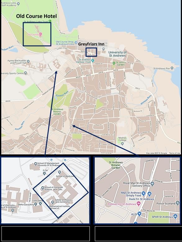
1Premier Inn to Old Course Hotel – 20-25 min walk
Greyfriars Inn to Old Course Hotel – 10 min walk
2Old Course Hotel Ground Floor Plan
3Meeting Arrangements
The whole meeting, including all lunches and the conference dinner, will be held at the Old Course
Hotel. Sign posts are placed at various points around the hotel to guide you to various locations.
Registration desk
This is located in the Hamilton Grand. Sandra Murray and Holly Fleming will be available for registra-
tion from 08.15 am on Wednesday 22nd May, 08.00 am on Thursday 23rd May, and 09.00 am on Fri-
day 24th May.
Talks
All talks are held in The Hall of Champions.
Coffee/tea breaks are just outside The Hall of Champions.
Posters
Poster boards will be in The Hall of Champions: we ask all those presenting a poster to please check
their poster number in this booklet and place their poster up by 14.15 on Wednesday 22nd. There will
be poster prizes awarded for the best posters presented by early career researchers (e.g. PhD stu-
dents and post docs).
Lunch
Lunch will be served in the Sands Restaurant of the Old Course Hotel.
Welcome Reception
The Welcome Reception will take place in the Ballroom on Wednesday 22nd at 18.30.
Conference Dinner
There will be pre-dinner drinks served in the Conservatory at 19.00.
The conference dinner and ceilidh will be held in the main Ballroom of the Old Course Hotel, com-
mencing at 19.20 sharp. A seating plan will be provided, please check this prior to the meal. The
seating plan will be available outside the Hall of Champions on Thursday afternoon.
Photos
Please note that photos and videos of may be taken during the meeting for publicity and marketing
purposes.
4SHORT PROGRAMME
Wednesday 22nd (full day)
8.15 - 9.00 Registration (coffee and pastries)
9.00 - 9.10 Welcome Kishan Dholakia
9.10 - 10.00 Plenary
Chair: Kishan Dholakia
Brian Pogue Dartmouth College, USA
Optical Guidance in Surgery & Radiation Therapy
10.00 - 10.50 SPIE Plenary
Chair: Kishan Dholakia
Andrew Brown SPIE, USA
An SPIE View of Trends in Biophotonics
10.50 - 11.10 Discussion
11.10 - 11.30 Coffee
11.30 - 13.00 GLOBAL HEALTH AND INFECTION
Chair: Brian Wilson and Dennis Matthews
11.30 - 12.00 Andrew Blaikie University of St Andrews, UK
The Arclight - "Less is More" - a medical diagnostic tool for low re-source countries
12.00 - 12.30 Beth Mills University of Edinburgh, UK
The development of fluorescence-based point-of-care diagnostics – design considerations for use in
low resource settings
12.30 - 13.00 Juergen Popp IPHT, Jena, Germany
Photonics for Infection
13.00 - 13.15 Discussion
13.15 - 14.15 Lunch, Sands Restaurant
Exhibition and Posters
14.15 - 15.15 ICOB Hot Topics Session
Chair: David Sampson
Thomas Krauss University of York, UK
Nanophotonic biosensors-clever photonics in a small package
Malini Olivo A*STAR, Singapore
Skin inflammation imaging using Raster Scanning Optoacoustic Imaging and its quantitative
analysis
Kirill Larin University of Houston, USA
Translational dynamic optical coherence elastography
Isla Barnard University of St Andrews, UK
Simulating Light-Tissue Interactions with MCRT
5SHORT PROGRAMME
15.15 - 16.15 Breakout: Global Health and Infection
Discussion Chairs: Dennis Matthews, Brian Wilson and Juergen Popp
16.15 - 17.15 Poster and Exhibition Session: with coffee and refreshments
17.15 - 18.30 Free time
18.30 - 20.00 Welcome Reception, Ballroom
Canapes and Drinks will be served
Thursday 23rd (full day)
8.00 - 8.30 Registration (coffee and pastries)
8.30 - 10.00 ENVIRONMENT, FOOD AND DRINK
Chair: Juergen Popp
8.30 - 9.00 Andrew Abell CNBP, University of Adelaide, Australia
Light activated molecular switches in chemical biology
9.00 - 9.30 Kate Bechtel Triple Ring Technologies, USA
Bridging the gap: what researchers can do to better the chances of successful transition from
prototype to product
9.30 - 10.00 Oliver Valet mibic GmbH & Co. KG, Germany
Industrial and Academic Applications of a Smart Single Microbe Raman Test Platform
10.00 - 10.15 Discussion
10.15 - 10.30 Coffee
10.30 - 11.30 Breakout: Environment, Food and Drink
Discussion Chair: Juergen Popp
11.30 - 12.15 FUTURE TRENDS IN BIOPHOTONICS (1)
Chair: Halina Rubinsztein-Dunlop
11.30 - 11.45 Kishan Dholakia University of St Andrews, UK
Future perspectives for imaging at depth
11.45 - 12.15 David Sampson University of Surrey, UK
Polarisation-sensitive optical coherence tomography – here it comes again
12.15 - 13.15 Lunch, Sands Restaurant
Exhibition and Posters
6SHORT PROGRAMME
13.15 - 14.45 FUTURE TRENDS IN BIOPHOTONICS (2)
Chair: Halina Rubinsztein-Dunlop
13.15 - 13.45 Chris Xu Cornell University, USA
Deep and fast multiphoton microscopy
13.45 - 14.15 Daniele Faccio University of Glasgow, UK
Deep-imaging with time-of-flight diffusive optical tomography
14.15 - 14.45 Andy Yun Massachusetts General Hospital, Boston, USA
Laser particles for multiplexed cell tagging
14.45 - 15.00 Discussion
15.00 - 15.15 Coffee
15.15 - 16.00 Breakout: Future Trends in Biophotonics
Discussion Chair: Kishan Dholakia
16.00 - 17.30 Translation and Entrepreneurship Session
Panel:
Ignatius Rasiah National University of Singapore, Singapore
Dennis Matthews UC Davis, USA
Brian Wilson University of Toronto, Canada
Kate Bechtel Triple Ring Technologies, USA
19.00 Pre-dinner drinks, Conservatory
19.20 Conference Dinner, Ballroom
After dinner speaker: Miles Oglethorpe Historic Environment Scotland
Bridging the past with the future
Friday 24th (Half day, ends after lunch)
9.00 - 9.30 Registration (coffee and pastries)
9.30 - 11.00 SHAPING PHOTONICS FOR NEUROSCIENCE
Chair: Malini Olivo
9.30 - 10.00 Silvia Paracchini University of St Andrews, UK
Shedding light on language related disorders
10.00 - 10.30 Halina Rubinsztein-Dunlop University of Queensland, Australia
Sculpted light for quantitative imaging of nano and microsystems
10.30 - 11.00 Malte Gather University of St Andrews, UK
Microresonators and nanolasers to explore the biomedical world
11.00 - 11.15 Discussion
11.15 - 11.30 Coffee
7SHORT PROGRAMME
11.30 - 12.30 Breakout: Shaping photonics for neuroscience
Discussion Chair: Malini Olivo
Time to discuss and prepare white papers
12.30 Concluding remarks
Lunch, Sands Restaurant
8TALK ABSTRACTS
Wednesday 22nd (full day) years. Unfortunately only very few practitioners in low and mid-
dle-income countries (LMICs) have these essential tools. If they
Brian Pogue Dartmouth College, USA do own devices they are typically ‘hand-me-downs’ from well-
minded donors but often not functional being dependent upon
Optical Guidance in Surgery & Radiation Therapy hard to find and expensive consumables such as bulbs and batter-
ies. Sadly the vast majority of cases of vision and hearing impair-
The process of imaging medical treatments today is dominated by ment are however found in LMICs where access to diagnostic
optical devices which are used at the point of care, in settings tools is least. The Lancet Commission’s report on Technologies
such as surgery and endoscopy. These procedure-based tools are for Global Health has consequently recommended greater focus
used together with radiologic devices to capture unique contrast on ‘frugal technologies’ designed for the needs of LMICs rather
features that help guide medical decisions about tissue removal than the markets of wealthy countries. We describe the inspira-
and tissue response to therapy. In the developments in technolo- tion, development and potential of the Arclight a novel low cost
gies and nanotechnologies within the world of Optics in Medicine ‘frugal’ diagnostic tool for prevention of vision and hearing im-
have made major advances, such as image-guided spectroscopy pairment.
during surgery, as well as surgical guidance navigation tools, and
now radiologic guidance tools. Examples from each will be used
to highlight innovations in translational research that have gone Beth Mills University of Edinburgh, UK
from concept through to clinical trials, and now through to multi-
center trial use. Translation beyond the initial feasibility phase The development of fluorescence-based point-of-care
involves the type of R&D which only companies can accomplish, diagnostics – design considerations for use in low
and so partnerships with companies in translational research has resource settings
been paramount, and examples in surgical guidance can show
The widespread availability of low-cost, yet robust LEDs and
this. Translation through a start-up company, DoseOptics LLC, will
CMOS cameras, coupled with the advancement of pathogen-
be highlighted in which this pathway has enabled testing and
specific fluorescent imaging agents augment the development of
deployment of a fundamentally new technology to image radia-
frugal diagnostic platforms. Learning from wide-field optical en-
tion dose delivery in real time.
doscopy technology developed within the Proteus project for
interrogating the pathology of the distal lung in real-time (Pro-
Andrew Brown SPIE, USA teus.ac.uk), we are working with partners in India to expand our
repertoire of optical point-of-care diagnostic platforms for aetiol-
An SPIE View of Trends in Biophotonics ogies designated (by them) as important for rapid diagnosis with-
in their communities. These conditions include skin, corneal and
Photonics West/BiOS is the largest annual biomedical optics con-
urinary tract infections. Designing and implementing technologies
ference and exhibition in the world and has seen a 24% increase
in rural primary care settings globally comes with challenges at
in the number of papers presented there over the last five years.
the technical, financial and personnel levels. Often these re-
All indications are that this rapid growth of biophotonics research
quirements are overlooked by technologists, leading to the un-
and development will continue, driven by an increasingly broad
der-performance of many devices. Through stakeholder engage-
range of light-based applications to healthcare, and the growing
ment in India we have come to acquire a detailed understanding
market penetration of cost-effective photonics-based diagnostic
of end-user requirements as we develop our technologies to
and therapeutic medical devices. These devices range from ad-
ensure their applicability within the field.
vanced gene sequencing systems to wearables capable of real-
time monitoring of physiological health parameters. In this
presentation, we highlight some of the more exciting biomedical Juergen Popp IPHT, Jena, Germany
optics trends that are evident from recent BiOS conferences. We
also discuss some of the market challenges that are inherent in Photonics for Infection
realizing the promise of biophotonics for better health.
Infectious diseases are one of the major reasons of deaths
worldwide. Successful treatment of infection relies on a timely
Andrew Blaikie University of St Andrews, UK identification of the infectious pathogen and its antibiotic re-
sistance pattern to select the appropriate antibiotic treatment as
The Arclight - "Less is More" - a medical diagnostic tool early as possible. Here, we will highlight the potential of photonic
for low resource countries approaches for the fast bedside identification of pathogens to-
gether with their antibiotic resistances. The challenges to over-
There are estimated to be 285 million visually and 360 million come the valley of death to apply such novel photonic microbial
hearing impaired people in the world with the majority of cases analysis approaches for clinical routine requires novel infrastruc-
considered preventable or treatable if diagnosed promptly. Oph- tures. To reach this goal, we established the research campus
thalmoscopes and otoscopes are typically designed for markets InfectoGnostics to safeguard the transfer from fundamental re-
of wealthy countries and are complex, heavy and expensive with search into diagnostic systems.
their basic design remaining relatively unchanged for over 100
9TALK ABSTRACTS
ICOB Hot Topics Session Kirill Larin University of Houston, USA
Thomas Krauss University of York, UK Translational dynamic optical coherence elastography
Nanophotonic biosensors-clever photonics in a small The biomechanical properties of tissues can be dramatically al-
package tered by various diseases, such as keratoconus for the cornea of
the eye and systemic sclerosis for the skin. Therefore, the ability
Resonant nanophotonic concepts offer interesting opportunities to measure tissue biomechanical properties could provide critical
for sensing and imaging applications. Our work focuses on guided information for assessing its health and detecting disease etiolo-
mode resonances (GMRs), because they are easy to excite with gy as well as monitoring disease progression. Here, I will present
out-of plane illumination. By chirping the grating, we have been pilot results in development of noncontact dynamic optical co-
able to integrate the sensing and the readout function. Using herence elastography (OCE) technique to evaluate the biome-
nanohole arrays in silicon, we have obtained remarkable results, chanical properties of the cornea and skin of healthy subjects and
including a very high surface sensitivity which is comparable to those affected by diseases.
that of the plasmonic equivalent. Finally, we are now implement-
ing common-path interferometric methods which further in-
crease the available sensitivity. As a result, our detection limit is Isla Barnard University of St Andrews, UK
now approximately 1e-6 refractive index units, which is compa-
Simulating Light-Tissue Interactions with MCRT
rable to much more sophisticated approaches yet still compatible
with a handheld device. Monte Carlo radiative transfer (MCRT) methods use localised
scattering and absorption probabilities to describe the path of
photon packets through a medium, and are are ideally suited to
Malini Olivo A*STAR, Singapore
simulating radiation transfer through complex structures. An
Skin inflammation imaging using Raster Scanning ongoing collaboration between the School of Physics and Astron-
omy, St Andrews, and the Photobiology department at Ninewells
Optoacoustic Imaging and its quantitative analysis
Hospital & Medical School, Dundee, has developed expertise in
According to a recent clinical survey in Singapore, 1 in 5 kids and modelling light-skin interactions using modified astronomical
1 in 10 adults is affected by chronic skin inflammation condition MCRT computer codes. One such project, initiated by a clinician,
such as Eczema or Atopic dermatitis. This has caused poor quality investigates spectrally resolved skin penetration depths achieved
of life of those affected and substantial economic burden on Sin- by different phototherapy radiation sources.
gapore healthcare system. Currently, eczema is qualitatively
scored based on clinical questionnaires and it does not reflect the
any sub skin surface characteristics such as morphology, vascular
architecture and changes in epidermis thickness. Non-invasive
imaging techniques for assessing this change in skin vascular
Thursday 23rd (full day)
structures could potentially serve as an objective indicators to
characterize its severity that can help in developing an effective Andrew Abell CNBP, University of Adelaide
treatment procedure. Herein, for the first time, we present the Australia
preliminary results from an ongoing clinical study at Singapore
National Skin Center, using non-invasive raster scanning optoa- Light activated molecular switches in chemical biology
coustic mesoscopy (RSOM) imaging approach, which can combine The problem with biological probes and sensors is that they lack
the deep tissue interrogation of ultrasound and high contrast of an ability to be switched on and off and to present dual function-
optical techniques. Using RSOM imaging, we could objectively ality, such as the delivery of a therapeutic response following
characterize the severity of eczema by visualizing the skin mor- sensing or extended imaging capacity. Organic fluorescent probes
phology and vascular pattern changes in the dermis and sub-
also generally lack photostability required for extended intracel-
dermis of patients enabling a quantification of inflammation in a lular imaging. A hybrid nanomaterial is reported with an organic
label free manner. fluorescent probe bound to a nanodiamond for concurrent and
extended cell-based imaging and ratiometric detection of hydro-
gen peroxide. Far-red fluorescence of the nanodiamond offers
continuous imaging without photobleaching, while green fluores-
cence of the attached probe detects hydrogen peroxide on de-
mand. This nanosensor allows extended bio-imaging not possible
with a standalone organic fluorescent probe. Recent work is also
presented on developing photoswitchable antibiotics and prote-
ase inhibitors; sensors for detecting metal ions, Glutathione, NO,
and protein-protein interactions; and also surface bound drug
delivery systems and molecular devices.
10TALK ABSTRACTS
Kate Bechtel Triple Ring Technologies, USA Kishan Dholakia University of St Andrews, UK
Bridging the gap: what researchers can do to better the Something cool in physics on the day
chances of successful transition from prototype to product
Optically based methods for imaging have emerged as very pow-
While most researchers dream of seeing their technology utilized erful routes for biomedical discovery. In this talk I will describe
by those who need it, many would rather not be involved in as- routes for deeper penetration into tissue that allow the recovery
pects of commercialization unrelated to their PhD – regulatory of wide field images yet minimise photodamage. I will review
pathway, reimbursement/revenue strategy, cost of goods, etc. some emergent areas in Biophotonics and focus on the use of
However, much work remains in the area of science that is often temporal focusing for multiphoton imaging at depth. This ap-
overlooked. This presentation focuses on the critical gaps that proach enables us to retrieve images through scattering media in
exist when a technology is released too early from the lab and the absence of any form of aberration correction.
what researchers can do to fill those gaps and better position the
technology for development.
David Sampson University of Surrey, UK
Oliver Valet mibic GmbH & Co. KG, Germany Polarisation-sensitive optical coherence tomography –
here it comes again
Industrial and Academic Applications of a Smart Single
Polarisation-sensitive optical coherence tomography (PS-OCT)
Microbe Raman Test Platform
has a long history, dating back to the early 1990s when the field
Non-growth based methods for the phenotypic investigation of of OCT began. Alteration of the polarisation state of light by bio-
microorganisms provide rapid and label free direct access to logical tissue is an appealing source of contrast, and polarised
identity determination, vital metabolic functions and the study of light microscopy has an even longer history, and continues to
microbial interactions with substances. We present academic and evolve today. PS-OCT probes the presence and arrangement of
industrial applications of our software-controlled fully automated fibrous structures, such as collagen or muscle cells, and the shape
modular instrument concept GRAM-RAY + HESSE-X. For industrial of scatterers, such as cells. It is complicated by the influence of
applications, the liquid handling system with centrifuge (HESSEX) the propagation path on the contrast, and the complex physics of
is used for reproducible sample preparation from complex matri- polarised light-soft tissue interactions. Translational applications
ces. A wine producer detects living yeast and harmful organisms have been pursued for some time in glaucoma, tumour, and
in wine and controls up to 300 samples per day with HESSE-X and burns assessment and wound monitoring. Why has PS-OCT, then,
the micro Raman GRAM-RAY unit. A manufacturer of cooling not made more of a mark to date? In this talk, we will describe
lubricants is investigating the effectiveness of novel biocides by what has changed recently, and why we expect polarisation-
high-throughput in vivo tests. Academic applications use the based contrast to demonstrate great advances over the coming
highest sensitivity in single cell identification for the investigation years.
of zoonoses and detection of pathogens from beverages and
food. Vibriones such as Colera are living, difficult-to-cultivate
germs (VBNC) and these can be reliably detected with GRAM-RAY
Chris Xu Cornell University, USA
without a previous cultivation step. Their metabolism is exam- Deep and fast multiphoton microscopy
ined by stable isotopes Raman (SIRM). In addition to liquid han-
dling and centrifugation of the samples, HESSE-X also produces Multiphoton microscopy is the go-to technique for high spatial
silver nano colloids in situ. With the GRAM-RAY system, the via- resolution, deep imaging in scattering biological tissue. Multipho-
bility of bacteria can be directly investigated using SERS. The suit- ton imaging will likely play an essential role in understanding how
ability of potential antibiotics for specific bacteria was deter- the brain works at the level of neural circuits. In this talk, the
mined within only 1 hour. By visualizing the interaction of an fundamental challenges of deep tissue, high-resolution optical
antibiotic with the bacteria membrane, the mechanisms of anti- imaging are discussed. New technologies for in vivo structural
biotic resistance are elucidated. and functional imaging of mouse brain using long wavelength
excitation and three-photon microscopy will be presented. We
will illustrate the requirements for imaging the dynamic neuronal
activity at the cellular level over a large area and depth in awake
and behaving animals, and show applications where 3-photon
microscopy outperforms conventional 2-photon microscopy in
both signal strength and image contrast. Finally, we will discuss
several future directions, including adaptive optics and new laser
sources, to further improve the imaging depth and speed in bio-
logical tissues.
11TALK ABSTRACTS
Daniele Faccio University of Glasgow, UK Halina Rubinsztein-Dunlop University of
Queensland, Australia
Deep-imaging with time-of-flight diffusive optical
tomography Sculpted light for quantitative imaging of nano and
microsystems
An outstanding challenge in optical medical imaging is the ability
to image small objects or regions that a embedded in deep tissue, Use of spatial light modulators enables unprecedented control
where by “deep” here we refer to samples that are 5-20 cm thick. and highest versatility of light. We can now structure or sculpt
The scattering coefficients of typical biological (e.g. brain) tissue light in such a way that it enables control of matter, studies of
imply that with these thicknesses we are in the diffusive regime light-matter interactions, development of new and improved
where light propagates similarly to heat. Moreover, over these imaging techniques and many other applications. It is used in
distances ballistic or snake photon are essentially undetectable many fields and at scales ranging from nano to microsystems,
and cannot be used for imaging. Different approaches must from quantum physics to studies of complex biological systems.
therefore be searched for that typically rely on computational The use of sculpted light can vastly improve image resolution as
inversion to reconstruct an image of embedded objects. We re- well as enable applications of much stronger optical forces to the
port on a novel computational technique that can image deeply system under study. It provides valuable tool to studies in bio-
embedded objects using light transmitted through the opaque photonics.
material and measured using picosecond time-resolving single-
photon cameras.
Malte Gather University of St Andrews, UK
Andy Yun Harvard Medical School and MGH Microresonators and nanolasers to explore the
Boston, USA biomedical world
Laser particles for multiplexed cell tagging Optical resonators provide one of the most accurate rulers
known to mankind. In this talk I will summarize our recent work
Laser particles that have a size of an optical wavelength in all on translating this capability to studies of cells and tissue. In one
three dimensions and emit distinct narrowband spectra are new, example, we use ultra-soft elastic Fabry-Perot micro-resonators
promising optical probes for biomedical applications. We present as substrates for cells and interferometrically detect the forces
biocompatible semiconductor laser disks suitable for tracking cells apply to these – with piconewton precision and at video-rate
many cells for imaging and single-cell analysis. speed. This has enabled direct observation of biophysical pro-
cesses relevant to cancer invasion, immune cell migration, kidney
failure, cardiac contraction and others. In another example, we
integrated nanoscale lasers into live cells and used dense spectral
multiplexing of multiple lasers to tag and track individual cells
Friday 24th (Half day, ends at lunch) within large cell populations and over extended periods of time.
We recently developed this further to perform in vivo intracellu-
Silvia Paracchini University of St Andrews, UK lar sensing, e.g. to measure contractility in the beating heart.
Shedding light on language related disorders
Language-related disorders like dyslexia affect 5-10% of children
and are caused by a significant genetic component (~70%). So far,
very few underlying susceptibility genes have been identified and
for those it is very difficult to dissect the role in diseases. We
apply a range of assays based on the properties of light to under-
stand the role of genes associated to disorders during neurode-
velopment. These assays are allowing us to study tissues-specific
patterns of expression since the very early phases of develop-
ment; to fine-tune expression regulation and study the down-
stream effects of lower/higher expression; and study the effect of
genes on the mechanical properties of cells. Gene function char-
acterization is a major challenge for translational research de-
rived from genetic discoveries. These assays provide new ave-
nues to advance the field of complex trait genetics.
12POSTER LIST
POSTER LIST
P1 Investigating the forces in heterogeneous cultures of cancer cells through resonant photonics
Dinesh Kumar, University of St Andrews
P2 Super-resolution label-free non-linear techniques for bio-imaging
Peter Johnson, University of Southampton
P3 Interferometric sensing with guided-mode resonances
Isabel Barth, University of York
P4 Artificial neural networks for light delivery through complex media
Alex Turpin, University of Glasgow
P5 Label-free optical hemogram of granulocytes enhanced by artificial neural networks
Roopam Gupta, University of St Andrews
P6 Highly sensitive label-free biosensing with all-dielectric nanohole arrays
Donato Conteduca, University of York
P7 Label-free imaging for light sheet microscopy
Niall Hanrahan, University of Southampton
P8 Podocyte injury elicits loss and recovery of cellular forces
Paul Reynolds, University of St Andrews
P9 Light-sheet microscopy for histopathology
Stella Corsetti, University of St Andrews
P10 Organic light-emitting diode photostimulation for optogenetics
Andrew Morton, University of St Andrews
P11 Spectral unmixing of oxygen abundance for dye-free retinal angiography
Tomasz Tkaczyk, Rice University
P12 Spatially offset optical coherence tomography
Philip Wijesinghe, University of St Andrews
P13 TRAFIX: Multiphoton imaging through scattering media
Adrià Escobet Montalbán, University of St Andrews
P14 Through Bottle Sensing using Advanced Raman Geometries
Holly Fleming, University of St Andrews
P15 Semiconductor intracellular nanolasers
Alasdair Fikouras, University of St Andrews
P16 Human salivary Raman fingerprint as biomarker for the diagnosis of Amyotrophic Lateral Sclerosis
Cristiano Carlomagno, Fondazione Don Gnocchi
P17 Spectrally and spatially resolved depth penetration achieved by phototherapy lamps
Isla Barnard, University of St Andrews
13POSTER LIST
P18 Microlaser-based contractility sensing in single cardiomyocytes and whole hearts
Marcel Schubert, University of St Andrews
P19 Fabrication of lasing networks for enhanced light-matter interaction
Soraya Carlos Caixeiro, University of St Andrews
P20 Multiphoton excitation in light sheet microscopy
Federico Gasparoli, University of St Andrews
P21 Podocyte injury elicits loss and recovery of podocyte cellular forces
Nils Kronenberg, University of St Andrews
P22 High-throughput analysis of individual protein aggregates in human cerebrospinal fluid
Juan Varela, University of St Andrews
P23 Quantitative phase and polarisation endoscopy applied to detection of early oesophageal
tumourigenesis
George Gordon, University of Nottingham
P24 Tissue recognition in gastro intestinal tract using diffuse reflectance spectroscopy
Siddra Maryam, Tyndall National Institute
P25 Real-time imaging of cellular forces with piconewton precision
Andrew Meek, University of St Andrews
P26 Shining a light on mesoscale signalling in the vascular endothelium
Larry Fitzpatrick, Durham University
P27 Measurement and analysis of invadopodia forces in 2D and 3D environments
Eleni Dalaka, University of St Andrews
P28 Numerical model of laser tissue ablation and thermal injury
Lewis McMillan, University of St Andrews
P29 Numerical comparison of robustness of multimode and multicore fibre sensitivity against fibre
bending
Madhu Veettikazhy, Technical University of Denmark
P30 Pushing the limits of label-free non-linear techniques for bio-imaging
Sumeet Mahajan, University of Southampton
P31 MAGNIFI - Margin assessment using global non-invasive fluorescence imaging
Khushi Vyas, Imperial College London
P32 Direct measure of the effect of Isoniazid on M. smegmatis using acoustic trapping combined with
wavelength modulated Raman spectroscopy.
Vincent Baron, University of St Andrews
P33 iPlacenta - tackling the challenge of imaging vasculature endoscopically
Lukas Markwalder, University of Dundee
P34 Multiparameter susceptibility test based on hydrodynamic trapping of individual E. coli
Giampaolo Pitruzzello, University of York
14NOTES
15NOTES 16
You can also read