IntRIS 2019 International Retinal Imaging Symposium 2019 - Saturday, March 16, 2019 - UCLA CME
←
→
Page content transcription
If your browser does not render page correctly, please read the page content below
UCLA Stein Eye Institute
and Doheny Eye Institute
present the
International
Retinal Imaging
Symposium
2019
IntRIS 2019
Saturday, March 16, 2019
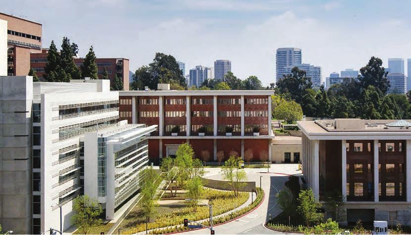
California NanoSystems Institute
at UCLA
570 Westwood Plaza
Los Angeles, CA 90095
www.cme.ucla.eduT
he International Retinal Imaging Symposium (IntRIS 2019) will feature
a series of lectures devoted to advanced retinal imaging and the
newest developments in this exciting field. Innovative systems such as
fundus autofluorescence, ultra-widefield imaging, spectral domain and swept
source optical coherence tomography (OCT), and OCT angiography will all be
covered. A groundbreaking session on artificial intelligence and deep learning
will also be highlighted in the program. IntRIS 2019 will include a full day
of lectures by many of the world’s experts in retinal imaging who will speak
on recent innovations in retinal imaging that have occurred in this rapidly
advancing field.
Our world-renowned faculty will aim to familiarize course participants with
the newest evolving technologies and will guide and instruct our registrants
in the application of these advanced systems and in the interpretation of
novel and challenging imaging findings. This will help our participants better
manage their patients with macular and retinal disorders and achieve better
patient outcomes in their practices.
We welcome your participation at IntRIS 2019 which promises to provide
insight and understanding in retinal imaging and showcase the integral
importance of innovative retinal imaging in the evaluation and management
of retinal disease.
David Sarraf, M.D.
K. Bailey Freund, M.D.
SriniVas Sadda, M.D.DIRECTORS David Sarraf, M.D. SriniVas Sadda, M.D. K. Bailey Freund, M.D. SPEAKERS Thomas Ach, M.D. Won Ki Lee, M.D. Francesco Bandello, M.D. Belinda Leong, M.D. Jiwon Baek, M.D. Brandon Lujan, M.D. Steven Bailey, M.D. William Mieler, M.D. Paul Bernstein, M.D., Ph.D. Eduardo Navajas, M.D. Barbara Blodi, M.D. Francesco Pichi, M.D. Enrico Borrelli, M.D. Maximillian Pfau, M.D. Vittorio Capuano, M.D. Rony Preti, M.D. Usha Chakravarthy, M.D. Giuseppe Querques, M.D., Ph.D. Gemmy Cheung, M.D. Tushar Ranchod, M.D. Netan Choudhry, M.D. Kasra Rezaei, M.D. Christine Curcio, Ph.D. Richard Rosen, M.D. Rosa Dolz Marco, M.D., Ph.D. Philip Rosenfeld, M.D., Ph.D. Amitha Domalpally, M.D. SriniVas Sadda, M.D. Chantal Dysli, M.D. David Sarraf, M.D. Amani Fawzi, M.D. Steffen Schmitz-Valckenberg, M.D. Anibal Francone, M.D. J. Sebag, M.D. K. Bailey Freund, M.D. Eric Souied, M.D., Ph.D. James Fujimoto, Ph.D. Richard Spaide, M.D. Alain Gaudric, M.D. Giovanni Staurenghi, M.D. Isaac Gendelman, M.D. Natarajan Sundaram, M.D. Frank Holz, M.D. Nadia Waheed, M.D. Michael Ip, M.D. Ruikang Wang, Ph.D. Yali Jia, Ph.D. Lawrence Yannuzzi, M.D. Jesse Jung, M.D. Young Hee Yoon, M.D., Ph.D. Jennifer Kang-Mieler, Ph.D. Seung-Young Yu, M.D., Ph.D. Pearse Keane, M.D. Suzanne Yzer, M.D., Ph.D. Eung Suk Kim, M.D. Yuhua Zhang, Ph.D. MODERATORS Alexander Brucker, M.D. Giuseppe Querques, M.D., Ph.D. Barbara Blodi, M.D. Philip Rosenfeld, M.D., Ph.D. Gemmy Cheung, M.D. SriniVas Sadda, M.D. Christine Curcio, Ph.D. David Sarraf, M.D. Amani Fawzi, M.D. Lawrence Singerman, M.D. K. Bailey Freund, M.D. Richard Spaide, M.D. Lee Jampol, M.D. Lawrence Yannuzzi, M.D. Bruno Lumbroso, M.D.
IntRIS 2019 Program
6.15 – 6.45 AM	REGISTRATION AND                         ANATOMY AND IMAGING
                    CONTINENTAL BREAKFAST                Moderator: Christine Curcio, Ph.D.
INNOVATIONS: DEEP LEARNING                               9.10 – 9.15 	SS-OCT En-Face Analysis of the
Moderator: SriniVas Sadda, M.D.                                           Vitreous Cavity
7.00 – 7.10	MEDnet, a Neural Network for                                 Belinda Leong, M.D.
                 Automated Detection of Avascular        9.15 – 9.20 	En Face OCT Identification of New
                 Area in OCT Angiography                                  Foveal Anatomical Landmark
                 Yali Jia, Ph.D.                                          David Sarraf, M.D.
7.10 – 7.20	The Moorfields-Deep Mind                    9.20 – 9.30 	Hemodynamic Response of the Three
                 Collaboration: An Update on the                          Retinal Capillary Plexuses in Dark
                 First Results                                            Adaptation and Flicker Stimulation
                 Pearse Keane, M.D.                                       using OCT Angiography
7.20 – 7.25 	Artificial Intelligence for                                 Amani Fawzi, M.D.
                 Morphology-Based Function               9.30 – 9.40 	Topographic Distribution of
                 Predictions                                              Choriocapillaris Flow Deficits in
                 Steffen Schmitz-Valckenberg, M.D.,                       Healthy Eyes
                 Maximillian Pfau, M.D.                                   SriniVas Sadda, M.D.
7.25 – 7.30 	Validation of Medios: An Offline           9.40 – 9.45 	Classification and Guidelines
                 Smartphone-Based AI in Automated                         for Widefield Imaging
                 Diabetic Retinopathy Screening                           Recommendations from the
                 using the Non Mydriatic Fundus on                        International Widefield Imaging
                 Phone (NM FOP)                                           Study Group
                 Natarajan Sundaram, M.D.                                 Netan Choudhry, M.D.
7.30 – 7.40      Discussion                              9.45 – 10.00     Discussion
INNOVATIONS: IMAGING I                                   PATHOLOGY AND IMAGING
Moderator: Richard Spaide, M.D.                          Moderator: K. Bailey Freund, M.D.
7.40 – 7.45 	Standard Retinas in Quantified             10.00 – 10.10 	Cuticular Drusen: Multimodal
                 Fundus Autofluorescence Imaging                          Imaging and Clinico-Pathological
                 – Prerequisite to Discriminate the                       Correlation
                 Diseased Retina                                          Lawrence Yannuzzi, M.D.
                 Thomas Ach, M.D.                        10.10 – 10.20 	Histologic Correlates of Optical
7.45 – 7.55 	Macular Pigment: A New Imaging                              Coherence Tomography Signatures
                 Tool for Retinal and Neurological                        in Geographic Atrophy Secondary to
                 Diseases                                                 Age-Related Macular Degeneration
                 Giovanni Staurenghi, M.D.                                Christine Curcio, Ph.D.
7.55 – 8.05 	Clinical Applications of Fluorescence      10.20 – 10.30 	Type 1 Aneurysmal NV (Polypoidal
                 Lifetime Imaging Ophthalmoscopy                          Choroidal Vasculopathy) in Age-
                 (FLIO)                                                   Related Macular Degeneration
                 Paul Bernstein, M.D., Ph.D.                              Rosa Dolz Marco, M.D., Ph.D.
8.05 – 8.10      Discussion                              10.30 – 10.40    Discussion
INNOVATIONS: IMAGING II                                  10.40 – 11.00 BREAK
Moderator: Bruno Lumbroso, M.D.                          NON-NEOVASCULAR AMD
8.10 – 8.15      Rapid Array Capture Technology          Moderator: Philip Rosenfeld, M.D., Ph.D.
                 Tushar Ranchod, M.D.                    11.00 – 11.10 	Natural History, Morphology and
8.15 – 8.20 	High Magnification Imaging in                               Impact on Photoreceptors of
                 Research and Clinical Practice                           Subretinal Drusenoid Deposits in
                 Frank Holz, M.D., Chantal Dysli, M.D.                    Age-Related Macular Degeneration
8.20 – 8.25 	Tracer Kinetic Model Optimized for                          Yuhua Zhang, Ph.D.
                 Dynamic Fluorescein Angiography in      11.10 – 11.20 	Reticular Pseudodrusen: Prevalence
                 Early Diabetic Retinopathy                               and Risk of Late Age-Related
                 Jennifer Kang-Mieler, Ph.D.                              Macular Degeneration
8.25 – 8.35      Discussion                                               Amitha Domalpally, M.D.
INNOVATIONS: IMAGING III                                 11.20 – 11.30 	Precursors and Development
Moderator: Amani Fawzi, M.D.                                              of Geographic Atrophy with
                                                                          Autofluorescence Imaging
8.35 – 8.45 	Advances in Ultrahigh Speed SS-OCT
                                                                          Barbara Blodi, M.D.
                 and Ultrahigh Resolution SD-OCT
                 Technologies for Assessing Vascular     11.30 – 11.40    Discussion
                 and Photoreceptor Impairment            NON NEOVASCULAR AMD
                 James Fujimoto, Ph.D.                   Moderator: Barbara Blodi, M.D.
8.45 – 8.55 	Attenuation Correction Assisted            11.40 – 11.50 	Impact of Bleaching on
                 Automatic Segmentation for                               Photoreceptors in Different
                 Assessing Choroidal Thickness and                        Intermediate AMD Phenotypes
                 Vasculature with Swept-Source OCT                        Enrico Borrelli, M.D.
                 Ruikang Wang, Ph.D.                     11.50 – 12.00 	Changes in the Choriocapillaris in
8.55 – 9.00 	Correlation of Quantitative                                 Dry AMD: Longitudinal Data
                 Measurements with Diabetic Disease                       Nadia Waheed, M.D.
                 Severity Utilizing Multiple En Face     12.00 – 12.10 	Relationship Between
                 OCTA Images                                              Choriocapillaris Flow Deficits
                 Jesse Jung, M.D.                                         Around Geographic Atrophy and
9.00 – 9.10      Discussion                                               Enlargement Rates Based on Swept
                                                                          Source OCT Imaging
                                                                          Philip Rosenfeld, M.D., Ph.D.
                                                         12.10 – 12.20    Discussion
March 16, 2019
NEOVASCULAR AMD 4.10 – 4.15 Sectoral Analysis of Choriocapillaris
Moderator: Lawrence Yannuzzi, M.D. Flow Deficits in Diabetic Retinopathy
12.20 – 12.25 Pigment Epithelium Detachment using OCT-Angiography
Dimple as a Result of Hyperreflective Nadia Waheed, M.D.,
Sub-Retinal Membrane Isaac Gendelman, M.D.
Vittorio Capuano, M.D. 4.15 – 4.30 Discussion
12.25 – 12.30 Detection and Follow-up of DIABETES II
Non-Exudative Choroidal Moderator: Lee Jampol, M.D.
Neovascularization in Age-Related 4.30 – 4.40 Simulating Vascular Leakage on
Macular Degeneration with Optical Optical Coherence Tomography
Coherence Tomography Angiography Angiography
Steven Bailey, M.D. William Mieler, M.D.
12.30 – 12.35 OCTA-Guided Laser Therapy for 4.40 – 4.45 Progressive Retinal
Choroidal Neovascular Membranes Neurodegeneration and
Eric Souied, M.D., Ph.D. Microvascular Change in Diabetic
12.35 – 12.40 Discussion Retinopathy: A Longitudinal
NEOVASCULAR AMD Study Using Optical Coherence
Moderator: David Sarraf, M.D. Tomography Angiography
Seung-Young Yu, M.D., Ph.D.,
12.40 – 12.50 Predicting Treatment Response and
Eung Suk Kim, M.D.
Visual Acuity in Wet AMD Based on
Quantitative Analysis of OCT 4.45 – 4.50 Ultra-Wide Field OCTA for Evaluation
Michael Ip, M.D. of Different Stages of Diabetic
Retinopathy
12.50 – 1.00 New Proposal for the
Kasra Rezaei, M.D.
Pathophysiology of Type 3
Neovascularization as Based on 4.50 – 5.00 Assessment of Retinal Non-Perfusion
Multimodal Imaging Findings in Diabetic Retinopathy under
Richard Spaide, M.D. Anti-VEGF Therapy using Swept-
Source Wide-Field OCT-Angiography
1.00 – 1.10 Can AMD Treat Itself?
Compared to Ultra-Wide-Field
K. Bailey Freund, M.D.
Fluorescein Angiography
1.10 – 1.20 Discussion Alain Gaudric, M.D.
1.20 – 2.30 LUNCH BREAK 5.00 – 5.10 Discussion
PACHYCHOROID RETINAL VASCULAR
Moderator: Gemmy Cheung, M.D. Moderator: Giuseppe Querques, M.D., Ph.D.
2.30 – 2.40 Staging of Pachychoroid Spectrum 5.10 – 5.20 Gauging Perfusion Abnormality:
Disease Graphic Identification of Subnormal
Seung-Young Yu, M.D., Ph.D. Retinal Capillary Density Using
2.40 – 2.50 Choroidal Morphology under Normative OCTA Deviation Mapping
Pachydrusen Richard Rosen, M.D.
Won Ki Lee, M.D., Jiwon Baek, M.D. 5.20 – 5.30 Cilioretinal Artery Hypoperfusion
2.50 – 3.00 ICGA and SD-OCT Evaluation of and its Association with Paracentral
Aneurysmal Lesions in PCV Acute Middle Maculopathy
Gemmy Cheung, M.D. Francesco Pichi, M.D.
3.00 – 3.10 Choroidal Thickness in Eyes with 5.30 – 5.40 Multimodal Imaging Including
Choroidal Neovascularization due to Optical Coherence Tomography
Multifocal Choroiditis/Punctate Inner Angiography of Tamoxifen
Choroidopathy Retinopathy: Similarity with Macular
Giuseppe Querques, M.D., Ph.D. Telangiectasia Type 2
3.10 – 3.20 Multimodel Imaging Including OCT-A Young Hee Yoon, M.D., Ph.D.
in Staphyloma Induced Serous 5.40 – 5.45 Discussion
Maculopathy VITREORETINAL INTERFACE
Suzanne Yzer, M.D., Ph.D. Moderator: Lawrence Singerman, M.D.
3.20 – 3.35 Discussion
5.45 – 5.50 Structure and Function in Lamellar
DIABETES I
Macular Holes
Moderator: Alexander Brucker, M.D.
J. Sebag, M.D.
3.35 – 3.45 OCTA Features of the Macular 5.50 – 5.55 En-Face OCT and OCT Angiography
Microcirculation in Eyes without of Inner Retinal Dimples after
OCT Findings of DME or Diabetic Internal Limiting Membrane Peeling
Retinopathy. for Full Thickness Macular Hole
Usha Chakravarthy, M.D. Eduardo Navajas, M.D.
3.45 – 3.55 Morphofunctional Analysis of the 5.55 – 6.00 Detection of Neurosensory
Retina in Type 1 Diabetic Patients Retinal Detachment Complicating
without Complications after 30 Years Degenerative Retinoschisis by Ultra-
of Disease Wide-Field Fundus Autofluorescence
Francesco Bandello, M.D. Imaging
3.55 – 4.05 Dynamic Structural and Functional Anibal Francone, M.D.
Assessment of Macular Ischemia 6.00 – 6.10 Discussion
Brandon Lujan, M.D. 6.10 – 6.40 RECEPTION
4.05 – 4.10 Prevalence of Superficial and Deep
Retinal Capillary Plexus Ischemia
in Different Stages of Diabetic
Retinopathy
Rony Preti, M.D.COURSE OBJECTIVES
At the conclusion of this course, participants will be able to:
• Integrate advanced OCT imaging, including en face OCT and OCT angiography, into clinical practice
to better diagnose and manage patients with macular disease including macular degeneration and
diabetic retinopathy
• Integrate advanced retinal imaging including fundus autofluorescence and wide field angiography
into clinical practice to better evaluate and manage patients with retinal disease including macular
degeneration and diabetic retinopathy
• Gain knowledge of the new imaging findings of macular diseases including age-related macular
degeneration and diabetic retinopathy as well as other less common retinal disorders
• Gain knowledge of the anatomy of the retinal capillary system and its importance in macular
diseases using advanced retinal imaging
• Gain knowledge of the Müller cell and its importance in macular diseases using advanced retinal
imaging
• Gain insight and understanding of the concept of deep learning and its potential impact on research
and clinical care in the field of retinal imaging
ACCREDITATION
The Office of Continuing Medical Education, David Geffen School of Medicine at UCLA, is accredited by
the Accreditation Council for Continuing Medical Education to provide continuing medical education
for physicians.
The Office of Continuing Medical Education, David Geffen School of Medicine at UCLA, designates this
continuing medical education activity for a maximum of 9.5 AMA PRA Category 1 CreditsTM. Physicians
should only claim credit commensurate with the extent of their participation in the activity.
UCLA CONFLICT OF INTEREST DISCLOSURE
The FDA has issued a concept paper which classifies commercial support of scientific and educational
programs as promotional unless it can be affirmed that the program is “truly independent” and
free of commercial influence. In addition to independence, the FDA requires that non-promotional,
commercially supported education be objective, balanced, and scientifically rigorous. The policy further
states that all potential conflicts of interest of the CME staff and faculty be fully disclosed to the
program’s participants. In addition, the Accreditation Council for Continuing Medical Education policy
mandates that the provider adequately manage all identified potential conflicts of interest prior to the
program. We, at UCLA, fully endorse the letter and spirit of this concept.
PARKING AND DIRECTIONS
From the 405 freeway, exit on Wilshire Blvd. East. Proceed on Wilshire Blvd. to Westwood Blvd. and
make a left. Proceed north on Westwood Blvd., turn right on Charles E. Young Drive South and continue
½ a block. Turn left into Lot 9. There will be a parking attendant selling permits from 5:45-7:45AM.
If you arrive outside of this time frame, please visit the parking kiosk on Westwood Plaza to pay for
your parking permit. Participants are responsible to pay for their own parking charges at a rate of $12
per vehicle, cash only.
ACCOMMODATIONS
HILGARD HOUSE HOTEL PALOMAR LOS ANGELES - WESTWOOD
927 Hilgard Avenue 10740 Wilshire Blvd.
Los Angeles, California, 90024 Los Angeles, California 90024
Within walking distance Reservations: (310) 475-8711
Reservations: (310) 208-3945
W Los Angeles - WESTWOOD HOTEL
HOTEL ANGELENO 930 Hilgard Avenue
170 N. Church Lane Los Angeles, California, 90024
Los Angeles, California, 90049 Within walking distance
Reservations: (310) 476-6411 Reservations: (310) 208-8765
UCLA TIVERTON HOUSE UCLA Meyer & Renee Luskin Conference Center
900 Tiverton Avenue 425 Westwood Plaza
Los Angeles, CA 90024 Los Angeles, California 90095
Within walking distance Reservations: (855) 522-8252
Reservations: (310)794-0151
Pacific Retina Club (PRC)
Please also join us for Pacific Retina Club on Friday, March 15, 2019 from 12:00PM to 9:30PM at the California
NanoSystems Institute at UCLA. The Pacific Retina Club was established to bring together retina specialists from the
across the United States to discuss interesting retinal cases and exchange knowledge and expertise regarding the
evaluation and management of common and rare retinal diseases. PRC registration is a separate fee from the IntRIS
2019 registration. Please go to www.cme.ucla.edu/courses and click on “Pacific Retina Club” to register.E189-2
REGISTRATION
UCLA Stein Eye Institute and
Doheny Eye Institute present the
International
Retinal Imaging
IntRIS 2019
March 16, 2019
Symposium 2019
Registration Fee:
Please make checks payable to UC Regents or pay by credit card
(complete form below)
Saturday, March 16, 2019
IntRIS 2019 Registration Fee: $350.00
Name____________________________________________________
Degree ___________________________________________________
Address__________________________________________________
City _______________________________ State____ Zip__________
Telephone ______________________ Fax _______________________
E-Mail ___________________________________________________
Last four digits of Social Security Number. X X X – X X – __ __ __ __
MasterCard Visa American Express Discover
_______________________________________________________
Card Holder’s Name
_______________________________________________________
Card Number
_______________________________________________________
Expiration Date
_______________________________________________________
Signature
Cancellation Policy:
A handling fee of $75 will be deducted from each cancelled registration.
No refund for cancellation after February 15, 2019.
Submit registration to:
UCLA Office of Continuing Medical Education
IntRIS 2019
10920 Wilshire Blvd., Suite 1060, Los Angeles, CA 90024
Telephone: (310) 794-2620
or register online at www.cme.ucla.eduUCLA Office of Continuing Medical Education NONPROFIT
David Geffen School of Medicine at UCLA ORGANIZATION
U.S. POSTAGE
405 Hilgard Avenue, MC 29 PAID
Los Angeles, California 90095-6938 UCLAYou can also read