Morphological Description of Extrinsic Muscles of the Thoracic Limb in a Specimen of Pudu (Pudu puda) - International Journal of Morphology
←
→
Page content transcription
If your browser does not render page correctly, please read the page content below
Int. J. Morphol.,
39(2):366-370, 2021.

          Morphological Description of Extrinsic Muscles of the
          Thoracic Limb in a Specimen of Pudu (Pudu puda)

                              Descripción Morfológica de la Musculatura Extrínseca del
                              Miembro Torácico de un Espécimen de Pudú (Pudu puda)

             Pamela Morales Muñoz; Catalina Arriagada Valdés; Jorge Sánchez Oñate & Rodemil Medina Puentes

MORALES, M. P.; ARRIAGADA, V. C.; SÁNCHEZ, O. J. & MEDINA, P. R. Morphological description of extrinsic muscles of the
thoracic limb in a specimen of pudu (Pudu puda). Int,. J. Morphol., 39(2):366-370, 2021.

        SUMMARY: The pudu (Pudu puda) is classified as an artiodactyl of the Cervidae family. It is a native species found in Argen-
tina and Chile. It is estimated that its population has been substantially reduced due to several causes, such as loss of forests, predation,
hunting, and vehicle accident. Therefore, this species is protected due to its vulnerable conservation status. The extrinsic muscles of the
thoracic limb have great importance in the biomechanical functionality of the suspension of the limb, neck, and head, as well as participating
in the movement of the thoracic wall and thoracic limb. The objective of the present study is to describe the extrinsic musculature of the
thoracic limb of a specimen of pudu, comparing the results with those described for domestic ruminants in the classical anatomical
bibliography. Basic procedures: All extrinsic muscles were analyzed, describing shape, distribution, origin, and insertion. The results
indicate that the pudu specimen has anatomical characteristics similar to domestic ruminants; however, some differences should be
considered. Main findings: The brachiocephalicus muscle has an evident clavicular intersection that separates it into cleidobrachialis,
cervicalis, and mastoideus pars. The pectoralis superficialis muscle has two distinct and independent pars, and the pectoralis profundus
muscle has a partial fusion with the latissimus dorsi and cutaneous trunci muscles. The subclavius muscle is small and has an elongated
shape and goes deep into the cephalic vein, just at the point it is a tributary of the external jugular vein. The results of this study present
specific anatomical features of Pudu puda providing novel reference information and expanding scientific knowledge of this scarcely
studied wild species.

        KEY WORDS: Description; Extrinsic Muscles; Thoracic Limb; Pudu.

INTRODUCTION

        The pudu is an artiodactyl of the Cervidae family. It                      The pudu has a low and robust body, with an
is estimated that the current total population is less than                approximate size of 90 cm long, 40 cm high and weighing
10,000 individuals. It is mainly threatened by territorial                 10 kilogram in its adult state. Males have two small, straight
fragmentation, loss of forests causing their habitat to                    and sharp white antlers, which renew each year and vary in
disappear, poaching, vehicle accidents, and predator attack,               size between 6.3 and 9 cm in height (Bro-Jørgensen, 2008).
where the growing numbers of abandoned dogs that turn                      The pudu is small in size and has a characteristic morphology
wild, represent an important cause of the decreasing numbers               that allows it to move easily and stealthily in the undergrowth
of these specimens (Weber & González, 2003).                               (Eldridge et al., 1987).

         The pudu is an endemic species of Chile and Argen-                       Musculature is essential for the survival of every
tina. It has been in a vulnerable state since 1996 as indicated            animal, especially in wild species that are predated as in
in the Regulation of Species Classification (Decreto 151,                  the case of the pudu. Specifically, the extrinsic muscles of
2007), as it also belongs to the red list of the International             the thoracic limb are fundamental in the biomechanics of
Union for Conservation of Nature (IUCN) and is included                    the limb suspension, supporting the tensile forces of the
in Appendix I of the Convention on International Trade in                  limb, neck, and head. In addition, due to the absence of the
Endangered Species of Wild Fauna and Flora (CITES)                         clavicle bone and because the articulation of the scapula
(Weber & González; Silva-Rodríguez et al., 2010).                          with the thorax is classified as synsarcosis, the extrinsic

Escuela de Medicina Veterinaria, Facultad de Recursos Naturales y Medicina Veterinaria, Universidad Santo Tomás, Chile.

366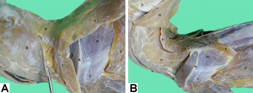
MORALES, M. P.; ARRIAGADA, V. C.; SÁNCHEZ, O. J. & MEDINA, P. R. Morphological description of extrinsic muscles of the thoracic limb in a specimen of pudu (Pudu puda).
                                                            Int. J. Morphol., 39(2):366-370, 2021.

muscles are responsible for linking the thoracic limb to the                              their insertion into the head, according to the direction their
rest of the body (Sisson & Grossman, 1982; Pellegrino et                                  fibers take to reach the insertion site. This description is
al.; 1998). Accordingly, the location of the center of gravity                            very similar to that described in domestic ruminants,
of the body shows that the thoracic limb supports more                                    although Sisson & Grossman indicate that in domestic
weight than the pelvic limb, both in season and during                                    ruminants, the division between the pars cleidobrachialis,
movement (Pellegrino et al.; König & Liebich, 2005).                                      pars cervicalis, and pars mastoidea is unclear. In the case
                                                                                          of the pudu specimen, the clavicular intersection is clearly
       There is currently little scientific information on the                            evident and corresponds to the place of origin of all
anatomical characteristics of the pudu species (Morales-                                  portions. In addition, the pars cervicalis has a wide fusion
Muñoz et al., 2020). Therefore, it is necessary to generate                               with the m. omotransversarius, a situation not described
novel scientific studies providing specific morphological                                 in domestic ruminants; however, König & Liebich, indicate
knowledge of this species, thereby equally contributing in                                that in the case of the horse, there is a muscular fusion of
reducing its threat and vulnerability (Sánchez et al., 2017;                              the pars mastoidea with the m. omotransversarius (Figs.
Saldivia & Villegas, 2019). Consequently, the objective of                                1, 3A, 3B, 4 and 5).
the present study is to describe the extrinsic muscles of the
thoracic limb of a specimen of pudu (Pudu puda).                                          M. pectoralis superficialis: The presence of two portions
                                                                                          is clearly observed as described in domestic ruminants.
                                                                                          These portions correspond to the descendens and
MATERIAL AND METHOD                                                                       transversus portions, which are very distinguishable and
                                                                                          independent in the pudu specimen, contrary to what occurs
                                                                                          in domestic ruminants, in which this muscle is poorly
        We worked with a specimen of Pudu puda donated                                    divided and differentiated (Sisson & Grossman). Both
by the Servicio Agrícola y Ganadero (SAG) of the Ministry                                 portions originate from the sternum until they are inserted
of Agriculture of Chile. The dissection was carried out in                                into the humerus bone. The transversus portion, despite
the Animal Anatomy Laboratory of Santo Tomás                                              being thin, is quite developed in contrast to that indicated
University of the School of Veterinary Medicine in Talca,                                 by Sisson & Grossman in cattle and small ruminants, in
Chile. The specimen corresponded to an adult female with                                  which it is thin and weakly developed (Fig. 5).
size of 40 cm high and 63 cm long. The animal was
euthanized due to the presentation of lumbar fracture in                                  M. pectoralis profundus: It originates from the sternum
the lumbar region between L3-L4 possibly caused by                                        and is then lateralized to insert into the greater tubercle of
outrage. The body was immediately fixed using a fixative-                                 humerus (Figs. 1, 2A, 2B and 5). This muscle is larger
preservative solution. The solution was introduced by                                     than the m. pectoralis superficialis and is partially fused
filling through the common carotid artery. Subsequently,                                  with the m. latissimus dorsi, with some muscle fibers also
the body was kept refrigerated during the entire descriptive                              fused to m. cutaneous trunci, a characteristic not described
process. Routine dissection instruments were used to                                      in the classical anatomical bibliography for domestic
dissect each muscle that forms the shoulder girdle,                                       ruminants (Dyce et al.). Deep in this muscle, the axillary
describing shape, origin and insertion.                                                   hollow associated with the brachial plexus is observed, as
                                                                                          well as arteries and veins from the region.

RESULTS AND DISCUSSION                                                                    M. subclavius: this muscle was not well-developed, si-
                                                                                          milar to that described in other ruminants, except in the
                                                                                          goat, in which it is described as very prominent (Sisson &
M. brachiocephalicus: It is divided in three portions,                                    Grossman). It was also found to have an elongated shape
which correspond to the pars cleidobrachialis, pars                                       unlike domestic ruminants, in which a trapezoidal shape
cervicalis, and pars matoidea. These portions are described                               is present (Sisson & Grossman). The muscle goes deep
in domestic ruminants. The cleidobrachialis pars originates                               into the cephalic vein, just at the point is a tributary of the
from the clavicular intersection until its insertion into the                             external jugular vein (Fig. 5), information that is not
crest of the humerus bone. The clavicular intersection is                                 described in the classical anatomical literature for domestic
observed as a cranial fibrous line to the scapulohumeral                                  ruminants (Ashdown & Done, 2011; Dyce et al.). Its
joint, which corresponds to the vestige of the clavicle bone,                             muscle fibers are cranial and superficial to the m. pectoralis
a similar situation to that described in other animals (Dyce                              superficialis and pectoralis profundus. It originates from
et al., 2012). The pars cervicalis and pars mastoidea also                                the sternum to the scapular fascia, similar to that described
originate from the clavicular intersection, then ascend until                             in domestic ruminants (Figs. 4 and 5).
                                                                                                                                                                          367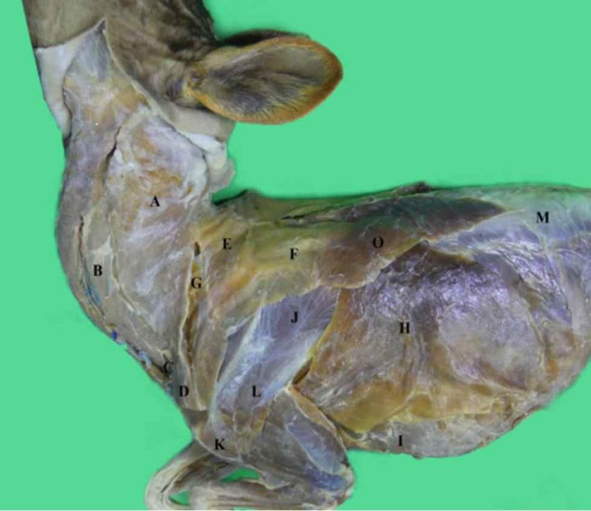
MORALES, M. P.; ARRIAGADA, V. C.; SÁNCHEZ, O. J. & MEDINA, P. R. Morphological description of extrinsic muscles of the thoracic limb in a specimen of pudu (Pudu puda).
Int. J. Morphol., 39(2):366-370, 2021.
M. Omotransversarius: This muscle is partially superficial only partial fusion is described in domestic ruminants (Sisson
to the m. infraspinatus (Morales-Muñoz et al.). It is highly & Grossman). Due to the wide fusion that it presents with the
fused to the m. brachiocephalicus (Fig. 3A), similar to the m. brachiocephalicus, it was only possible to observe the region
situation described in the horse (König & Liebich); however, of insertion at the level of the scapular spine and acromion.
Its other insertion was observed in conjunction with the pars
cervicalis of the m. brachiocephalicus and not in the cervical
region as indicated for domestic ruminants. Likewise, it was
located toward the proximal an aponeurotic fusion with the
m. trapezius, a situation that also differs from that described
for domestic ruminants (Figs. 1, 3A and 4).
M. trapezius: This muscle is superficial to the m.
rhomboideus, m. supraspinatus, and m. infraspinatus (Mora-
les-Muñoz et al.). It has a pars cervicalis and a pars thoracica
likewise described in ruminants and other domestic animals
(Ashdown & Done; Dyce et al.). It originates in the dorsal
raphe of the neck and thorax, and its insertion is observed in
the spine of the scapula, accordingly with the distribution
described in domestic ruminants (Gloobe, 1989) (Figs. 1, 2A
and 4).
M. rhomboideus: This muscle is observed deep in the m.
Fig. 1. Left side view of a pudu. A= M. Brachiocephalicus, pars
brachiocephalicus and m. trapezius and was well-developed
cervicalis; B= M. Brachiocephalicus, pars mastoidea; C= M.
Brachiocephalicus, intersection clavicularis; D= M. in the studied specimen. As in other ruminant species, it has
Brachiocephalicus, pars cleidobrachialis; E= M. Trapezius, pars two portions, the pars cervicalis that is inserted into the spinal
cervicalis; F= M. Trapezius, pars thoracica; G= M. processes of the cervical vertebrae and the pars thoracica that
Omotransversarius; H: M. Latissimus dorsi; I= M. Pectoralis is inserted into the spinous processes of the first thoracic
profundus; J= M. Infraspinatus; K= M. Deltoideus; pars acromialis; vertebrae and the scapular cartilage (Gloobe; Popesko, 1998)
L= M. Deltoideus, pars scapularis, M= Thoracolumbar fascia. (Figs. 2B and 3B).
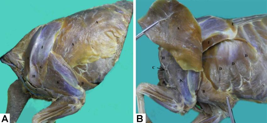
Fig. 2.A. 2B. Left side view of the scapular and brachial region of a pudu. M. Trapezius, m. Omotransversarius and m. Cutaneous trunci
were removed. B. Have been sectioned and rejected m. Brachiocephalicus and m. Pectoralis profundus. A= M. Latissimus dorsi; B= M.
Pectoralis profundus; C= M. Brachiocephalicus, pars cleidobrachialis; D= M. Serratus ventralis, pars thoracica; E= M. Rhomboideus; F=
M. Teres major; G= M. Deltoideus, pars scapularis; H= Deltoideus, pars acromialis; I= M. Supraspinatus; J= M. Infraspinatus; K= M.
Triceps brachii.
368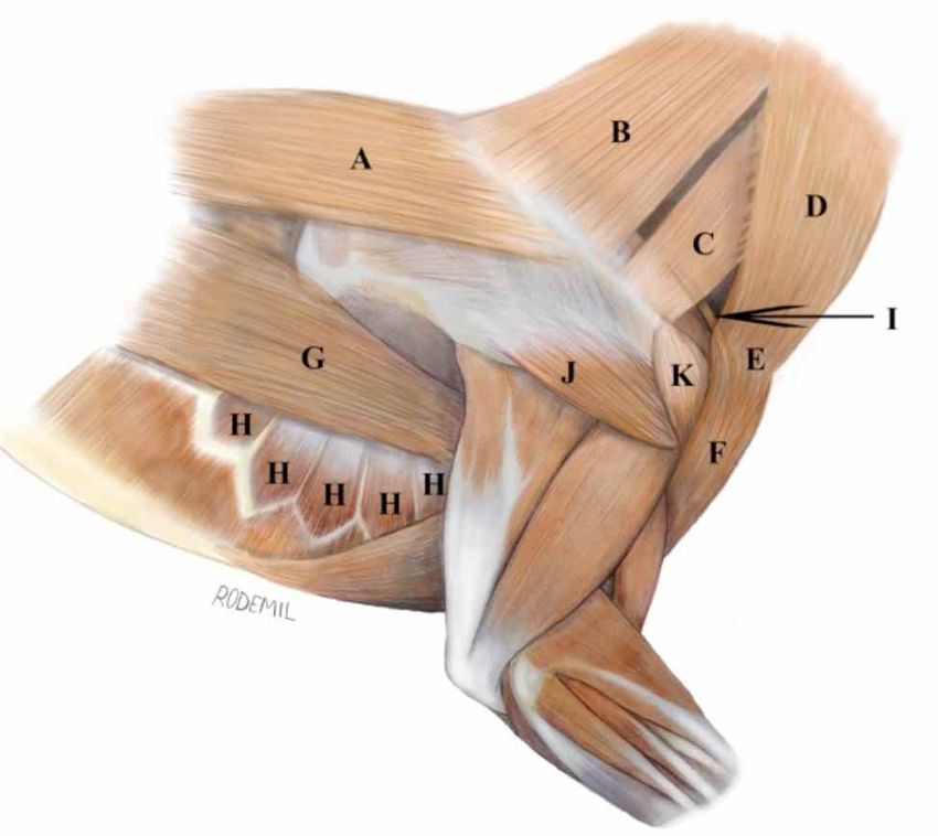
MORALES, M. P.; ARRIAGADA, V. C.; SÁNCHEZ, O. J. & MEDINA, P. R. Morphological description of extrinsic muscles of the thoracic limb in a specimen of pudu (Pudu puda).
                                                             Int. J. Morphol., 39(2):366-370, 2021.

Fig. 3. A: Left side view of the cervical and scapular region of a pudu. B: The m. Trapezius and m. Omotransversarius have been
removed. A= M. Trapezius, pars thoracica, B= M. Trapezius, pars cervicalis; C= M. Omotransversarius; D= M. Brachiocephalicus, pars
cervicalis (observe fusion of the muscles when taking with the gripper); E= M. Latissimus dorsi; F= M. Rhomboideus, pars cervicalis
and pars thoracica, inserted in scapular cartilage; G= M. Serratus ventralis cervicis; H= M. Infraspinatus; I= M. Deltoideus, pars scapularis;
J= M. Supraspinatus; K= Spine of the scapula bone.

                                                                                           Fig. 5. Illustration of the cervical, scapular, brachial, and thoracic
                                                                                           regions of a pudu. Right ventral view. A= M. Brachiocephalicus,
                                                                                           pars cervicalis; B= M. Brachiocephalicus, intersectio clavicular;
                                                                                           C= M. Brachiocephalicus, pars cleidobrachialis; D= M. Pectoralis
Fig. 4. lllustration of the cervical, scapular, brachial, and thoracic                     superficialis, pars descendens; E: M. Pectoralis superficialies, pars
regions of a pudu. Right side view. A= M. Trapezius, pars thoracica;                       transversus; F= M. Pectoralis profundus; G= M. Serratus ventralis,
B= M. Trapezius, pars cervicalis; C= M. Omotransversarius; D=                              pars thoracica; H= M. Latissimus dorsi; I= M. Subclavius; J=
M. Brachiocephalicus, pars cervicalis; E= M. Brachiocephalicus,                            External jugular vein.
intersection clavicular; F= M. Brachiocephalicus, pars
cleidobrachialis; G= M. Latissimus dorsi; H= M. Serratus ventralis,
                                                                                           Serratus ventralis: This muscle is located medial to the
pars thoracica; I= M. Subclavius; J= M. Deltoideus, pars scapularis;
K= M. Deltoideus, pars acromialis.                                                         scapula with a serrated shape. It is well-developed and is
                                                                                           divided into a pars thoracica that originates from the second
                                                                                           to the ninth rib and is inserted into the serrata face of the
M. latissimus dorsi: This muscle is observed deep in the                                   scapula. It is located deep in the M. latissimus dorsi. The
m. cutaneus trunci. It has an asymmetrical and flattened shape                             pars cervicalis originates in the transverse processes of the
and originates from the thoracolumbar fascia. Then, it is                                  third to the seventh cervical vertebrae and is widely inserted
directed cranially and ventrally, reaching its insertion into                              into the scapular cartilage until it reaches the serrata face of
the tuberosity of the teres major of the humerus bone. In                                  the scapula. It is less developed than the thoracic portion,
general, it is very similar to that described in domestic                                  thereby not differing from that described in the classical
ruminants (Popesko; Ashdown & Done; Dyce et al.) (Figs.                                    bibliography for domestic ruminants (Shively; Popesko)
1, 2A, 2B, 3A, 3B and 4). M.                                                               (Figs. 2B, 4 and 5).
                                                                                                                                                                           369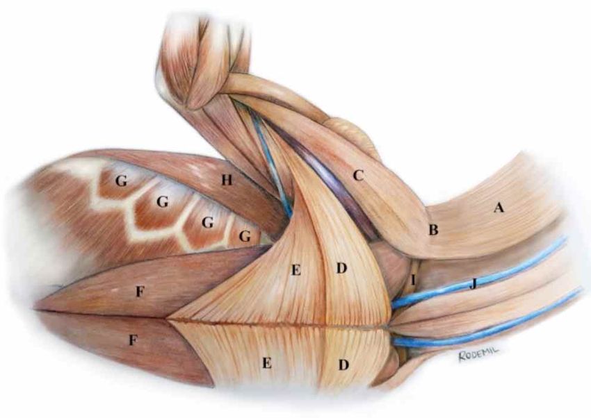
MORALES, M. P.; ARRIAGADA, V. C.; SÁNCHEZ, O. J. & MEDINA, P. R. Morphological description of extrinsic muscles of the thoracic limb in a specimen of pudu (Pudu puda).
                                                             Int. J. Morphol., 39(2):366-370, 2021.

CONCLUSIONS                                                                                REFERENCES

       The descriptive study of the extrinsic muscles of the                               Ashdown, R. R. & Done, S. H. Atlas en Color de Anatomía Veterinaria.
                                                                                               Rumiantes. 2nd ed. Madrid, Elsevier, 2011.
thoracic limb of a specimen of pudu (Pudu puda) allows us
                                                                                           Bro-Jørgensen, J. Dense habitats selecting for small body size: a comparative
to conclude that the structures are similar to those described                                 study on bovids. Oikos, 117(5):729-37, 2008.
for domestic ruminants. However, some differences should                                   Decreto 151. Oficializa Primera Clasificación de Especies Silvestres Se-
be considered, both in the fusion of some muscles and in the                                   gún su Estado de Conservación. Santiago de Chile, Ministerio Secre-
                                                                                               taria General de la Presidencia, BCN Legislación Chilena, Biblioteca
development and shape of muscle structures. The great fusion
                                                                                               del Congreso Nacional de Chile, 2007. Disponible en: http://
between various extrinsic muscles joins the block work                                         www.conaf.cl/cms/editorweb/transparencia/marco_normativo/DTO-
function and stability of thoracic limbs, as well as the neck                                  151_24-MAR-2007.pdf
and head. Consequently, the present data will be useful for                                Dyce, K. M.; Sack, W. O. & Wensing, C. J. Anatomía Veterinaria. 4th ed.
                                                                                               Ciudad de México, El Manual Moderno, 2012.
veterinarians working in wildlife, zoos, and animal
                                                                                           Eldridge, W. D.; MacNamara, M. M. & Pacheco, N. V. Activity Patterns
rehabilitation centers, providing novel reference data                                         and Habitat Utilization of Pudus (Pudu puda) in South-Central Chile.
regarding the morphological features of Pudu puda, thereby                                     En: Wemmer, Ch. M. (Ed.) Biology and Management of the Cervidae.
contributing to the knowledge of this scarcely studied vul-                                    Washington D. C., Smithsonian Institution Press, 1987. pp. 352-70.
                                                                                           Gloobe, H. Anatomía Aplicada del Bovino. San José, Instituto Interameri-
nerable species.
                                                                                               cano de Cooperación para la Agricultura (IICA), 1989.
                                                                                           König, H. & Liebich, H. Anatomía de los Animales Domésticos. Tomo 1.
                                                                                               2nd ed. Madrid, Médica Panamericana, 2005.
MORALES, M. P.; ARRIAGADA, V. C.; SÁNCHEZ, O. J. &                                         Morales-Muñoz, P.; Arriagada-Valdés, C. & Sánchez-Oñate, J.
MEDINA, P. R. Descripción morfológica de la musculatura ex-                                    Morphological and morphometric description of thoracic limb intrinsic
trínseca del miembro torácico de un espécimen de pudú (Pudu                                    myology in a specimen of Pudu (Pudu puda). Int. J. Morphol., 38(1):91-
puda). Int. J. Morphol., 39(2):366-370, 2021.                                                  5, 2020.
                                                                                           Pellegrino, F.; Tangelson, C.; Galliano, L.; Trevisan, L.; Sánchez, G. &
         RESUMEN: El pudu (Pudu puda) se clasifica como un                                     Puricelli, F. Homologation criterion between the scapular and pelvic
                                                                                               waists and their associated structures. Rev. Chil. Anat., 16(1):75-82,
artiodáctilo de la familia Cervidae. Es una especie nativa que se
                                                                                               1998.
encuentra en Argentina y Chile y se estima que su población se ha                          Popesko, P. Atlas de Anatomía Topográfica de los Animales Domésticos.
reducido sustancialmente debido a varias causas, tal como la pér-                              Tomo III. Pelvis y Miembros. 2ª ed. Barcelona, Masson, 1998.
dida de bosques, depredación, caza y accidentes de vehículo. De-                           Saldivia, M. & Villegas, F. Anatomical description of the bone segments
bido a lo anterior esta especie está protegida por su estado de con-                           that make up the skull of the Pudu puda Species. Int. J. Morphol.,
servación vulnerable. Los músculos extrínsecos del miembro                                     37(1):167-73, 2019.
torácico tienen gran importancia en la funcionalidad biomecánica                           Sánchez, O. J.; Morales, M. P. & Medina, P. R. Anatomical description of
de la suspensión del miembro, el cuello y la cabeza, además de                                 pelvic limb myology and its topographic relationship with vascular
                                                                                               and nervous systems in Pudú (Pudu puda). Int. J. Morphol., 35(4):1370-
participar en el movimiento de la pared torácica y el miembro
                                                                                               6, 2017.
torácico. El objetivo del presente estudio es describir la muscula-                        Silva-Rodríguez, E. A.; Verdugo, C.; Aleuy, O. A.; Sanderson, J. G.; Orte-
tura extrínseca del miembro torácico de un ejemplar de pudu, com-                              ga-Solís, G. R.; Osorio-Zúñiga, F. & González-Acuña, D. Evaluating
parando los resultados con los descritos para rumiantes domésti-                               mortality sources for the Vulnerable pudu Pudu puda in Chile:
cos en la bibliografía anatómica clásica. Se analizaron todos los                              implications for the conservation of a threatened deer. Oryx, 44(1):97-
músculos extrínsecos, describiendo la forma, distribución, origen                              103, 2010.
e inserción. Los resultados indican que el espécimen de pudu tiene                         Sisson, S. & Grossman, J. Anatomía de los Animales Domésticos. 5ª ed.
características anatómicas similares a las de los rumiantes domés-                             Barcelona, Masson, 1982.
                                                                                           Weber, M. & González, S. Latin American deer diversity and conservation:
ticos; sin embargo, deben tenerse en cuenta algunas diferencias.
                                                                                               A review of status and distribution. Écoscience,10(4):443-54, 2003.
Hallazgos principales: El músculo braquiocefálico tiene una inter-
sección clavicular evidente que lo separa en pars cleidobraquial,
cervical y mastoideo. El músculo pectoral superficial tiene dos
porciones distintas e independientes, y el músculo pectoral pro-                           Corresponding author:
fundo tiene una fusión parcial con los músculos latissimus dorsi y                         Pamela Morales Muñoz
cutáneo del tronco. El músculo subclavio es pequeño y tiene una                            Universidad Santo Tomás
forma alargada y se ubica profundamente a la vena cefálica, justo                          Avda. Carlos Schorr 255
en el punto en el que es un afluente de la vena yugular externa.                           Talca - CHILE
Los resultados de este estudio presentan características anatómi-
cas específicas de Pudu puda proporcionando información de re-
ferencia novedosa y ampliando el conocimiento científico de esta                           E-mail: pmoralesm@santotomas.cl
especie silvestre escasamente estudiada.

       PALABRAS CLAVE: Descripción; Músculos extrínse-                                     Received: 22-09-2020
cos; Miembro torácico; Pudu.                                                               Accepted: 26-11-2020

370
You can also read