Comparison of two novel swept source optical coherence tomography devices to a partial coherence interferometry based biometer
←
→
Page content transcription
If your browser does not render page correctly, please read the page content below

www.nature.com/scientificreports
OPEN Comparison of two novel
swept‑source optical coherence
tomography devices to a partial
coherence interferometry‑based
biometer
Tommy C. Y. Chan1,2, Marco C. Y. Yu1,3, Vivian Chiu1, Gilda Lai1, Christopher K. S. Leung1,4,5 &
Poemen P. M. Chan1,4*
To evaluate the repeatability and agreement of corneal and biometry measurements obtained with
two swept-source optical coherence tomography (SSOCT) and a partial coherence interferometry-
based device. This is a cross-sectional study. Forty-eight eyes of 48 patients had three consecutive
measurements for ANTERION (Heidelberg Engineering, Germany), CASIAII (Tomey, Japan) and
IOLMaster500 (Carl Zeiss Meditec, USA) on the same visit. Mean keratometry (Km), central
corneal thickness (CCT), anterior chamber depth (ACD) and axial length (AL) were recorded.
Corneal astigmatic measurements were converted into vector components—J0 and J45. Intra-
device repeatability and agreements of measurements amongst the devices were evaluated using
repeatability coefficients (RCs) and Bland–Altman plots, respectively. All devices demonstrated
comparable repeatability for Km (p ≥ 0.138). ANTERION had the lowest RC for J0 amongst the devices
(p ≤ 0.039). Systematic difference was found for the Km and J0 obtained with IOLMaster500 compared
to either SSOCTs (p ≤ 0.010). The ACD and AL measured by IOLMaster500 showed a higher RC
compared with either SSOCTs (p < 0.002). Systematic difference was found in CCT and ACD between
the two SSOCTs (p < 0.001), and in AL between ANTERION and IOLMaster500 (p < 0.001), with a mean
difference of 1.6 µm, 0.022 mm and 0.021 mm, respectively. Both SSOCTs demonstrated smaller
test–retest variability for measuring ACD and AL compared with IOLMaster500. There were significant
disagreement in keratometry and AL measurements between the SSOCTs and PCI-based device; their
measurements should not be considered as interchangeable.
The advancement of surgical techniques and intraocular lens (IOL) design has greatly improved the outcomes
of cataract surgery but also raised patients’ expectations for refractive-error free vision. Accurate measurements
of axial length (AL), corneal power, astigmatic axis and other anterior segment parameters are crucial for IOL
power calculation1, especially with the growing popularity of toric IOL implantation to correct preexisting
corneal astigmatism at the time of cataract surgery. The IOLMaster (Carl Zeiss Meditec, Jena, Germany) was
the first commercially available optical biometry device and has been used widely for IOL power calculation2,3.
The IOLMaster500 is based on the principle of partial coherence interferometry (PCI) and have demonstrated
accurate measurement for IOL power calculation in both routine and complicated cataract cases4,5.
Anterior segment ocular coherence tomography (OCT) has the advantage of high tissue penetration, high
scanning speed and has the ability to identify both anterior and posterior surfaces with high r epeatability6. OCT
is emerging as a new modality for corneal tomographic analysis7. The development of swept-source OCT allows
quick, one-station, detailed imaging of the cornea, the anterior chamber, as well as the anterior and posterior
1
Department of Ophthalmology and Visual Sciences, The Chinese University of Hong Kong, Hong Kong Eye
Hospital, Kowloon, Hong Kong, People’s Republic of China. 2Department of Ophthalmology, Hong Kong
Sanatorium & Hospital, Hong Kong, People’s Republic of China. 3Singapore Eye Research Institute, Singapore
National Eye Centre, Singapore, Singapore. 4Hong Kong Eye Hospital, Hong Kong, SAR, People’s Republic
of China. 5Department of Ophthalmology, The University of Hong Kong, Hong Kong, People’s Republic of
China. *email: poemen@gmail.com
Scientific Reports | (2021) 11:14853 | https://doi.org/10.1038/s41598-021-93999-8 1
Vol.:(0123456789)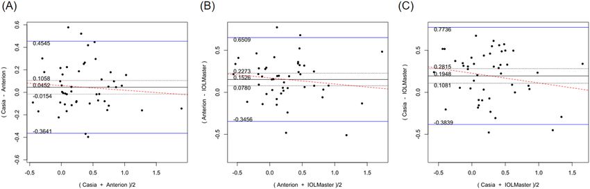
www.nature.com/scientificreports/
surfaces of the lens with improved image resolution and scan speed8,9. Since the first generation CASIA SS-1000
(Tomey, Nagoya, Japan), the technology continues to improve. For instance, compared with CASIA SS-1000,
the new CASIAII offers a faster scan-speed (50,000 vs. 30,000 A-scans/s) and a higher transverse resolution (800
A-scans/B-scan vs. 256 A-scans/B-scan) for 360° imaging of the anterior chamber angle using 18 evenly-spaced
radial scans over 36 angle locations. The CASIAII (introduced in 2016–2017) and the ANTERION (introduced in
2019, Heidelberg Engineering, Heidelberg, Germany) are two swept-source OCTs that were developed recently.
The purpose of this study was to compare the repeatability and agreement between ANTERION, CASIAII and
IOLMaster500 for measurements of keratometry and anterior chamber depth (ACD). Central corneal thickness
(CCT) measurements obtained from ANTERION and CASIAII were compared. Axial length measurements
obtained from IOLMaster500 and ANTERION were also compared.
Methods
This was a cross-sectional study conducted at The Chinese University of Hong Kong, Department of Ophthal-
mology and Visual Science between June and September 2019. Eyes that had previous ocular surgery (including
corneal refractive surgery and lens extraction) were excluded. Apart from cataract, eyes with ocular disease,
including corneal pathologies (e.g. cornea ectasia, pterygium), infectious disease (e.g. infective keratitis, viral
conjunctivitis), and/or problems with dry eyes were excluded. Patients with myopia of more than − 6.0 D and
visual acuity of worse than Snellen 6/12 were also excluded. Written informed consents were obtained from
all subjects. The study was conducted in accordance with the ethical standards stated in the 2013 Declaration
of Helsinki and approved by Hong Kong Kowloon Central Research Ethics Committee with written informed
consent obtained.
IOLMaster500. The IOLMaster500 (Carl Zeiss Meditec, Jena, Germany) utilizes the principle of PCI to
measure the AL. It evaluates the keratometry with a six-point telecentric technique and an image-based slit lamp
system for ACD measurements. It does not provide lens thickness nor CCT measurement. It measures AL from
the anterior corneal surface to the retinal pigmented epithelium along the line of sight.
CASIAII swept‑source OCT. The CASIAII swept-source OCT (Tomey, Nagoya, Japan) is a form of Fou-
rier-domain OCT that utilizes a swept-source wavelength of 1310 nm as the light source and a photodetector
to detect wavelength-resolved interference s ignal9, with improved in image resolution, scan speed, width, and
depth8,9. At a scan speed of 50,000 A scan/s and an axial resolution of < 10 µm, it allows multiple high-resolution,
up to 256 cross-sectional images of the entire anterior segment and angle to be captured within 3 s. The maxi-
mum scan width is 16 mm with a scan depth of 13 mm10. It also provides corneal topography and automatic
measurement software. With the measuring mode of corneal map and lens biometry, 16 radial cross-sectional
images, with 800 A-scan per line sampling, a scan width of 16 mm and a scan depth of 11 mm can be delivered
with a scan speed of 0.3 s. It only images the anterior segment with no AL measurement.
ANTERION swept‑source OCT. The ANTERION swept-source OCT (Heidelberg Engineering, Heidel-
berg, Germany) is another form of Fourier-domain OCT that offers a fast scan-speed of 50,000 A-sans/second.
It utilizes a 1300 nm light source to offer an axial resolution of < 10 µm. Compared with the CASIAII, it provides
a wider scan width (up to 16.5 mm wide) and a deeper scan depth range (14 ± 0.5 mm)11. This allows visualiza-
tion of detail corneal, anterior chamber, angle, and lens (both anterior and posterior surfaces). The four different
in-built imaging Apps—Cornea App, Cataract App, Metrics App, and Imaging App—allows a comprehensive
examination of the anterior segment imaging, corneal topography and tomography, anterior segment biometry,
IOL calculation, and AL measurement in a single scan. With the ANTERION Cornea App, 65 radial B-scan
images (256 A-scans per B-scan) are acquired in less than 1 s and the data can generate corneal maps of 8 mm
in diameter12. It measures the distance between the anterior corneal surface and the retinal pigment epithelium,
along the line of sight, as the AL12.
Imaging and measurements. One randomly selected eye of each subject was imaged by all three instru-
ments. Each eye was scanned three times for each instrument to obtain clear images of the anterior and posterior
corneal surfaces. The sequence of measurement recording between the ANTERION, CASIAII, and IOLMas-
ter500 was not fixed. The time elapsed between measurement devices included a short break for the patient to
relax for tear film recovery and avoid fatigue. All measurements were performed by a single experienced techni-
cian (G.L.) and were taken under dim room illumination. Patients were asked to blink in between consecutive
scans to produce an optically smooth tear film, thereby improving the reflectivity of the cornea. During the
imaging, the subjects were asked to fixate at an internal fixation target. To avoid lid artifact, the technician would
retract the upper and lower lids of the participant while taking the imaging. To ensure stable corneal conditions,
patients were asked to withhold soft contact lens wear for 2 weeks before the evaluation; none of our patients
wore hard contact lens.
Power vector analysis was conducted for obtaining vectors along the 0° and 45° meridians according to the
following equation: J0 = (− [Ksteep − Kflat]/2 cos 2α), and J45 = (− [Ksteep − Kflat]/2 sin 2 α), for comparison
in a Cartesian coordinate system13. Ksteep, Kflat, and α represent the steep keratometry, flat keratometry, and
axes values, respectively. J0 represents the astigmatic component along the vertical meridian (with-the-rule or
against-the-rule astigmatism), while J45 represents oblique astigmatism. Fellow eye data were flipped to avoid
neutralization of the J45 vector component when both eyes data were included for comparison.
R 3.2.5 (R Foundation, Vienna, Austria) was used for statistical analysis. Repeatability coefficients (RCs) was
used to evaluate the repeatability of measurements obtained by the IOLMaster500, CASIAII and ANTERION.
Scientific Reports | (2021) 11:14853 | https://doi.org/10.1038/s41598-021-93999-8 2
Vol:.(1234567890)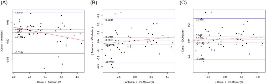
www.nature.com/scientificreports/
p-value for pairwise comparison in RC
ANTERION vs ANTERION vs CASIAII vs
Parameters Devices Mean (± SD) RC (95% CI) CASIAII IOLMaster500 IOLMaster500
0.2345 (0.2010–
ANTERION 43.878 ± 1.415
0.2681)
0.2484 (0.2125–
Km (D) CASIA II 43.851 ± 1.440 0.600 0.138 0.254
0.2843)
0.3148 (0.2666–
IOLMaster500 43.902 ± 1.456
0.3629)
0.2003 (0.1716–
ANTERION 0.305 ± 0.457
0.2289)
0.3217 (0.2752–
J0 (D) CASIA II 0.376 ± 0.422 0.002 0.039 0.361
0.3682)
0.5448 (0.4614–
IOLMaster500 0.207 ± 0.469
0.6282)
0.2232 (0.1913–
ANTERION − 0.002 ± 0.180
0.2551)
0.2115 (0.1809–
J45 (D) CASIA II 0.029 ± 0.196 0.980 0.245 0.072
0.2420)
0.3336 (0.2825–
IOLMaster500 − 0.024 ± 0.226
0.3846)
0.0311 (0.0264–
ANTERION 2.899 ± 0.597
0.0352)
0.0260 (0.0227–
ACD (mm) CASIA II 2.912 ± 0.586 0.216 < 0.001 < 0.001
0.0298)
0.1947 (0.1652–
IOLMaster500 2.932 ± 0.563
0.2241)
2.1759 (1.8648–
ANTERION 543.17 ± 32.20
2.4869)
CCT (µm) 0.575 – –
2.3223 (1.9868–
CASIA II 543.02 ± 33.45
2.6579)
0.0154 (0.0130–
ANTERION 23.683 ± 1.137
0.0178)
AL (mm) – 0.002 –
0.0260 (0.0221–
IOLMaster500 23.881 ± 1.323
0.0299)
Table 1. Repeatability outcomes for biometric measurements obtained using the ANTERION, CASIA II and
IOLMaster500. Km mean keratometry, J0 J0 vector components of astigmatism, J45 J45 vector components of
astigmatism, ACD anterior chamber depth, AL axial length, CCTcentral corneal thickness, RC repeatability
coefficient. 10.9% of the 5 significant results out of the 14 tests are expected to be false discoveries due to
random error as determined by the false discovery rate.
RC is defined as the 95% confidence limit of the difference of measurement between examinations, which is
equal to 1.96 2 × pooledtest − retestvariance . A high RC value represents a low test–retest variability, and
vice versa14.
Comparison of RCs for parameters between the CASIAII vs ANTERION, ANTERION vs IOLMaster500,
and CASIAII vs IOLMaster500 were evaluated by empirical bootstrap resampling with 2000 replicates. The first
attempted measure of each subject is evaluated. Bland–Altman plots were used to assess the agreement between
measurements of the two devices. Differences between the measurement values were plotted against the mean
values of the measurements, and the 95% limits of agreement (LoA), which is equal to the mean difference
± 1.96 × SD, were evaluated. Systematic differences of each parameter between the two devices were compared
using the t-test. Proportional bias was investigated by linear regression of the difference in values measured by
the two devices.
Based on test for equality of two within-subject variances in a parallel design, a sample of size 45/46/45 is
needed to detect a difference in variance in terms of ratio of 1.6/1.8/2 at 5% level of significance with power of
60%/80%/90%15. A p value less than 0.05 was considered statistically significant. False discovery rate, which
measures the percentage of false discovery due to random error, was evaluated for multiple statistical tests with
the threshold of p-value < 0.05 16.
Results
A total of 48 eyes of 48 patients were included. The mean age was 57.6 ± 14.8 years. Table 1 summarizes the repeat-
ability outcomes of the parameters obtained from ANTERION, CASIAII, and IOLMaster500. These include the
mean keratometry (Km), J0 and J45 vector components of astigmatism, ACD (measured from epithelium to
anterior lens surface), CCT (for the two OCT devices only), and AL (for ANTERION and IOLMaster500 only).
Both ANTERION and CASIAII demonstrated comparable test–retest repeatability for measurements of Km and
CCT (p ≥ 0.138). The J0 vector component of astigmatism measured by CASIAII showed a significantly greater
RC (p = 0.002) compared with the J0 measured by the ANTERION. The repeatability of the J45 vector component
Scientific Reports | (2021) 11:14853 | https://doi.org/10.1038/s41598-021-93999-8 3
Vol.:(0123456789)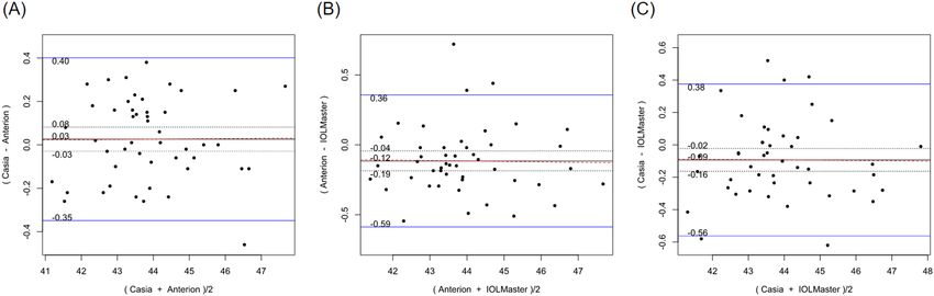
www.nature.com/scientificreports/
Systematic difference Proportional bias
Parameters Mean differences 95% LoA p Scaling difference R2 p
CASIA II vs ANTERION
Km (D) 0.026 − 0.349 to 0.401 0.351 0.001 7.33 × 10⁻⁵ 0.954
J0 (D) 0.045 − 0.364 to 0.455 0.140 − 0.041 0.008 0.557
J45 (D) 0.016 − 0.256 to 0.289 0.419 0.058 0.007 0.581
ACD (mm) 0.022 − 0.032 to 0.077 < 0.001 − 0.016 0.116 0.018
CCT (µm) − 1.625 − 5.278 to 2.028 < 0.001 0.012 0.045 0.149
ANTERION versus IOLMaster500
Km (D) − 0.115 − 0.588 to 0.357 0.002 − 0.003 3.41 × 10⁻⁴ 0.902
J0 (D) 0.153 − 0.346 to 0.651 < 0.001 − 0.072 0.018 0.374
J45 (D) 0.044 − 0.398 to 0.485 0.189 − 0.274 0.058 0.104
ACD (mm) 0.002 − 0.260 to 0.263 0.932 0.037 0.026 0.278
AL (mm) − 0.021 − 0.055 to 0.013 < 0.001 0.007 0.260 < 0.001
CASIA II versus IOLMaster500
Km (D) − 0.093 − 0.562 to 0.376 0.010 − 0.002 9.55 × 10⁻⁵ 0.948
J0 (D) 0.195 − 0.384 to 0.774 < 0.001 − 0.116 0.032 0.232
J45 (D) 0.067 − 0.348 to 0.482 0.035 − 0.201 0.039 0.185
ACD (mm) 0.024 − 0.235 to 0.283 0.219 0.020 0.008 0.547
Table 2. Systematic differences and proportional biases between different devices. Km mean keratometry, J0 J0
vector components of astigmatism, J45 J45 vector components of astigmatism, ACD anterior chamber depth,
CCTcentral corneal thickness, 95% LoA 95% limit of agreement. 9.8% of the 10 significant results out of the 28
tests are expected to be false discoveries due to random error as determined by the false discovery rate.
of astigmatism were similar across all the devices (p ≥ 0.072). RCs were greater for the measurements obtained
from IOLMaster500 compare with those obtained by either OCTs although significant differences were found
only in ACD and AL measurements (p ≤ 0.002).
Table 2 shows the systematic differences and the 95% LoA between ANTERION versus CASIAII, ANTERION
versus IOLMaster500, and CASIAII versus IOLMaster500 measurements. Systematic differences were found in
CCT and ACD between ANTERION and CASIAII (p < 0.001). However, only the ACD measurement showed a
proportional bias (p = 0.018) and not the CCT measurement (p = 0.149). There were systematic differences for the
Km and the J0 vector component between either OCT devices and the IOLMaster500. The mean difference of Km
between ANTERION and IOLMaster500 was − 0.115 D (p = 0.002) (span of 95% LoA was 0.945 D [range: − 0.588
to 0.357 D]; maximum absolute 95% LoA was 0.588 D) and the mean differences of J0 was 0.153 D (p < 0.001)
(span of 95% of LoA was 0.997 D [range: − 0.346 to 0.651 D]; maximum absolute 95% LoA was 0.651 D). There
was no proportional bias between the two devices (p ≥ 0.104). The mean differences of Km, J0 and J45 vectors of
astigmatism between CASIAII and IOLMaster500 were − 0.093 D (p = 0.010) (span of 95% of LoA was 0.908 D
[range: − 0.532 to 0.376 D]; maximum absolute 95% LoA was 0.532 D), 0.195 D (p < 0.001) (span of 95% of LoA
was 1.158 D [range: − 0.384 to 0.774 D]; maximum absolute 95% LoA was 0.774 D), and 0.067 D (p = 0.035) (span
of 95% of LoA was 0.830 D [− 0.348 to 0.482 D]; maximum absolute 95% LoA was 0.482 D), respectively. None
of these comparisons showed any proportional bias between the two devices (p ≥ 0.185). As for the AL, which
was only measured by ANTERION and IOLMaster500, there was a significant mean difference of − 0.021 mm
(p < 0.001) (span of 95% of LoA was 0.068 mm [range: − 0.055 to 0.013 mm]; maximum absolute 95% LoA was
0.055 mm) between the two devices; a proportional bias was observed (p < 0.001). The Bland–Altman plots of
the parameters measured by the three devices were shown in Figs. 1, 2, 3, 4, 5, and 6.
Discussion
Our study compared the two anterior segment swept-source OCT devices (CASIAII and ANTERION) with the
widely used, PCI-based IOLMaster500 for measurements of the corneal topography, ACD, and AL. Previously, we
had also compared the performance of CASIAII and ANTERION for the measurement of the angle p arameters17.
Other relevant studies that were recently published included comparison between ANTERION and IOLMaster
50018,19, Tomey CASIA SS-1000 and ANTERION20,21, IOLMaster700 and CASIAII22, and amongst three swept-
source OCT devices (CASIAII, ANTERION, and IOLMaster700)23.
In the current study, comparison between ANTERION and IOLMaster500 showed that the swept-source
OCT had superior repeatability in measuring AL. There was also a significant systematic difference and propor-
tional bias between ANTERION and IOLMaster500 for AL measurements. AL measured by the ANTERION
were shorter compared to that measured by the IOLMaster500 although a mean difference of 0.021 mm may
not be clinically significant given that a difference in AL of 0.030 mm would only result in a spherical error of
approximately 0.1 D in eyes with average AL and corneal curvature1. This finding correlates with the recent
publication that compared ANTERION and IOLMaster50018. The new IOLMaster700 (Carl Zeiss Meditec, Jena,
Germany) also bases on swept-source OCT to measure AL. Some studies found no difference for AL measure-
ments between the IOLMaster500 and 7 0024,25, whereas others reported significant difference between the two
Scientific Reports | (2021) 11:14853 | https://doi.org/10.1038/s41598-021-93999-8 4
Vol:.(1234567890)www.nature.com/scientificreports/
Figure 1. Bland–Altman plots showing the pairwise agreement between ANTERION vs CASIAII (A),
ANTERION vs IOLMaster500 (B), and CASIAII vs IOLMaster500 (C) for mean keratometry (Km).
Figure 2. Bland–Altman plots showing the pairwise agreement between ANTERION vs CASIAII (A),
ANTERION vs IOLMaster500 (B), and CASIAII vs IOLMaster500 (C) for J0 vector component of astigmatism.
Figure 3. Bland–Altman plots showing the pairwise agreement between ANTERION vs CASIAII (A),
ANTERION vs IOLMaster500 (B), and CASIAII vs IOLMaster500 (C) for J45 vector component of astigmatism.
devices26,27. Recent study that compared ANTERION and IOLMaster 700 (both are swept-source OCT based
devices) showed excellent agreement in measuring A L23. Swept-source OCT device had demonstrated a superior
ability to perform measurement compared with PCI-based device in the case of dense cataract and posterior
subcapsular cataract28. The ability to use a longer wavelength than that used by PCI (780 nm) can reduce light
scattering from opaque media, allowing greater penetration through a severe cataract. However, our patients had
an axial length in the normal range and a relatively good vision (at least 6/12), the performance of these devices
for eyes with extreme AL and dense cataract requires further study.
Scientific Reports | (2021) 11:14853 | https://doi.org/10.1038/s41598-021-93999-8 5
Vol.:(0123456789)www.nature.com/scientificreports/
Figure 4. Bland–Altman plots showing the pairwise agreement between ANTERION vs CASIAII (A),
ANTERION vs IOLMaster500 (B), and CASIAII vs IOLMaster500 (C) for anterior chamber depth (ACD).
Figure 5. Bland–Altman plots showing the pairwise agreement between ANTERION vs CASIAII for central
corneal thickness (CCT).
The ACD obtained with the ANTERION and CASIAII showed similar repeatability. There was a mean dif-
ference of 0.022 mm with a span of 95% LoA of 0.109 mm and a maximum absolute 95% LoA of 0.077 mm.
The ACD measurements obtained with the ANTERION were shorter than CASIAII. Such difference may not
affect IOL power calculation because a difference of 0.100 mm in ACD would only lead to an approximately
0.15 D change in refraction in eyes with an average AL and corneal curvature1. Either swept-source OCTs device
provided more repeatable measurements of ACD compared to the IOLMaster500. There is no statistically signifi-
cant systematic difference or proportional bias between the ACD of IOLMaster500 compared with either OCT.
However, the span of 95% LoA were as much as 0.5 mm (and a maximum absolute 95% LoA of 0.263 mm) when
compared with either ANTERION and CASIAII, and this can be clinically significant for IOL power calculation.
Comparison between IOLMaster500 and a different swept-source OCT biometer (OA-2000, Tomey, Nagoya,
Japan) also found a wide span of 95% LoA (0.41 mm) in ACD measurements29. Unlike the OCTs that measure
the ACD from OCT image that is aligned with the optical axis of the eye, the principle of IOLMaster500 for
measuring ACD is based on an optical section through the anterior chamber using a lateral slit beam illumina-
tion technique, which can be prone to operator e rrors26.
Although CCT measurement is not crucial for IOL power calculation, it is important for eyes planning for
corneal refractive surgery. The CCT obtained with the two swept-source OCTs demonstrated similar repeatability
but a significant systematic difference of 1.6 µm with a span of 95% LoA of 7.3 µm (and a maximum absolute
95% LoA of 5.3 µm). A much wider span of 95% LoA of 35 µm has been found between two other swept-source
OCT-based biometers, IOLMaster 700 and ARGOS (Movu, Komaki, Japan)30. The reason for the differences
could be the inconsistent measurement location and different measurement strategies between the different
swept-source OCTs.
Scientific Reports | (2021) 11:14853 | https://doi.org/10.1038/s41598-021-93999-8 6
Vol:.(1234567890)www.nature.com/scientificreports/
Figure 6. Bland–Altman plots showing the pairwise agreement between ANTERION vs IOLMaster500 for
axial length (AL).
The Km measurements demonstrated comparable repeatability for all three devices. However, the Km meas-
ured by IOLMaster500 showed a systematic difference when compared with either ANTERION or CASIAII,
implying between-device disagreement. The Km measured by IOLMaster500 was significantly steeper than
ANTERION (0.115 D) and CASIAII (0.093 D). It has been suggested that a Km difference of 0.20 D between
the devices is sufficient to give different optimized constants for IOL power calculation31. Intraocular lens con-
stant optimization based on the difference in magnitude in Km values has been proposed. Karunaratne et al.
demonstrated that the keratometry readings of the IOLMaster and Pentacam (Oculus, Wetzlar, Germany) were
not equivalent, but their keratometry readings were similarly effective in IOL power calculations after constant
optimization32. Similarly, although our findings showed that the Km measured by IOLMaster500 significantly
differs from either swept-source OCTs, such disagreement could be refined by readjustment of intraocular lens
constant in clinical practice.
As for the J0 and J45 astigmatic vector measurements, the J0 measured by ANTERION demonstrated a
significantly lower value of RC compared with CASIAII, and a similar RC for J45. Although the RC of J0 and
J45 obtained with IOLMaster500 did not show a significant difference compared with either OCT (apart from
a significantly higher RC for J0 when compared with the ANTERION), there is a trend of higher RC values for
the vector components obtained with the IOLMaster500. The between-instrument agreement of IOLMaster500
and either OCT for the astigmatic vector measurements was poor. There was a systematic difference for the J0
obtained with the IOLMaster500 compared with either the ANTERION or CASIAII. A systematic difference
was also found for the J45 between the IOLMaster500 and CASIAII. These observations are clinically impor-
tant, given that the accuracy of astigmatism measurements is crucial for the implantation of toric IOL. Our data
suggested that ANTERION demonstrated superior repeatability for the measurement of J0, which represents
with-the-rule and against-the-rule astigmatism, and its agreement with the IOLMaster500 was poor. In practice,
it is important to perform corneal topography when before implanting a toric IOL in cataract surgery since
irregular corneal astigmatism, which is not correctable by toric IOL, has been shown to increase with increasing
age33. It is likely that the difference between PCI-based and swept-source OCT-based devices exists because of
the different methods used to analyze the mires of spots reflected from the cornea. The IOLMaster500 measures
keratometry from the anterior cornea in the 2.5 mm zone using only 6 spots of light projected on the c ornea24
is different from swept-source OCTs that utilizes multiple evenly spaced radial B-scans for the measurement24.
Therefore, the astigmatic axis measured by the swept-source OCT and the PCI-based IOLMaster500 should not
be considered interchangeable. It remains to be elucidated how the differences of the measurements would affect
the predicted residual astigmatism after toric IOL implantation.
This study has several limitations. We did not grade the severity of cataract in our patients. A previous study
showed that swept-source OCT-based device was more effective in obtaining biometric measurements in eyes
with posterior subcapsular and dense nuclear cataract26. Our study did not recruit patients with visual acuity
worse than Snellen 6/12. Our study did not investigate the refractive outcome after cataract surgery; we, there-
fore, could not determine the refractive prediction errors because of the measurement differences in IOL power
calculation. Further study is required to verify how the findings in the current study that may affect the cataract
surgical outcome in clinical practice.
In conclusion, both ANTERION and CASIAII demonstrated excellent repeatability in biometry and corneal
measurements. Favorable agreement was demonstrated in keratometry and its astigmatic axis measurements
between the two swept-source OCTs. On the other hand, IOLMaster500 demonstrated lower repeatability for
Scientific Reports | (2021) 11:14853 | https://doi.org/10.1038/s41598-021-93999-8 7
Vol.:(0123456789)www.nature.com/scientificreports/
ACD and AL measurements. There was significant disagreement in keratometry and AL measurements between
swept-source OCT and PCI-based devices.
Received: 19 December 2020; Accepted: 28 June 2021
References
1. Norrby, S. Sources of error in intraocular lens power calculation. J. Cataract Refract. Surg. 34, 368–376 (2008).
2. Chen, Y. A., Hirnschall, N. & Findl, O. Evaluation of 2 new optical biometry devices and comparison with the current gold standard
biometer. J. Cataract Refract. Surg. 37, 513–517 (2011).
3. Kaswin, G., Rousseau, A., Mgarrech, M., Barreau, E. & Labetoulle, M. Biometry and intraocular lens power calculation results
with a new optical biometry device: Comparison with the gold standard. J. Cataract Refract. Surg. 40, 593–600 (2014).
4. Olsen, T. Improved accuracy of intraocular lens power calculation with the Zeiss IOLMaster. Acta Ophthalmol. Scand. 85, 84–87
(2007).
5. Kunavisarut, P., Poopattanakul, P., Intarated, C. & Pathanapitoon, K. Accuracy and reliability of IOL master and A-scan immersion
biometry in silicone oil-filled eyes. Eye (Lond.) 26, 1344–1348 (2012).
6. Fan, R., Chan, T. C., Prakash, G. & Jhanji, V. Applications of corneal topography and tomography: A review. Clin. Exp. Ophthalmol.
46, 133–146 (2018).
7. Radhakrishnan, S. et al. Real-time optical coherence tomography of the anterior segment at 1310 nm. Arch Ophthalmol. 119,
1179–1185 (2001).
8. Liu, S., Yu, M., Ye, C., Lam, D. S. & Leung, C. K. Anterior chamber angle imaging with swept-source optical coherence tomography:
An investigation on variability of angle measurement. Invest. Ophthalmol. Vis. Sci. 52, 8598–8603 (2011).
9. Lai, I. et al. Anterior chamber angle imaging with swept-source optical coherence tomography: Measuring peripheral anterior
synechia in glaucoma. Ophthalmology 120, 1144–1149 (2013).
10. Tomey. (2016). Fourier Domain OCT CASIA2: 3D swept source OCT. https://tomey.de/images/product_flyer/CASIA2_br_w.pdf.
(Accessed 15 March 2020).
11. Teussink, M.M., Donner, S., Otto, T., Williams, K., Tafreshi, A. (2019). State-of-art commercial Spectral Domain and Swept-Source
OCT technologies and their clinical applications in ophthalmology. Heidelberg Enginnering Academy 1–19. https://b usine ss-l ounge.
heidelbergengineering.com/gf/en/products/anterion/anterion/publications/#publications. (Accessed 12 July 2019).
12. Asam, J. S., Polzer, M., Tafreshi, A., Hirnschall, N. & Findl, O. Anterior Segment OCT: High Resolution Imaging in Microscopy and
Ophthalmology (Springer, 2019).
13. Thibos, L. N., Wheeler, W. & Horner, D. Power vectors: An application of Fourier analysis to the description and statistical analysis
of refractive error. Optom. Vis. Sci. 74, 367–375 (1997).
14. Bland, J. M. & Altman, D. G. Measurement error. BMJ 312, 1654 (1996).
15. Chow, S. C., Chao, J., Wang, H. & Lokhnygina, Y. Sample Size Calculation in Clinical Research 3rd edn. (Chapman & Hall/CRC,
2018).
16. Benjamini, Y. & Hochberg, Y. Controlling the false discovery rate: A practical and powerful approach to multiple testing. J. R. Stat.
Soc. Series B (Methodol.) 57, 289–300 (1995).
17. Chan, P. P. et al. Anterior chamber angle imaging with swept-source optical coherence tomography: Comparison between CASIAII
and ANTERION. Sci. Rep. 10, 18771 (2020).
18. Kim, K. Y., Choi, G. S., Kang, M. S. & Kim, U. S. Comparison study of the axial length measured using the new swept-source optical
coherence tomography ANTERION and the partial coherence interferometry IOL Master. PLoS ONE 15, e0244590 (2020).
19. Schiano-Lomoriello, D., Hoffer, K. J., Abicca, I. & Savini, G. Repeatability of automated measurements by a new anterior segment
optical coherence tomographer and biometer and agreement with standard devices. Sci. Rep. 11, 983 (2021).
20. Gjerdrum, B., Gundersen, K. G., Lundmark, P. O. & Aakre, B. M. Repeatability of OCT-based versus scheimpflug- and reflection-
based keratometry in patients with hyperosmolar and normal tear film. Clin. Ophthalmol. 14, 3991–4003 (2020).
21. Pardeshi, A. A. et al. Intradevice repeatability and interdevice agreement of ocular biometric measurements: A comparison of two
swept-source anterior segment OCT devices. Transl. Vis. Sci. Technol. 9, 14 (2020).
22. Fisus, A. D., Hirnschall, N. D. & Findl, O. Comparison of 2 swept-source optical coherence tomography-based biometry devices.
J. Cataract Refract. Surg. 47, 87–92 (2021).
23. Oh, R., Oh, J. Y., Choi, H. J., Kim, M. K. & Yoon, C. H. Comparison of ocular biometric measurements in patients with cataract
using three swept-source optical coherence tomography devices. BMC Ophthalmol. 21, 62 (2021).
24. Srivannaboon, S., Chirapapaisan, C., Chonpimai, P. & Loket, S. Clinical comparison of a new swept-source optical coherence
tomography-based optical biometer and a time-domain optical coherence tomography-based optical biometer. J. Cataract Refract.
Surg. 41, 2224–2232 (2015).
25. Kunert, K. S. et al. Repeatability and agreement in optical biometry of a new swept-source optical coherence tomography-based
biometer versus partial coherence interferometry and optical low-coherence reflectometry. J. Cataract Refract. Surg. 42, 76–83
(2016).
26. Akman, A., Asena, L. & Gungor, S. G. Evaluation and comparison of the new swept source OCT-based IOLMaster 700 with the
IOLMaster 500. Br. J. Ophthalmol. 100, 1201–1205 (2016).
27. Yang, J. Y., Kim, H. K. & Kim, S. S. Axial length measurements: Comparison of a new swept-source optical coherence tomography-
based biometer and partial coherence interferometry in myopia. J. Cataract Refract. Surg. 43, 328–332 (2017).
28. McAlinden, C. et al. Axial length measurement failure rates with biometers using swept-source optical coherence tomography
compared to partial-coherence interferometry and optical low-coherence interferometry. Am. J. Ophthalmol. 173, 64–69 (2017).
29. Huang, J. et al. Repeatability and interobserver reproducibility of a new optical biometer based on swept-source optical coherence
tomography and comparison with IOLMaster. Br. J. Ophthalmol. 101, 493–498 (2017).
30. Sabatino, F., Matarazzo, F., Findl, O. & Maurino, V. Comparative analysis of 2 swept-source optical coherence tomography biom-
eters. J. Cataract Refract. Surg. 45, 1124–1129 (2019).
31. Ozyol, P. & Ozyol, E. Agreement between swept-source optical biometry and Scheimpflug-based topography measurements of
anterior segment parameters. Am. J. Ophthalmol. 169, 73–78 (2016).
32. Karunaratne, N. Comparison of the Pentacam equivalent keratometry reading and IOL Master keratometry measurement in
intraocular lens power calculations. Clin. Exp. Ophthalmol. 41, 825–834 (2013).
33. Hayashi, K., Kawahara, S., Manabe, S. & Hirata, A. Changes in irregular corneal astigmatism with age in eyes with and without
cataract surgery. Invest. Ophthalmol. Vis. Sci. 56, 7988–7998 (2015).
Author contributions
P.P.C. and T.C.C. wrote the main manuscript text. G.L. operated the instruments and performed the measure-
ments. V.C. and G.L. collected the data and performed data entry. M.Y. and P.P.C. performed statistical analysis
Scientific Reports | (2021) 11:14853 | https://doi.org/10.1038/s41598-021-93999-8 8
Vol:.(1234567890)www.nature.com/scientificreports/
and prepared the Figs. 1, 2, 3, 4, 5 and 6. C.K.L. provided the design of the study and coordinated the team work.
All authors reviewed the manuscript.
Competing interests
CL has received research support in the form of instrument and speaker honorarium from Tomey (Japan) and
Heidelberg Engineering (Germany).
Additional information
Correspondence and requests for materials should be addressed to P.P.M.C.
Reprints and permissions information is available at www.nature.com/reprints.
Publisher’s note Springer Nature remains neutral with regard to jurisdictional claims in published maps and
institutional affiliations.
Open Access This article is licensed under a Creative Commons Attribution 4.0 International
License, which permits use, sharing, adaptation, distribution and reproduction in any medium or
format, as long as you give appropriate credit to the original author(s) and the source, provide a link to the
Creative Commons licence, and indicate if changes were made. The images or other third party material in this
article are included in the article’s Creative Commons licence, unless indicated otherwise in a credit line to the
material. If material is not included in the article’s Creative Commons licence and your intended use is not
permitted by statutory regulation or exceeds the permitted use, you will need to obtain permission directly from
the copyright holder. To view a copy of this licence, visit http://creativecommons.org/licenses/by/4.0/.
© The Author(s) 2021
Scientific Reports | (2021) 11:14853 | https://doi.org/10.1038/s41598-021-93999-8 9
Vol.:(0123456789)You can also read