Isolated epiglottic rhinosporidiosis: a rare case report - International Journal of ...
←
→
Page content transcription
If your browser does not render page correctly, please read the page content below

International Journal of Otorhinolaryngology and Head and Neck Surgery
Das A et al. Int J Otorhinolaryngol Head Neck Surg. 2020 Oct;6(10):1903-1905
http://www.ijorl.com                                                                       pISSN2454-5929 | eISSN2454-5937

                                                          DOI: http://dx.doi.org/10.18203/issn.2454-5929.ijohns20204199
Case Report

                 Isolated epiglottic rhinosporidiosis: a rare case report
                           Aurobinnda Das, Rajat Kumar Dash*, Kamalini Bepari

  Department of ENT, VIMSAR, Burla, Sambalpur, Odisha, India

  Received: 04 July 2020
  Revised: 07 August 2020
  Accepted: 07 September 2020

  *Correspondence:
  Dr. Rajat Kumar Dash,
  E-mail: rajatdash84@gmail.com

  Copyright: © the author(s), publisher and licensee Medip Academy. This is an open-access article distributed under
  the terms of the Creative Commons Attribution Non-Commercial License, which permits unrestricted non-commercial
  use, distribution, and reproduction in any medium, provided the original work is properly cited.

   ABSTRACT

   Rhinosporidiosis is a chronic granulomatous disease, caused by Rhinosporidium seeberi. More than 70% of cases are
   nasal. Usually extranasal rhinosporidiosis is associated with nasal rhinosporidiosis. Isolated extra nasal variety of
   laryngeal and tracheal rhinosporidiosis are very rare, 7 cases has detected till date. A 45 years male of LSES with
   habit of pond bath presented to ENT OPD, VIMSAR, Burla, with chief complain of intermittent blood vomiting for
   last 30 days, associated with foreign body sensation in throat without any dysphagia or dyspnea. On ILE, there is
   polypoidal pinkish mass studded with white spots found at lingual surface of epiglottis. Ant and post rhinoscopic
   examination found to be normal. UGIE guided biopsy shows rhinosporiodic mass. Under GA, DL had done mass was
   excised and base cauterised with bipolar cautery and send for HPE. HPE confirmed the diagnosis. Post operative
   follow up upto 10 months showed no recurrence. Epiglottic rhinosporidiosis may be one of the differential diagnosis
   of epiglottic growths especially in endemic zone. Laryngeal involvement of rhinosporidiosis has diagnostic and
   therapeutic challenges, due to the potential risk of bleeding, aspiration and recurrence.

   Keywords: Rhinosporidiosis, Epiglottis, Isolated, Extranasal

INTRODUCTION                                                         Medical treatment is ineffective. Surgical excision is
                                                                     mode of treatment, however recurrence is common. HPE
Rhinosporidiosis is a chronic granulomatous disease,                 confirm the clinical diagnosis.
caused by Rhinosporidium seeberi.1 It belongs to the
class protoctistan mesomycetazoa.2 Most common in                    CASE REPORT
Southern India and Srilanka.3 Contaminated stagnant
fresh water is an important source of infection.4,5 It               Forty five years married Hindu male, farmer by
mostly affects nasal mucosa, ocular conjunctiva and                  occupation from Jharsuguda district, presented to ENT
nasopharynx.1,6-9 More than 70% of cases are nasal.1                 OPD, VIMSAR, Burla, with chief complain of
Usually extra nasal rhinosporidiosis is associated with              intermittent blood vomiting for last 30 days, associated
nasal rhinosporidiosis. Extra nasal variety of laryngeal             with foreign body sensation in his throat. He had no
and tracheal rhinosporidiosis are very rare.8,10,11 Six cases        complained of difficulty in swallowing or breathing.
has been detected involving larynx till date.7,10-13 Our             There was no history of previous rhinosporidiosis. He
case is the seventh reported case involving larynx . More            had no history of bleeding disorder or chronic disease
prevalence in male of the age group of 15 to 50 years                like TB. He belongs to LSES (low socio economic status)
with low SES. Male to female ratio of 4:1.14 Most of                 and has habit of pond bath. On examination higher
these patients have history of pond bath. Mostly present             function and vitals of the patient found to be normal. He
as polypoidal pinkish nasal mass studded with white                  had mild pallor and thin body built. Face and dorsum of
spots with intermittent blood tinged nasal discharge.5               nose found to be normal (Figure 1). Oral cavity, posterior

                 International Journal of Otorhinolaryngology and Head and Neck Surgery | October 2020 | Vol 6 | Issue 10 Page 1903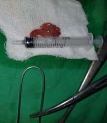
Das A et al. Int J Otorhinolaryngol Head Neck Surg. 2020 Oct;6(10):1903-1905

pharyngeal wall examination found to be normal. On ILE
(indirect laryngeal examination), there is polypoidal
pinkish mass studded with white spots found at lingual
surface of epiglottis. Ant and post rhinoscopic
examination found to be normal. UGIE (upper
gastrointestinal endoscopy) guided biopsy shows
rhinosporiodic mass (Figure 2). Then patient was planned
for surgical excision. Under GA (general anaesthesia),
DL (direct laryngoscopy) was done. Attachment of mass
to the lingual surface of epiglottis identified (Figure 3).
The mass was excised and base cauterised with bipolar.
The excised mass was send for HPE (Figure 4).
Histopathological examination confirmed the diagnosis               Figure 4: Intra OP picture showing site of attachment.
(Figure 5). Patient was kept nil per oral with Ryles tube
feeding and prophylactic antibiotic for 3 days. HPE
confirm the clinical diagnosis. Post opwrative follow-up
upto 12 month showed no recurrence (Figure 6).

                                                                       Figure 5: Histopathological examination finding.

    Figure 1: Patient showing no facial deformity.

                                                                        Figure 6: Follow up laryngeal endoscopic photo
                                                                            showing no recurrence after 12 month.

                                                                    DISCUSSION

 Figure 2: Upper gastrointestinal endoscopy finding.                Rhinosporidiosis mostly affects nose and nasopharynx,
                                                                    other sites such as conjunctiva, palate, lip, epiglottis,
                                                                    larynx, trachea, skin, vulva and bone may also be
                                                                    affected.6 More than 70% of cases are nasal.1 Extra nasal
                                                                    rhinosporidiosis is usually associated with nasal
                                                                    rhinosporidiosis. Extra nasal variety of laryngeal &
                                                                    tracheal rhinosporidiosis are very rare, 7 cases has been
                                                                    detected till date including our case. However isolated
                                                                    epiglottic rhinosporidiosis is very rare. Patient seeks
                                                                    medical attention late as compared to nasal variety. Pond
                                                                    bathing with injury at extra nasal mucosa, may
                                                                    predispose to extra nasal variety. Proper history &
                                                                    oropharyngeal, laryngeal examination may help to reach
                                                                    the diagnosis. Histopathology study confirms the
                                                                    diagnosis. Excision with wide base cauterisation is the
                Figure 3: Excised mass.

                International Journal of Otorhinolaryngology and Head and Neck Surgery | October 2020 | Vol 6 | Issue 10 Page 1904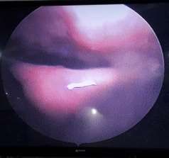
Das A et al. Int J Otorhinolaryngol Head Neck Surg. 2020 Oct;6(10):1903-1905

treatment of choice Intermittent follow up with post                5.    Dhingra PL, Dhigra S. Cholesteatoma and chronic
operative dapsone may be given to reduce recurrence.                      suppurative otitis media. In: Diseases of the ear, nose
Differential diagnosis is carcinoma, squamous papilloma,                  and throat. 5th ed. Amsterdam: Elsevier; 2014:174.
ductal epiglottic cyst, fibroepithelial polyps, tubercular          6.    Pal S, Chakravarti S, Das PC. Cytodiagnosis of
mass.15                                                                   extranasal rhinosporidiosis. J Lab Physicians.
                                                                          2014;6(2):80-3.
CONCLUSION                                                          7.    Daharwal A, Banjara H, Singh D, Gupta A, Singh S.
                                                                          Laryngeal rhinosporidiosis. Journal of Laryngology
As a rare case and asymptomatic for long time, the case                   & Voice. 2011;1(1):30-2.
may be missed by physician in first instant, hence all              8.    Madan J, Yolmo D, Gopalkrishnan S, Saxena SK.
suspected case should be examined and investigated in                     Rhinosporidiosis of upper airway and trachea. J
the line of rhinosporidiosis, especially in endemic zone.                 Laryngol Oto. 2010;124:1139-41.
Epiglottic rhinosporidiosis may be one of the important             9.    Mahmud S, Haque R, Almamun A, Alam R. A
differential diagnosis of epiglottis growths. Laryngeal                   clinico pathological study of Rhinosporidiosis.
involvement of rhinosporidiosis may possess diagnostic                    Bangladesh Journal of Otorhinolaryngology. 2015;
and therapeutic challenges, due to the potential risk of                  21(2):94-6.
bleeding, aspiration and recurrence.                                10.   Pillai OS. Rhinsporidiosis of the larynx. J Laryngo
                                                                          Otol. 1974;(3):277-80.
Funding: No funding sources                                         11.   Kumar S, Mathew J, Cherian V, Rozario R, Kurien
Conflict of interest: None declared                                       M. Laryngeal Rhinosporidiosis:Report of a rare case.
Ethical approval: Not required                                            Ear, nose & throat Journal. 2004;83(8):568-70.
                                                                    12.   Banarjee SB, Sarkar A, Mukherjee S, Bhowmik A.
REFERENCES                                                                Laryngeal rhinosporidiosis. J India Med Asso.
                                                                          1996;94(4):148-50.
1.   Watkinson JC, Clarke RW. Scott Brown’s                         13.   Mathew JS, Padhy S, Lata S, Balachander H,
     Otorhinolaryngology Head Neck Surgery. 8th ed.                       Gopalakrishnan S. Case report: Tele-laryngoscopy-
     Florida: CRC Press; 2018:215-6.                                      guided     flexible   fiberoptic     intubation     for
2.   Herr RA, Ajello L, Taylor JW, Arseculeratne SN,                      laryngeal     rhinosporidiosis.    Anesth      analog.
     Mendoza       L.     Phylogenetic    analysis    of                  2010;110(4):1066-8.
     Rhinosporidium seeberi 18S small-subunit ribosomal             14.   Rath R, Baig S, Debata T. Rhinosporidiosis
     DNA groups this pathogen among members of the                        presenting as an oropharyngeal mass:A clinical
     protoctistan Mesomycetozoa clade. Journal of                         predicament. J Nat Sci Bio Med. 2015;6(1):241-5.
     Clinical Microbiology. 1999;37:2750-4.                         15.   Tresley J, Saraf EL, Sargi Z. Epiglottic masses
3.   Arsecularatne SN, Ajello L. Rhinosporidium seeberi.                  identified on CT imaging. Neuroradiol J.
     In: Ajello L, Hay RJ, eds. microbiology and                          2015;28(3):247-53.
     microbial infections. 4th ed. London: Arnold; 1998:
     67-73.
4.   Shina A, Phukan JP, Bandhyopadhyag G, Sengupta                  Cite this article as: Das A, Dash RK, Bepari K.
     S, Bose K, Mondal RK, et al. Clinico pathological               Isolated epiglottic rhinosporidiosis: a rare case report.
     study of Rhinosporidiosis. Journal of Cytology.                 Int J Otorhinolaryngol Head Neck Surg 2020;6:1903-
     2012;29(4):246-9.                                               5.

                International Journal of Otorhinolaryngology and Head and Neck Surgery | October 2020 | Vol 6 | Issue 10 Page 1905
You can also read