Journal of Clinical & Experimental Immunology - Opast Online ...
←
→
Page content transcription
If your browser does not render page correctly, please read the page content below
ISSN: 2475-6296

Research Article                                      Journal of Clinical & Experimental Immunology
Evolutive Immunologic and Toxicologic Approach in Some Neuroinflamatory and
Degenerative Disease like SM, DA, PD
Luisetto M1*, Akram Muhamad2, Gaber Ibrahim3, Behzad Nili Ahmadabadi4, Farhan Ahmad Khan5, Ahmed Yesvi Rafa6
and Oleg yurevich latish7
1
 Applied pharmacologist independent researcher, IMA academy                *Corresponding author
pharmacology branch Italy                                                  Luisetto M, Applied pharmacologist independent researcher, IMA academy
Department of Eastern Medicine, Government College, Faisalabad
2                                                                          pharmacology branch Italy.
University, Pakistan
                                                                           Submitted: 09 Apr 2020; Accepted: 22 Apr 2020; Published: 18 May 2020
Associate professor, Department of Zoology, Alexandria University, Egypt
3

4
 Pharm D/PhD Innovative Pharmaceutical Product Development
Specialist, USA
Professor and Head Department of Pharmacology Government
5

Medical College Shahdol, Madhya Pradesh
6
 Founder and President, Yugen Research Organization; Western Michigan
University, MI, USA
PRESIDENT of IMA RU
7

           Abstract
           In order to better, understand some neurologic process is fundamental to use an evolutionary approach. Imaging can
           help in measuring efficiency of brains wasting system in the various subject. The brain glimphatic systems is well
           studied today but an accurate measure of the real efficiency of the system is needed. Aim of this work is to submit to the
           researcher a working method to measure this parameter to verify if possible to use the brain glymphatic system as new
           therapeutics strategy.

Keywords: Evolutionary, Immunology, Imaging, Neurology,                    of PD over the centuries. The apparently low prevalence of PD
Pathology, Toxicology, New Therapeutic Strategies, DA, PD, ALS             before the twentieth century may be related to the shorter life
                                                                           expectancy and survival compared to present times. Changes in
Introduction                                                               lifestyle over the course of human history might also account for the
We start this work whit a simply PARADOX: observing that sepia             increasing burden of PD. Our hunter-gatherers ancestors invested
officinal is not show dementia. The different species of animals           large energy expenditure on a daily basis, a prototypical physical
show various rate of DA dementia and related PD Parkinson                  way of life for which our genome remains adapted. Technological
disease: According Pedro J. Garcia-Ruiz et al.                             advances have led to a dramatic reduction of physical exercise.
                                                                           Since the brain release of neuro-trophic factors (including brain-
“There are 2 central premises to this evolutionary view of Parkinson       derived neuro-trophic factor) is partially exercise related, the marked
disease. First, PD is a specific human disease. Second, the prevalence     reduction in exercise may contribute to the increasing prevalence of
of PD has increased over the course of human history. Several lines        PD. Many neurological diseases can be found in non-human
of evidence may explain why PD appears to be restricted to the             mammals both acquired and hereditary (such as myelopathy, brain
human species. The major manifestations of PD are the consequence          tumors, epilepsy, muscular dystrophy, and narcolepsy, to mention a
of degeneration in the dopamine-synthesizing neurons of the meso-          few). AD and PD are considered specific to Homo sapiens. While
striatal neuronal pathway. It is of note the enormous expansion of         there are useful animal models of PD including MPTP and alpha-
the human dopamine mes-encephalic neurons onto the striatum                synuclein-over-expressing transgenic mouse models, which may
compared with other mammals. Hence, an evolutionary bottleneck             recapitulate important clinical features of the human disorders,
was reached with the expansion of the massive nigro-striatal axonal        especially in aged monkeys, no spontaneous akinetic-rigid syndrome
arborization. This peculiar nigral overload may partly explain the         is known to occur in wild mammals including non-human primates.”
selective fragility of the human dopaminergic mes-encephalic
neuro-transmission and the unique presence of PD in humans. On             And Vernier P et al: “Parkinson’s disease is, to a large extent,
the other hand, several facts may explain the increasing prevalence        specific to the human species. Most symptoms are the consequence

    J Clin Exp Immunol, 2020                                  www.opastonline.com                                        Volume 5 | Issue 3 | 123
of the preferential degeneration of the dopamine-synthesizing Some question are really interesting:
cells of the meso-striatal-meso-cortical neuronal pathway. Reasons What is the role-played by the fact that CNS is a kind of
for that can be traced back to the evolutionary mechanisms that sancturaries? And the fact that loss of A complete lymphatic
shaped the dopamine neurons in humans. In vertebrates, dopamine- system? No limpho-nodes inside. And the role-played by BEE
containing neurons and nuclei do not exhibit homogenous even in the sense of from inside to outside barrier for toxic
phenotypes. In this respect, mes-encephalic dopamine neurons of molecule? Why in brain white matter is outside vs grey matter and
the substantia nigra and ventral tegmental area are characterized opposite in spinal cord? Is due to the progressive. Increase of
by a molecular combination (tyrosine hydroxylase, aromatic cortical surface? Why cervical region is so often involved in some
amino acid de-carboxylase, mono-amine oxidase, vesicular mono- neurodegenerative pathology? There is a specific vulnerability?
amine transporter, dopamine transporter--to name a few), which is And why in example cerebellum is not involved in initial stages of
not found in other dopamine-containing neurons of the vertebrate PD? What are the role played by endogenous toxic movens? And
brain. The size of these mes-encephalic DA nuclei is tremendously how is possible to depurate this kind of toxic condition?
expanded in humans as compared to other vertebrates.
Differentiation of the mes-encephalic neurons during development Material and Methods
depends on genetic mechanisms, which also differ from those of Whit an observational approach some relevant biomedical
other dopamine nuclei. In contrast, pathophysiological approaches literature are analyzed to produce a global conclusion related the
to PD have highlighted the role of ubiquitously expressed topics of this work. Al literature presented is presented in scientific
molecules such as a-synuclein, parkin, and microtubule-associated biomedical database like PUBMED. (1-49) after this review a
proteins. We propose that the peculiar phenotype of the dopamine global conclusion is produced.
mes-encephalic neurons, which has been selected during vertebrate
evolution and reshaped in the human lineage, has also rendered Results
these neurons particularly prone to oxidative stress, and thus, to From literature
the fairly specific neuro-degeneration of PD. evidence demonstrate “We have seen from literature the relationship existing between
that perturbed regulation of DAT-dependent dopamine uptake, systemic immune status and local situation like in brain tissue. We
DAT-dependent accumulation of toxins, dysregulation of TH can consider under a toxicological view this kind of influences in
activity as well as high sensitivity of DA mes-encephalic neurons order to re- consider some brain pathologies expecially if time age
to oxidants are key components of the neuro-degeneration process related. (Peaks-age classes more involved in some neurologic
of PD. This view points to the contribution of non-specific pathologies). Local flogosys and related immune reaction
mechanisms (alpha-synuclein aggregation) in a highly specific activation contribute in some brain pathology and this can be
cellular environment (the dopamine mes-encephalic neurons) and consider a sort of toxicological effect that must to be deeply
provides a robust framework to develop novel and rational investigated in order to discover the patho-genetic movens and
therapeutic schemes in PD.” innovative pharmacological strategies. Toxicology science can
add to immunology and pathology to have a more complete vision
In some neurological conditions is clear the role-played by brain in some brain patology in time evolution and strategic opportunities.
immune system and the global effect Due by BEE and other factor We have seen in example that using fingolimod we have a
like brain-washing system. Observing the complex mechanism reduction in linfocites activation and when discontinued this effect
involved in KURU disease is possible to verify the role played by reduced (like a discontinue of a toxic substantia). Dose related and
specific and A-specific immune system (Innate and Adaptative). time related. FINGOLIMOD Significantly improved relapse rates
The same observing the anatomy and physiology of CNS, BEE and end points measured on magnetic resonance imaging (MRI) in
and relate immune system: role played by astrocyte, microglia and objective way. Concepts as toxical doses, time of exposition,
other cells. The evolutionary approach make possible to verify cumulative dosage, kinetics, dynamics, metabolism Iatrogenic
evolution of CNS, BEE and related brain structure and the related ADME and other toxicological parameter can be usefully
selective vulnerability. Form invertebrates to superior vertebrates introduced also in neuro-immune toxicology to adeguately focus a
various physio -anatomical modifications emerged. The new need physio-pathogenetic phenomena. The results related to the
in movement and in environmental relationship request more references cited show a specific effect of a systemic drugs in a
complicated structure and the same from primitive invertebrates to local place as brain. We think that observing a specific side effect
the more complex superior vertebrates the same immune system of a drug can be a right method to clear some interference between
requested new strategy. Also regenerative of retina show great the immunologic status and some development disorder” [1].
difference in neuronal evolution in the various species. Neocortical
cognitive activities needed a new sovra-structure development. “Related to the reference findings presented in this review and
research work is crucial to submit to the researcher a new hypothesis
Archeo brain vs neo brain. But this sovra-structure was moves to related the anatomic decussatio pyramid and cervical tract in global
produce circon-voluntion in cortex to increase number of neuron. physio-metabolic status of Spinal cord. In same condition like
But the same this created some wasting system increased repeated head micro-trauma or in particular weakness of motor
problems? Why in example cerebellum is later interested by PD vs neuron this anatomic conformation can produce UN unbalance that
basal ganglia? Different neurodegenerative disease present can aggravate this situation. (See the higher frequencies related
different neuronal toxicity. Superior vertebrates need less cervical tract vs other in spinal cord). A fact to be take in
regenerative properties then more primitive organism. And if DA consideration even if is a normal anatomic conformation. The
affect more cognitive cortex PD the extrapyramidal first. Some experimental project proposed make possible to verify any
characteristic of brain evolution with new sovrastructures in old connection between an anatomic peculiarity (decussation pyramidi,
ones created vulnerability. cervical spinal cord tract) and an increased weakness of the
J Clin Exp Immunol, 2020 www.opastonline.com Volume 5 | Issue 3 | 124neuronal tracts. So in neuro- degenerative spinal cord disease is “Observing that some cases of CJD are due by peripheral exposition
relevant to observe the global topography of the lesions to verify if to prions and that KURU disease and BSE are transmitted by oral
some tract are more involved vs other to better understand the real intake of infected food we try to produce new theories in immune
reason .The evolutionary approach make possible to better systems and brain inter-connections. Can we consider KURU
understand the global process under a new light” [2]. infectious disease an instrument to verify interconnection between
immune cell out and in central nervous systems? Like a ERLIC
Related the result of this work is relevant for some neuro- magic bullet? Or an imaging tracer to follow the neuro immune
degeneretaive condition to verify the role played by brain process? Prions result neurotropic but other antigen are normally
glymphatic system as well as the effect played by some brain presented inside brain? And with what consequences? other brain
vascular inefficiencies and the role played by SOME TOXIC disease present the similar patho-genetic move’s immune system
catabolic substantia and their accumulation. An imbalances related? Transgenic modified mouses study showed that immune
between production and clearance of alfa sinuclein in the brain is system are involved in amplification and transmission to central
involved in PD evolution. The relationship with some kind of food nervous system (Lymph. B and follicular dendritic cell). In prions
and PD is not so high but what is to take in consideration is the disease we see species barrier and is necessary a molecular similarity
functionality of the brain clearance system that can be more between prions and endogenous PRP-C. Are present relationship
saturated is some kind of diet. Also interesting the way of influence between some brain disease as amyloidosis and other degenerative
in this system by body posture (is a dynamic process). Is possible disease like PD, DA and prions disease and other?
that 2 factors influence this process? Factor A: clearance efficiency
of the system in basal status (genetic factor, age) Factor B: In SM (a neuro inflammatory disease) is involved adaptative
Clearance efficiency in SATUTARED situation? (Environmental immunity: Lymphocyte T and B, While in other disease as
factor) and Global Function: A x B Alzheimer (a neurodegenerative pathology) is involved the innate
immunity (microglia- macrophage like activity, first immune
This result can be used to search new therapeutic strategies: or to control system in the central nervous systems). Observing the
reduce saturation condition of the system analyzed or new KURU disease, the time involved in presentation of
molecule that can improve the “washing” of this toxic endogenous symptomatology after intake of Prions and (a slow process) related
substantial or a combination of this [2]. A detox gel in mouse to the fast time In some cases involved in some neurotropic viruses
model of DA showed activity to suggest to develop this strategy, We can think to an passive vs active process by which immune
and the same observing epidemiological data related world PD systems transfer to the brain the toxic prions from GI system. In all
incidence compared to some diet it seen to show how reduce the this pathologies immune system play a relevant role (adaptive or
saturation of this system. As global conclusion of this work is innate) giving tissue damage and accumulation of bio products.
possible to say that a combination of this 2 strategies can produce Observing the global role played by immune system in some brain
an interesting clinical effect to be verified. (This combined strategy pathology under a specific toxicological aspect, we can think to
is a new association: Pharmacological effect, added to de toxicant other therapeutic strategies to improve the actual pharmacological
strategy). Four are the main factor involved: The toxic molecule to scenario. This paper is produces under a specific medicinal
be depurate, the carrier- vectors, the brain washing system, chemistry and pharmacological point of view” [4].
saturation condition (Environmental factor).
“In recent years, the existence of a mass transport system in the
In animal model the detox strategy produced effect, so is possible brain via cerebrospinal fluid (CSF) or interstitial fluid (ISF) has
to say that a toxicological approach can be a way to be walked [3- been suggested by many studies. The glymphatic system is
5]. A “total Aβ-42 by 30% in the group of mice that received the hypothesized to be a waste clearance system of the CSF through
detox gel when compared to the untreated group with a statistical the perivascular and interstitial spaces in the brain. Tracer studies
significance (p < 0.001)” is a good result to start. A new drug class have primarily been used to visualize or evaluate the waste
able to link and detox the toxicological molecule and to be carried clearance system in the brain, and evidence for this system has
out from brain structure using the glimphatic system through accumulated. The initial study that identified the glymphatic
adequate pharmacokinetics chemical group. So in drug design system was an in vivo tracer study in mice. In that study, fluorescent
activity: Drugs with 2 parts: a part that link the toxico molecule tracers were injected into the cisterna magna and visualized by
(de-tox), added to another part with Pharmacokinetics group for two-photon microscopy. MRI has also been used to evaluate
brain glimphatic system. Molecule whin the necessary bio- glymphatic function primarily with gadolinium-based contrast
tollerability, absence of toxicity and high kinetics properties (to agents (GBCAs) as tracers. A number of GBCA studies evaluating
arrive in site of actions and to be washed). High affinity, glymphatic function have been conducted using either intrathecal
sequestering ability, not immunogenicity, right molecular weight, or intravenous injections. Stable isotopes, such as 17 O-labeled
lipofilic- idrofilic balances right electrical charges, persistence of water, may also be used as tracers since they can be detected by
actions and other relevant molecular properties drive in the MRI. In addition to tracer studies, several other approaches have
research of this new drugs. BEE is a crucial protective structure of been used to evaluate ISF dynamics within the brain, including
CNS since from outside of it but it can be also crucial in the outside diffusion imaging. Phase contrast evaluation is a powerful method
Transfer of toxic- catabolic products. A deep knowledge in this for visualizing flow within the CSF space. In order to evaluate the
dynamics make possible to search innovative approach in an movement of water within tissue, diffusion-weighted MRI
endogen toxicological condition. Keyword to be investigated: represents another promising technique, and several studies have
brain wasting molecule clearance, kinetics of the process, genetic, utilized diffusion techniques for the evaluation of the glymphatic
environmental factors, saturation process. The finding in animal system. This review will discuss the findings of these diffusion
model are interesting point to start. studies. Level of Evidence: 5 Technical Efficacy: Stage 3” [5].
J Clin Exp Immunol, 2020 www.opastonline.com Volume 5 | Issue 3 | 125“The glymphatic system is functional waste clearance path from for an adequate neuronal environment and fluid homeostasis.
the brain parenchyma through dynamic exchange of cerebrospinal Understanding the mechanisms of clearance systems in the eye
fluid with interstitial fluid (ISF). Impairment of glymphatic waste and the brain can help exploit fluid transport and potentially offer
clearance is involved in the development of neurodegenerative new targets for therapy to the visual system and beyond. We
conditions. Despite many recent studies investigating the describe and criticize how quantitative imaging can play a role in
glymphatic system, few studies have tried to use a mathematical evaluating different models of clearance systems.
model to describe this system, quantitatively. In this study, we aim
to model the glymphatic system from the kinetics of Gd-DTPA Challenges in imaging the clearance systems of the eye and the
tracer measured using MRI in order to: 1) map the glymphatic brain Apart from the physical factors from imaging techniques in
system path, 2) derive kinetic parameters of the glymphatic probing the mechanisms of clearance systems, CNS waste
system, and 3) provide quantitative maps of the structure and clearance appears to be affected by a number of physiological
function of this system. In the proposed model, the brain is factors such as wakefulness, anesthesia regimes, exercise, age,
clustered to similar regions with respect to the profile of contrast tracer delivery, and posture. CSF tracer influx appears to be
agent (CA) density measured by MRI. Then, each region is suppressed in awake subjects, increased after voluntary exercise,
described as a two-compartment kinetic model ‘derived from’ or and increased or decreased in sedated subjects depending on the
‘clears to’ its neighbors with local input function. We thus fit our anesthesia regimes. At a high anesthetic dose such as 3% isoflurane,
model to the local cerebral regions rather than to the averaged time general anesthesia may have a negative impact on the intracranial
signal curve (TSC) of the whole brain. The estimated parameters CSF circulation. This occurs not simply by inducing un-
showed distinctive differences between diabetes mellitus (DM) consciousness but also by additional mechanisms including
and control rats. The results suggest that in a typical DM brain the repression of nor-epinephrine release. To minimize the effects of
CSF bulk speed in the para-vasculature network is low. In addition, anesthesia on solute transport during imaging, anesthesia with
the resulting maps indicate that there may be increased binding dexmedetomidine and low-dose isoflurane has been proposed if
and decreased absorbing of large molecules in a diabetic compared awake imaging is not feasible.
with a non-diabetic brain. The important contribution of this work
was to fit the model to the local regions rather than to the averaged CNS waste clearance research often requires surgical procedures
time signal curve (TSC) of the whole brain. This enabled us to for the administration of tracers prior to image acquisition. In
derive quantitative maps of the glymphatic system from MRI” [6]. animal studies, imaging of lymphatic drainage in the eye typically
relies on intra-cameral, intra-vitreal, or subretinal contrast
Kristian Eide et al: “Pre-clinical research in rodents provides injection. For imaging brain waste clearance, intra-thecal
evidence that the central nervous system (CNS) has functional catheterization is desirable over intracranial administration since it
lymphatic vessels. In-vivo observations in humans, however, are eliminates the need for a craniotomy. SAS catheterization via the
not demonstrated. We here show data on CNS lymphatic drainage atlanto-occipital membrane allows catheter insertion into the
to cervical lymph nodes in-vivo by magnetic resonance imaging cisterna magna or down to the lower levels of the lumbar spinal
(MRI) enhanced with an intra-thecal contrast agent as a cord of rats. Longitudinal studies in rodents typically rely on
cerebrospinal fluid (CSF) tracer. Standardized MRI of the lumbar intra-thecal catheterization for the infusion of tracers into
intracranial compartment and the neck were acquired before and the CSF. MRI studies of human glymphatic function also rely on
up to 24–48 hours following intra-thecal contrast agent lumbar puncture and intrathecal administration of CSF tracers to
administration in 19 individuals. Contrast enhancement was ensure brain-wide CSF contrast enhancement and clearance. Intra-
radiologically confirmed by signal changes in CSF nearby inferior cerebro-ventricular tracer injection can also be used, yet cautions
frontal gyrus, brain parenchyma of inferior frontal gyrus, para- should be noted when performing invasive procedures for
hippocampal gyrus, thalamus and pons, and parenchyma of glymphatic studies, as it is reported that intra-striatal injections
cervical lymph node, and with sagittal sinus and neck muscle suppress glymphatic function.
serving as reference tissue for cranial and neck MRI acquisitions,
respectively. Time series of changes in signal intensity shows that Quantitation of the Clearance Systems in the Eye and
contrast enhancement within CSF precedes glymphatic the Brain
enhancement and peaks at 4–6 hours following intra-thecal Quantification of the complete CNS waste clearance system remains
injection. Cervical lymph node enhancement coincides in time challenging partly due to limitations in tracer choices for full eye or
with peak glymphatic enhancement, with peak after 24 hours. Our brain in vivo research, and in the resolution and specificity of non-
findings provide in-vivo evidence of CSF tracer drainage to invasive imaging techniques. Indirect measurements in distal
cervical lymph nodes in humans. The time course of lymph node locations, synchronization with physiological monitoring and
enhancement coincided with brain glymphatic enhancement rather biophysical modeling may help improve the understanding of the
than with CSF enhancement” [7]. relative contributions of individual components to the clearance
systems. Upon intraocular injection of radioactive tracers, we can
“Debilitating neuro-degenerative conditions, such as MS, AD and quantify the clearance in the trabecular meshwork and uveo-scleral
PD, are often presented with the accumulation of metabolic pathway separately by measuring the time progression of total
byproducts in brain tissues. Studies also suggest linkages between radioactivity in the plasma and lymphatic tissues, respectively using
ocular and cerebral diseases, yet the underlying mechanisms a gamma counter. Quantification of CSF flow can be accomplished
remain unclear. Unlike the rest of the body, the CNS does not with fluorescent tracers and microspheres infused into the cisterna
comprise lymphatic vasculature for metabolic waste removal. magna of mice and visualized by two-photon microscopy, while
Instead, several hypotheses have been proposed that rely on the simultaneously measuring pulse and respiration. Microsphere
complex but highly regulated clearance mechanisms responsible velocity measurements, electro-cardiograms, and spectral analyses
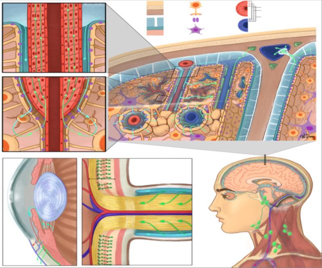
J Clin Exp Immunol, 2020 www.opastonline.com Volume 5 | Issue 3 | 126demonstrate that CSF flow more closely resembles the cardiac cycle Figure 1 Schematic of major clearance -systems in (A, B, C, F) the
than the respiratory cycle, suggesting that the primary driver of CSF brain and (D, E, F) the eye. (A, B, C) are representations of the
flow is perivascular pumping. The synchronization of diffusion- IPAD and glymphatic pathways; (A) is a cross-section of an
weighted MRI acquisition with electro-cardiogram also allows for arteriole and represents CSF flow (cyan arrows) from the SAS into
the characterization of the effect of arterial pulsatility on the the peri-arterial space, as well as ISF flow (green arrows) through
perivascular space and the surrounding fluid movement in terms of the smooth muscle basement membranes; (B) is a cross-section of
pseudo-diffusivity indices. an arteriole transitioning into a capillary, where CSF exits the peri-
arterial space via AQP4 water channels (purple) located on the
In terms of biophysical modeling, a two-compartment astrocytic end feet before mixing with ISF (cyan green arrow) and
pharmacokinetic model for GBCA-based glymphatic transport entering the smooth muscle basement membranes; (C) is a coronal
has been tested in rats with severe disruption in micro- and macro- cross-section through the head and represents the glymphatic
vasculature induced by diabetes mellitus. The contrast -pathway, dorsal mLVs, and CSF flow through an AG. CSF flows
concentration in 2 compartments (free and bound fractions) is from the SAS into peri-arterial spaces before flowing into the
represented by a system of differential equations describing brain parenchyma via AQP4 channels, mixing with ISF, and then
contrast dynamics among the arterial peri-vasculature and brain entering the perivenous space for drainage via a convective flow.
parenchyma. The model solution has a bi-exponential form whose Fluid from the SAS can then drain into the mLVs (green openings)
respective fractions and time constants allow the estimation of surrounding the SSS; (D) represents a cross-section of the anterior
contrast retention and loss to the perivascular space. This approach chamber of the eye. Cyan arrows represent production and flow of
has also been used to demonstrate the dependence of glymphatic aqueous humor from the ciliary body. Red arrow represents the
clearance on corporal position. Experiments showed reduced trabecular meshwork pathway where aqueous humor flows into
overall clearance in rats scanned in a prone position compared to the episcleral vein by passing through the Schlemm’s canal (blue
supine or right lateral decubitus positions. These results were opening). Green arrow represents the uveoscleral pathway where
verified with fluorescence and radiolabeled tracer imaging, aqueous humor flows through the interstitial trabeculae of the
showing comparatively greater CSF influx in the supine and right ciliary bodies and enters the supra-choroidal space; (E) represents
lateral decubitus positions. a cross-section of the optic nerve head. Müller cells within the
retina, which share similar functions to astrocytes in the brain, are
The spontaneous rhythmic oscillations of vascular tone in the beds represented in dark green, with appendages that wrap around
of various tissues, including the cerebral tissues, known as retinal capillaries (red dots), constituting part of the blood-retinal
vasomotion, are central to the IPAD model for brain waste barrier. The broad stripe behind the Müller cells represents the
clearance. This model encompasses a complex system of INL. The optic nerve is surrounded by SAS through which CSF
equations, representing vasomotion-induced intramural peri- flows into the optic nerve; (F) is a diagram of CSF flow within the
arterial flow through the poro-elastic basement membrane, SAS, originating from the choroid plexus (orange) within the
coupled with the elastic response of the middle cerebral artery in ventricles, as well as dorsal and basal mLVs. These mLVs
the arterial wall model. This model has been proposed to explain travelling alongside the TS and SS, exiting out of the jugular
the mechanism of IPAD pathways through the basement foramen with the internal jugular vein, and draining into the deep
membranes“[8-10]. cervical lymph nodes. Pre-auricular, submandibular, superficial
cervical, deep cervical, and supraclavicular lymph nodes are
illustrated [1-5]. IPAD, intramural periarterial drainage; CSF,
cerebrospinal fluid; SAS, subarachnoid space; ISF, interstitial
fluid; AQP4, aquaporin-4; mLVs, meningeal lymphatic vessels;
AG, arachnoid granulation; SSS, superior sagittal sinus; INL,
inner nuclear layer; TS, transverse sinuses; SS, sigmoid sinuses
(FROM REFERENCE N. 10)
Experimental Hypotesys Project
In order to test single individual efficience of brain glimphatic
system is need to obtain basal level of normal individuals. (Animal
model? population data). It is necessary to use a right treaceant
medium for imaging scope and verify kinetics. Data organized by
age and other parameters
Discussion
Related all literature reported in reference and in the text is
possible to verify that: a)“AD and PD are considered specific to
Homo sapiens” GARZIA RUIZ b)“In animal model the detox
strategy produced effect, so is possible to say that a toxicological
approach can be a way to be walked. A “total Aβ-42 by 30% in the
group of mice that received the detox gel when compared to the
untreated group with a statistical significance (p < 0.001)” is a
good result to start [5]. A new drug class able to link and detox the
Figure 1: toxicological molecule and to be carried out from brain structure
J Clin Exp Immunol, 2020 www.opastonline.com Volume 5 | Issue 3 | 127using the glimphatic system through adequate pharmacokinetics 8. Pedro J Garcia-Ruiz, ImageAlberto J Espay (2017) Parkinson
chemical group” [20-35]. “CNS waste clearance appears to be Disease: An Evolutionary Perspective. Front Neurol 8: 157.
affected by a number of physiological factors such as wakefulness,
9. Vernier P1, Moret F, Callier S, Snapyan M, Wersinger C, et al.
anesthesia regimes, exercise, age, tracer delivery, and posture.
(2004) The degeneration of dopamine neurons in Parkinson’s
CSF tracer influx appears to be suppressed in awake subjects,
disease: insights from embryology and evolution of the
increased after voluntary exercise, and increased or decreased in
mesostriatocortical system. Ann N Y Acad Sci 1035: 231-249.
sedated subjects depending on the anesthesia regimes” [36-49].
10. Wenyu Deng, Crystal Liu, Carlos Parra, Jeffrey R Sims, Muneeb A
Conclusion Faiq, et al. (2020) Quantitative imaging of the clearance systems in
Related the topics of this works observing the literature reported and the eye and the brain. Quant Imaging Med Surg 10: 1-14.
other in biomedical database is clear. That imaging new technique
11. Abbott NJ (2013) Blood–brain barrier structure and function
are needed to adequately evaluate the brain washing system in order
and the challenges for CNS drug delivery. Journal of inherited
to verify. In example individual clearance efficacy towards various
metabolic disease 36: 437-449.
toxic movens (alfa synuclein, neuronal cell inclusion, Free radicals,
Immune molecule and other). 12. Abbott NJ, Patabendige AA, Dolman DE, Yusof SR, Begley
DJ (2010) Structure and function of the blood–brain barrier.
In all literature observed there is no a systematic measure of this Neurobiology of disease 37: 13-25.
parameter in groups o patients whit neurodegenegerative of
13. Achariyar TM, Li B, Peng W, Verghese PB, Shi Y, et al. (2016)
neuroinflamatory disease. A better quantitative measure of this
Glymphatic distribution of CSF-derived apoE into brain is
clearance make possible to find new therapeutic strategies.
isoform specific and suppressed during sleep deprivation.
Molecular neurodegeneration 11: 74.
Clarifications
This work have not any diagnostic or therapeutic intent, only produced 14. Brinker T, Stopa E, Morrison J, Klinge P (2014) A new look at
to submit to researcher new research hypothesys cerebrospinal fluid circulation. Fluids and Barriers of the CNS
11: 10.
Conflict of Interests: The authors declare that they have no competing 15. Damkier HH, Brown PD, Praetorius J (2013) Cerebrospinal
interests. fluid secretion by the choroid plexus. Physiological reviews
93: 1847-1892.
References
1. Luisetto M (2017) Brain response in some systemic immune 16. Davson H, Segal MB (1996) Physiology of the CSF and
condition-Toxicological aspects. Insights Clin Cell Immunol blood-brain barriers. Boca Raton: CRC 1.
1: 005-008. 17. Del Zoppo GJ, Moskowitz M, Nedergaard M (2016) The
2. Luisetto M, Farhan Ahmad K, Ahmed Yesvi R, Behzad Nili A, neurovascular unit and responses to ischemia. In Stroke
Ghulam Rasool M (2019) Decussatio Pyramid and Optical Elsevier 2016: 90-101.
Chiasm as an Interesting Example of Evolutionary Process 18. Di Terlizzi R, Platt S (2006) The function, composition and
Useful in Understanding Some Spinal Cord Phenomena? On J analysis of cerebrospinal fluid in companion animals: Part I–
Neur & Br Disord 3: 2019. Function and composition. The Veterinary Journal 172: 422-
3. Luisetto M, Khan FA, Muhamad A, Mashori GR, Ahmadabadi 431.
BN, et al. (2020) Brain washing systems and other circulating 19. Engelhardt B, Ransohoff RM (2012) Capture, crawl, cross:
factors in some neurological condition like Parkinson (Pd) and the T cell code to breach the blood–brain barriers. Trends in
vascular and diabetic dementia: How dynamics- saturation of immunology 33: 579-589.
clearance can act on toxic molecule? J Neurosci Neurol Disord 4:
001-013. 20. Hladky SB, Barrand MA (2016) Fluid and ion transfer across
the blood–brain and blood–cerebrospinal fluid barriers; a
4. Luisetto M, Behzad Nili-Ahmadabadi, Ghulam Rasool Mashori, comparative account of mechanisms and roles. Fluids and
Ahmed Yesvi Rafa, Ram Kumar Sahu, et al. (2018) “Brain and Barriers of the CNS 13: 19.
Immune System: KURU, a Strange Kind of Disease. An
Endogenous Toxicological Process Like?” EC Neurology 10: 21. Iliff JJ, Lee H, Yu M, Feng T, Logan J, et al. (2013) Brain-
613-625. wide pathway for waste clearance captured by contrast-
enhanced MRI. The Journal of clinical investigation 123:
5. Taoka T, Naganawa S (2020) Glymphatic imaging using MRI. 1299-1309.
J Magn Reson Imaging 51: 11-24.
22. Iliff JJ, Wang M, Liao Y, Plogg BA, Peng W, et al. (2012) A
6. Davoodi-Bojd E, Ding G, Zhang L, Li Q, Li L, et al. (2019) paravascular pathway facilitates CSF flow through the brain
Modeling glymphatic system of the brain using MRI. parenchyma and the clearance of interstitial solutes, including
Neuroimage 188: 616-627. amyloid β. Science translational medicine 4: 147ra111-
7. Per Kristian Eide, Svein Are Sirirud Vatnehol, Kyrre Eeg 147ra111.
Emblem, Geir Ringstad (2018) Magnetic resonance imaging 23. Jessen NA, Munk ASF, Lundgaard I, Nedergaard M (2015)
provides evidence of glymphatic drainage from human brain The glymphatic system: a beginner’s guide. Neurochemical
to cervical lymph nodes. Scientific Reports 8: 7194. research 40: 2583-2599.
J Clin Exp Immunol, 2020 www.opastonline.com Volume 5 | Issue 3 | 12824. Johnston M, Zakharov A, Papaiconomou C, Salmasi G, 37. Syková E, Nicholson C (2008) Diffusion in brain extracellular
Armstrong D (2004) Evidence of connections between space. Physiological reviews 88: 1277-1340.
cerebrospinal fluid and nasal lymphatic vessels in humans,
38. Tait MJ, Saadoun S, Bell BA, Papadopoulos MC (2008) Water
non-human primates and other mammalian species.
movements in the brain: role of aquaporins. Trends in
Cerebrospinal fluid research 1: 2.
neurosciences 31: 37-43.
25. Koh L, Zakharov A, Johnston M (2005) Integration of the
39. Thorne RG, Nicholson C (2006) In vivo diffusion analysis
subarachnoid space and lymphatics: is it time to embrace a
with quantum dots and dextrans predicts the width of brain
new concept of cerebrospinal fluid absorption? Cerebrospinal
extracellular space. Proceedings of the National Academy of
fluid research 2: 6.
Sciences 103: 5567-5572.
26. Kress BT, Iliff JJ, Xia M, Wang M, Wei HS, et al. (2014)
40. Thorne RG (2014) Primer on central nervous system structure/
Impairment of paravascular clearance pathways in the aging
function and the vasculature, ventricular system, and fluids of
brain. Annals of neurology 76: 845-861.
the brain. Drug Delivery to the Brain. Springer: New York
27. Kulik T, Kusano Y, Aronhime S, Sandler AL, Winn HR (2008) 2014: 685-706.
Regulation of cerebral vasculature in normal and ischemic
41. Thrane AS, Thrane VR, Nedergaard M (2014) Drowning
brain. Neuropharmacology 55: 281-288.
stars: reassessing the role of astrocytes in brain edema. Trends
28. Mathiisen TM, Lehre KP, Danbolt NC, Ottersen OP (2010) in neurosciences 37: 620-628.
The perivascular astroglial sheath provides a complete
42. Thrane VR, Thrane AS, Plog BA, Thiyagarajan M, Iliff JJ, et
covering of the brain microvessels: an electron microscopic
al. (2013) Paravascular microcirculation facilitates rapid lipid
3D reconstruction. Glia 58: 1094-1103.
transport and astrocyte signaling in the brain. Scientific
29. Murtha LA, Yang Q, Parsons MW, Levi CR, Beard DJ, et al. reports 3: 2582.
(2014) Cerebrospinal fluid is drained primarily via the spinal
43. Trevaskis NL, Kaminskas LM, Porter CJ (2015) From sewer
canal and olfactory route in young and aged spontaneously
to saviour—targeting the lymphatic system to promote drug
hypertensive rats. Fluids and Barriers of the CNS 11: 12.
exposure and activity. Nature Reviews Drug Discovery 14:
30. Nedergaard M (2013) Garbage truck of the brain. Science 781-803.
340: 1529-1530.
44. Xie L, Kang H, Xu Q, Chen MJ, Liao Y, et al. (2013) Sleep
31. Neuwelt EA, Bauer B, Fahlke C, Fricker G, Iadecola C, et al. drives metabolite clearance from the adult brain. science 342:
(2011) Engaging neuroscience to advance translational 373-377.
research in brain barrier biology. Nature Reviews Neuroscience
45. Zhang ET, Inman CB, Weller RO (1990) Interrelationships of
12: 169.
the pia mater and the perivascular (Virchow-Robin) spaces in
32. Neuwelt EA (2004) Mechanisms of disease: the blood-brain the human cerebrum. Journal of anatomy 170: 111.
barrier. Neurosurgery 54: 131-142.
46. Zhao Z, Nelson AR, Betsholtz C, Zlokovic BV (2015)
33. Pizzo ME, Thorne RG (2017) The extracellular and Establishment and dysfunction of the blood-brain barrier. Cell
perivascular spaces of the brain. In Brain edema 2017: 105- 163: 1064-1078.
127.
47. Zlokovic BV (2011) Neurovascular pathways to neurodegeneration
34. Prince EA, Ahn SH (2013) Basic vascular neuroanatomy of in Alzheimer’s disease and other disorders. Nature Reviews
the brain and spine: what the general interventional radiologist Neuroscience 12: 723.
needs to know. In Seminars in interventional radiology 30:
48. Luisetto M, Ahmadabadi BN, Rafa AY, Sahu RK, Cabianca L,
234-239.
et al. (2019) The turing machine theory for some spinal cord
35. Rennels ML, Gregory TF, Blaumanis OR, Fujimoto K, Grady and brain condition, A toxicological – antidotic depurative
PA (1985) Evidence for a ‘paravascular’ fluid circulation in approach. J Neurosci Neurol Disord 3: 102-134.
the mammalian central nervous system, provided by the rapid
49. Mauro Luisetto, G Ibrahim, Oleg latyschev, Muhammad
distribution of tracer protein throughout the brain from the
Akram (2019) The Evolution of the Nervous System as Model
subarachnoid space. Brain research 326: 47-63.
forSearch New Pharmacological Strategies in Human
36. Rennels ML, Blaumanis OR, Grady PA (1990) Rapid solute Neurological Condition. American Journal of Biomedical
transport throughout the brain via paravascular fluid pathways. Science & Research 5: 000960.
Advances in neurology 52: 431-439.
Copyright: ©2020 Luisetto M, et al. This is an open-access article distributed
under the terms of the Creative Commons Attribution License, which permits
unrestricted use, distribution, and reproduction in any medium, provided the
original author and source are credited.
J Clin Exp Immunol, 2020 www.opastonline.com Volume 5 | Issue 3 | 129You can also read