Juvenile Cataracts in West Highland White Terriers - Westie ...
←
→
Page content transcription
If your browser does not render page correctly, please read the page content below
Juvenile Cataracts in
                             West Highland White Terriers
                                       By Lindsey Buracker, DVM and John Robertson, VMD, PhD

I ntroduction
  Dogs have a very keen sense of vision, with an ability to see
in extreme conditions of light and darkness, and to be highly
                                                                         ability to heal. Consequently, the shape of the lens changes
                                                                         during life.

                                                                         There are three main components of the lens, namely the
perceptive of movement. Although we generally don’t think of
                                                                         capsule, surface layer or epithelium, and fibers. The capsule is
them as primal predators, Westies are born with these instincts
                                                                         a thickened smooth membrane made of collagen and produced
and need excellent vision. To make sense of what happens when
                                                                         by the lens epithelium and fibers. It completely surrounds the
cataracts develop, it is important to first have a solid understanding
                                                                         lens and has elastic properties, so when not under tension, the
of the anatomy and physiology of the eye.
                                                                         lens assumes a rounded shape. The epithelium is comprised of
                                                                         cells that elongate over time and are eventually transformed
The Anatomy of the Eye and                                               into lens fibers, which contain high concentrations of the
the Phenomenon of Vision                                                 protein crystalline. It is this protein that helps the lens refract
The eye is comprised primarily of two connected chambers.                and transmit light. The fibers are tightly packed and extend the
The smaller of the two chambers, called the anterior chamber, is         full length of the lens. Continual growth of the lens adds more
bounded by the transparent cornea at the front of the eye and the        elongated cells and fibers and produces an arrangement similar
lens posteriorly. The larger posterior chamber, which is bounded         to the layers of an onion (Magrane, 1972). Damage to any of the
externally by the tough outer layer called the sclera, contains the      components of the lens can result in a cataract.
lens, the gelatinous vitreous humor, and the retina. The retina is a
membrane comprised primarily of neural receptors that respond            The formation of the lens helps orchestrate the overall
to light and initiate the visual pathway. The eye also contains          development of the eye, as it forms relatively early and helps
blood vessels in the uveal tract and connective tissue.                  induce the formation of both chambers and other parts of the
                                                                         eye. This pivotal role of the lens in controlling development of
The lens is an amazing tissue. It is a tight clustering of               the eye is important for several reasons. First, if the lens does
specialized cells, enclosed in a capsule, located behind the             not properly form, this can affect the development of other parts
iris and in front of the vitreous body, held in place by fibers,         of the eye. Second, disease or defective gene expression that
the anterior vitreous face, and the iris (Magrane, 1972). The            occurs during pregnancy can significantly affect formation of
lens is normally quite flexible, and its shape is controlled by          the lens and, by extension, the development of healthy eyes.
small muscles and fibers that either tense or relax its edges. By        Third, the presence of cataracts at birth not only is indicative
changing its shape, the lens alters its refractive power to bring        of abnormalities in lens development, but also may signal
objects into focus, depending on whether they are near or far            the potential for problems elsewhere in the eye. Finally, it is
away. The lens grows in size with age, and requires nutrients that       important to remember that puppies are born with incompletely
reach it by diffusion through the aqueous and vitreous humors.           matured eyes and some of the process of development takes
It also can be damaged by injury and disease, and has a limited          place after birth. A good rule of thumb is that formation of the

                                               Common Clinical Findings
                                            Age When Recognized Varies
                                               Autosomal Recessive Trait
                               Common Ancestors or Selective Breeding
                              Examination by Veterinary Ophthalmologist
                                                                                                                         (Continued on page 5)

 4                                       www.westiefoundation.org                                                             Spring 2020
(Juvenile Cataracts from page 4)
present longer and may involve the entire lens. In some mature
cataracts, the cells in the lens have degenerated and liquefied.
This debris persists within the lens capsule.
Many cataracts are more common in older dogs than young
dogs, and develop in older dogs as a result of ocular disease
(e.g., glaucoma, panophthalmitis and uveitis), systemic disease
(e.g., diabetes mellitus), exposure to certain chemicals, as a
side effect of radiation therapy of the head and neck, or direct
penetrating trauma to the eye that damages the lens capsule and
lens cells. These cataracts are considered to be “acquired” as a
result of the initiating process.
Two specific ocular diseases associated with cataracts deserve
specific mention: uveitis and glaucoma. Uveitis, an inflammation
of the vascular (‘uveal’) layer of the eye, can be caused directly
by degeneration of the lens, in some cases. With the formation
and disruption of mature and hypermature cataracts, lens protein
can leak from inside the lens capsule into the anterior chamber,
spontaneously or as a result of trauma, and induce severe
inflammation.
The relationship between glaucoma and cataract formation
is complex. Glaucoma is a disease condition characterized
by elevated intraocular pressure. In some cases of glaucoma,
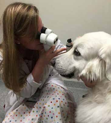
Figure 1 - Examination of a dog’s eyes using a specialized slit lamp ophthalmoscope.
interference with the production and drainage of fluids within
the eye is the primary disease process that increases intraocular
pressure. The increased pressure within the eye may damage
eye is complete by about 12 weeks of age, in most breeds of dog. the lens, resulting in the formation of cataracts. In other cases,
Dogs should have good visual acuity by this age. cataracts and other lens diseases may be a cause of glaucoma.
Vision is an interesting phenomenon. In essence, light energy For example, lenses with cataracts may become dislodged from
from the surroundings produces electrochemical changes in their normal fibrous connections and migrate into the pupil,
specialized nerve cells called rods and cones in the retina. These where they occlude the normal flow of fluid from the posterior
changes result in the generation of signals called ‘nerve action chamber to the anterior chamber. This essentially blocks the
potentials’ that are relayed to the brain, where they are processed drainage of fluid and pressure increases within the eye, resulting
and consciously appreciated as a vision (Magrane, 1972). The in glaucoma.
lens is a key part of the system that focuses and transmits light
to the retina so that signals that eventually produce vision are Juvenile Cataracts
received on the retina. Cataracts also may form as the result of a defect during
development of the eye. These cataracts, which are known
An Introduction to Cataracts as juvenile cataracts, either may form before birth or develop
Simply defined, a cataract is an irregularity and opacity in shortly after birth as the dog’s eyes mature. Juvenile cataracts
may be caused by the expression of defective genes and/or
the lens. In most cases, the cataract appears as a cloudy white
viral infections that occur during gestation or in newborns. In
discoloration in the lens. It is important to know, that cataracts
some breeds of dogs, the incidence of cataracts increases with
can affect only one or both eyes. When cataracts develop in
age; these cataracts are considered to be hereditary in origin.
the center of the lens, they will interfere with the path of light
Regardless of the cause, the outcome is the same – a decrease in
energy to the retina at the back of the eye, thereby impairing
visual acuity for the dog. Some inherited cataracts that appear
visual acuity. Cataracts often are classified as either ‘immature’
early in the dog’s life may result in blindness by the time the dog
or ‘mature,’ terms that refer to the developmental stage of the
is 3 years of age. Other late onset inherited cataracts often do not
cataract. Immature cataracts are newly formed and may occupy
interfere with vision and are identified before the dog reaches 8
only a portion of the lens, whereas mature cataracts have been
years of age.
(Continued on page 6)
Spring 2020 www.westiefoundation.org 5“
(Juvenile Cataracts from page 5)
Juvenile cataracts may form before birth or
develop shortly after birth as the dog’s eyes mature.”
Most inherited cataracts in dogs are inherited as autosomal Noticing Your Dog’s Eyes and Behavior
recessive traits, such as the mutation in heat shock transcription Owners and breeders are often the first to detect a problem with
factor gene, HSF4, which is responsible for recessively inherited a dog’s eyes and vision. Some common signs that something is
cataracts in Boston Terriers, Staffordshire Bull Terriers and not right include:
French Bulldogs (Mellersh et. al., 2006). Typically, dogs are
affected bilaterally, the cataracts are located in the posterior • The eyes, lids, and membranes of the eye just don’t look right;
region of the lens, and the rate at which they grow is highly there may be milky white/opaque discoloration, irregularities
variable. Interestingly, Muller and coworkers (2008) were in shape and size, or perhaps the eyes are inappropriately
not able to identify mutations in HSF4 in Dachshunds or proportioned to the dog’s head.
Enttlebucher Mountain Dogs with hereditary cataracts. • Puppies may bump into things in their path, and observations
that must be differentiated from just clumsiness or poor
Another mutation of the HSF4 gene affects Australian coordination.
Shepherds, but is different from the mutation in Boston Terriers, • Puppies appear reluctant to move about or are overly shy;
French Bulldogs and Staffordshire Bull Terriers (Mellersh et. most Westie puppies are pretty affable and playful.
al., 2009). This mutation is dominant, meaning that only a single • Puppies are reluctant to explore darkened areas.
copy is needed to predispose a dog to the disease. Fortunately, • Puppies appear to cue interactions based on hearing rather
not all dogs with this mutation develop cataracts, suggesting that than on both hearing and seeing.
one or more other gene interactions are involved in the process. If a problem with vision is suspected, your dog’s eyes should be
Many breeds of dogs appear to be predisposed to developing examined by your veterinarian.
juvenile cataracts, including the West Highland White Terrier.
To examine this concept, Oberbauer and colleagues recently Eye Examination by a Veterinarian
compared the prevalence of ten inherited disorders, including All thorough physical examinations of dogs include an
early onset cataracts, in purebred and mixed breed dogs in evaluation of the eyes. Most evaluations by veterinarians include
a study of more than 88,000 dogs. They determined that the common elements, and some of the routine evaluation is done
prevalence of early onset cataracts in most purebred groups with simple tests:
was not different from that in mixed breed populations. They
concluded that groups with higher specific disorders may have
• Evaluation of the gross appearance of both eyes, comparison
of one eye with the other and with the head in terms of size,
common ancestors or this could be an effect of selecting for
shape, coloration, tone, and integration with facial shape,
specific structural features (e.g., shape or size).
• Visual signal processing, based on the pupillary responses
Many puppies appear normal at birth, many do not show signs (constriction) to light shined in one eye. This is actually a
until six months to two years of age, and some may have the simple test of a complex process, as it tests whether light
cataracts appear after five years. Consequently, there is no way focused on the retina then creates a visual ‘signal’ that is then
to know if the puppy you are buying is going to develop juvenile transmitted via nerve fibers to the brain. At that point, the
cataracts. Fortunately, juvenile cataracts do not always lead to signal is interpreted as vision.
blindness. In many cases, the puppy or young dog still sees basic • The ability to track movement in a lighted area is assessed by
shapes, but they may be blurry. In some cases, the disease leads the dog’s responses to hand movements near the eyes. The
to the development of glaucoma. veterinarian will determine if the dog will blink in response
to movement near the eyes – assessing both visual perception
The only way to eradicate juvenile cataracts in dogs is for and the automatic blink response.
breeders to have both parents evaluated fully by a licensed • Tone (palpable firmness) of the eyes can be first evaluated by
veterinary ophthalmologist no more than a year before breeding. gently applying pressure through the lids. Most dogs do not
Because not all breeders do this, it is advisable to ask for eye mind this part of the examination and it helps determine if the
registry papers for both parents before agreeing to purchase a eyes are firm, but not too firm, and if there are irregularities or
puppy. pain,
• Ophthalmic evaluation of the anterior chamber, posterior
chamber, and intraocular structures (lens, iris, pupil, retina).
(Continued on page 7)
6 Spring 2020(Juvenile Cataracts from page 6)
Figures 2 - 4 - Example of the different ways that cataracts can appear in dogs.
The value to Westie owners in seeking regular evaluations of either sire or dam until the relationship between breeding and
their dogs should be evident. Health problems can be detected, cataract development can be clearly determined. Hereditary
diagnosed and most are treated effectively. Many eye problems cataracts were first identified as a significant problem in the
can be detected with the above approach. When more complex Miniature Schnauzer breed in the 1970s and 1980s. Following
treatments, such as surgery, are needed to treat problems and the leadership provided by breed associations, veterinarians
to correct defects, it makes sense to seek the services of a and research scientists, this autosomal recessive trait was
specialist, such as a veterinary ophthalmologist. These specialists identified and bred against, resulting in a substantial decrease
have the facilities and equipment needed for more extensive in the incidence of the disease in Miniature Schnauzers today.
diagnostic approaches and for treatment (Figure1). Because they By identifying those dogs that are carriers of the disease and
concentrate exclusively on treating diseases of the eye, they have not breeding them, juvenile cataracts can be controlled and
seen more cases, many of which are the more difficult ones, and eventually eliminated. The importance of keeping accurate
will be more familiar with the variety of abnormalities affecting breeding records and long-term follow-up information on litters
the lens (Figures 2 – 4). Veterinary opthalmologists are certified cannot be overstated.
by examination boards after years of advanced training and
experience treating diseases of the eye. Potential owners need to do an extensive “background check”
before purchasing a Westie from a breeder. These potential
Prevention of Cataracts Westie owners need to be sure there are accurate records for
Since we know that some types of cataracts have a hereditary each dam and sire, a solid bloodline, and no overt problems or
basis (Table 1), it is essential for dog breeders to keep thorough diseases noted in each litter from the time of whelping. If Westie
records of litters and diseases affecting each pup. Breeders owners and breeders work together, this disease will eventually
should keep in regular contact with the owners of pups from be eliminated from breeding stock.
their litters throughout the lives of these dogs. The presence of
cataracts in young dogs (less than 6 months old) and in multiple Treatment
dogs from the same breeding is very suggestive of an underlying Before dogs with cataracts undergo surgical treatment, it is
genetic problem. One caveat – if, during gestation, there is important to determine whether or not the dog is experiencing
evidence of ill-health in the dam, cataracts may be the result of any vision problems. In other words, if the cataracts are
damage to the developing puppies. Most experienced breeders relatively small and the dog is able to see sufficiently or can
are very aware of the need to keep pregnant dams well-nourished compensate for the impairment in vision, treatment isn’t
and free from exposure to potentially damaging viruses and needed. In some dogs, juvenile cataracts do not become more
chemicals in the environment. severe or do so very slowly. In other dogs, the severity of the
cataracts may change and other problems, such as glaucoma
In the event that a litter is delivered and one or more pups and inflammation of the eye, may develop. Consequently, it is
develop cataracts, the breeder has a responsibility to 1) seek advisable to have the dog’s eyes checked on a regular basis.
veterinary diagnosis and discuss treatment options for affected When cataracts interfere with vision, the dogs may have trouble
pups, 2) examine the breeding and pedigree of both dam and finding their way in their environment, locating food or water,
sire for similar problems (or the presence of other congenital and be reluctant to walk or run. In these cases, a visit to the
defects from this paired breeding), and 3) refrain from breeding veterinarian is warranted.
(Continued on page 8)
Spring 2020 7(Juvenile Cataracts from page 7)
Table 1. Inherited Cataracts In The Dog (Gelatt, 2008). time additional long term follow-up examinations will be
scheduled, since there remains a risk for complications.
Breed Age of Onset
Afghan Hound 6-12 Months Current Research About Juvenile
American Cocker Spaniel 6+ Months Cataracts in West Highland White
Terriers
Boston Terrier Congenital There has been a considerable amount of new information in the
Chesapeake Bay Retriever 1+ Years veterinary scientific literature in the past decade regarding the
pathogenesis, diagnosis and treatment of cataracts in dogs. The
German Shepherd 8+ Weeks following three recent studies would seem to be of most interest,
Golden Retriever 6+ Months as one concerns the clinical manifestations of the condition in
small breeds of dogs examined in Korea, one reviews the clinical
Labrador Retriever 6+ Months presentation of dogs with the disease in France, and the third
compares the prevalence of another important eye disease of
Miniature Schnauzer Congenital or 6+ Months
dogs covered in this eBook, keratoconjunctivitis sicca, in two
Old English Sheepdog Congenital populations of dogs after surgical treatment for cataracts.
Siberian Husky 6+ Months Park SA, Yi NY, Jeong MB, Kim WT, Kim SE, Chae JM,
Seo KM. Clinical manifestations of cataracts in small
Staffordshire Bull Terrier 6+ Months
breed dogs. Vet Ophthalmol. 2009 Jul-Aug;12(4):205-10.
Standard Poodle 1+ Years Because the majority of earlier clinical studies of cataracts in
dogs had involved middle and large breed dogs, this study was
Welsh Springer Spaniel Congenital performed to characterize the condition in small breed dogs
West Highland White presented to a veterinary teaching hospital in Korea. More than
Congenital 560 small breed dogs were included in this study, with the most
Terrier
frequently presented breeds being the Miniature/Toy Poodle (n
If cataracts are interfering with the dog’s vision, the treatment = 112, 20.0%), Yorkshire Terrier (n = 110, 19.6%), and Shih
requires surgical intervention by a veterinary ophthalmologist. Tzu (n = 95, 16.9%). The investigators noted that significantly
Before this is done, however, specialized tests including an ERG more female dogs were presented with cataracts than male dogs.
and ocular ultrasound will be performed to ensure that the dog’s The average age of affected dogs was 8.3 years, with Miniature/
retina is functioning normally. If the retina is not normal, the Toy Poodles and Yorkshire Terriers being significantly older and
end result of surgery is not likely to be an improvement in visual Miniature Schnauzers being significantly younger. While this
acuity. study focused primarily on the incidence of cataracts and their
clinical features, additional studies will need to be performed
The most commonly used surgical technique involves using to determine the prognosis associated with different types of
ultrasonic waves to transform the lens to a liquid, which then can treatments.
be removed through a small incision. This technique, which is
performed with the dog under general anesthesia, also is referred Donzel E, Arti L, Chahory S. Epidemiology and clinical
to as phacoemulsification. In many cases, an acrylic implant will presentation of canine cataracts in France: a retrospective
be inserted to replace the lens that has been removed. study of 404 cases. Vet Ophthalmol. 2016 Apr7. 1-9.
Although the prevalence of cataracts in dogs has been reported
Prognosis and Follow-up Care previously in North and South America and Korea, little
Although there is a risk of complications with any surgery, was known about the disease in Europe. Consequently, the
the short-term prognosis for the return of visual acuity after investigators undertook this study of more than 2,700 dogs
surgery exceeds 90%. The long-term outcome regarding restored presented for evaluation at a veterinary school in France.
or improved vision ultimately depends on the stage of the Of these dogs, 404 had cataracts; 54% were males and 46%
cataract at the time surgery is performed and other co-existing females. The mean age of all dogs with cataracts was 9 years,
conditions. A protective collar will be applied to prevent the and 54 breeds were represented. Of these, the Yorkshire Terrier
dog from scratching the eye and initially frequent eye drops was the only breed significantly overrepresented. The major
or lubricants will need to be administered. Typically, dogs that causes of cataracts in this population were breed predisposition,
undergo surgery are examined 1 week after surgery, at which aging, and progressive retinal atrophy.
(Continued on page 9)
8 Spring 2020(Juvenile Cataracts from page 8)
Gemensky-Metzler AJ, Sheahan JE, Rajala-Schultz
PJ, Wilkie DA, Harrington J. Retrospective study of
the prevalence of keratoconjunctivitis sicca in diabetic
and nondiabetic dogs after phacoemulsification. Vet
Ophthalmol. 2015 Nov;18(6):472-80.
Diabetes mellitus occurs commonly in dogs, and often is
complicated by the formation of cataracts. In fact, 50% of
diabetic dogs have been reported to develop cataracts within
6 months of being diagnosed with cataracts. When cataracts
interfere with visual acuity, phacoemulsification is used to
help improve vision. It has recently been determined that
tear production, as measured using the Schirmer tear test,
is significantly lower in diabetic dogs with cataracts than in
nondiabetic dogs with cataracts. Therefore, the investigators
hypothesized that keratoconjunctivitis sicca would be more
common in diabetic dogs after phacoemulsification. This
study, which involved 117 nondiabetic dogs and 118 diabetic intraocular lens implants following phacoemulsification and aspiration of
dogs, determined that the greatest risk for the development of cataracts in dogs. Vet Ophthalmol. 2009 Jan-Feb;12(1):13-21.
keratoconjunctivitis sicca for all dogs is during the first 2 weeks Klein HE, Krohne SG, Moore GE, Stiles J. Postoperative complications and
after surgery, and that the populations at greatest risk are small visual outcomes of phacoemulsification in 103 dogs (179 eyes): 2006-2008. Vet
Ophthalmol. 2011 Mar;14(2):114-20.
dogs, small diabetic dogs, and large dogs with preoperative
Schirmer tear test resultsYou can also read