Kaposi Sarcoma-Like Lesions Caused by Candida guilliermondii Infection in a Kidney Transplant Patient
←
→
Page content transcription
If your browser does not render page correctly, please read the page content below
Brief Report
https://doi.org/10.5021/ad.2021.33.1.91
Kaposi Sarcoma-Like Lesions Caused by Candida
guilliermondii Infection in a Kidney Transplant Patient
1 1 2 2 3,4 1
Soo-Jung Kim , Jung-Min Shin , Kang Wook Lee , Yeon-Sook Kim , Babar Rao , Young Lee
Departments of 1Dermatology and 2Internal Medicine, School of Medicine, Chungnam National University, Daejeon, Korea,
3
Department of Dermatology, Rutgers Robert Wood Johnson Medical School, Somerset, NJ, 4Department of Dermatology, Weill Cornell
Medical Center, New York, NY, United States
Dear Editor: deep fungal infection. The histopathological examination
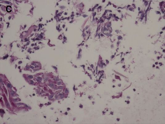
Candida guilliermondii is a uncommon newly emerging showed parakeratosis, pseudoepitheliomatous hyperplasia,
opportunistic pathogen found both in nature and as a and mixed cell infiltration in the dermis (Fig. 2A). Human
component of the normal human microbial flora1,2. In re- herpes virus 8 (HHV-8), Periodic acid-Schiff (PAS), and Go-
cent years, the rate of C. guilliermondii infections has been mori’s methenamine silver (GMS) staining was also per-
increasing, and which are associated mainly with onycho- formed. D-PAS and GMS staining showed multiple small
mycosis3. A case of Majocchi’s granuloma resulting from a yeast cells in the dermis (Fig. 2B, C) and HHV-8 staining
deep granulomatous dermatophyte infection and mimick- was negative. Tissue culture on Sabouraud agar showed
ing Kaposi sarcoma was reported4. However, a case of deep white, smooth, glabrous yeast-like colonies (Fig. 2D). To
dermal Candida infection mimicking Kaposi sarcoma has determine the strain of infected fungi, we did polymerase
not been reported yet. chain reaction (PCR) using internal transcribed spacer (ITS)
A 79-year-old male presented with multiple erythematous primers. We detected two bands in PCR results and per-
to black verrucous nodules on his right foot that appeared formed sequencing using ITS1 and ITS4 primers with ex-
1 month prior. The lesions were distributed in a sporotri- tracted PCR products (Fig. 2E). Sequencing results of PCR
choid lymphocutaneous pattern. The patient complained product in the lower band was analyzed by using BLAST
of tenderness and pain around the lesions, which were and confirmed C. guilliermondii. The patient was treated
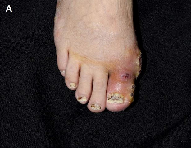
continuously discharging a exudate, forming a crust (Fig. with terbinafine, 125 mg daily, considering the renal func-
1A, B). In addition, the patient had onychomycosis, white tion and drug interaction with tacrolimus. After 4 months
and thick in appearance. The patient suffered from end-stage of treatment, the lesions improved dramatically (Fig. 1C, D).
renal disease due to diabetes mellitus. He had received a Superficial dermatomycosis is fairly prevalent in transplant
kidney transplant 3 months prior and was on immuno- recipients and its rate increases as patients receive massive
suppressive treatment with tacrolimus and steroids. immunosuppressive treatment. However, deep dermato-
As the patient was treated with immunosuppressive ther- mycosis involving the dermis and subcutaneous layer by
apy, we suspected the diagnosis of Kaposi sarcoma and dermatophytes and yeast, is relatively rare5. Deep derma-
tophytosis often presents with multiple nodules on lower
extremities, accompanied by superficial fungal infection5.
Received January 10, 2020, Revised February 7, 2020, Accepted for publi- There is no report of a similar case involving yeast, let
cation February 7, 2020
alone species as uncommon as C. guilliermondii. In our
Corresponding author: Young Lee, Department of Dermatology, Chungnam
National University Hospital, 282 Munhwa-ro, Jung-gu, Daejeon 35015, case, the patient presented with deep dermal dermatomy-
Korea. Tel: 82-42-280-7700, Fax: 82-42-280-8459, E-mail: resina20@cnu.ac.kr cosis with suppurative granuloma, resembling Kaposi sar-
ORCID: https://orcid.org/0000-0001-9205-1785 coma, caused by C. guilliermondii. The patient had severe
This is an Open Access article distributed under the terms of the Creative onychomycosis of toenails, which we suspected to be the
Commons Attribution Non-Commercial License (http://creativecommons.
org/licenses/by-nc/4.0) which permits unrestricted non-commercial use, cause of deep dermatomycosis.
distribution, and reproduction in any medium, provided the original work In conclusion, this case demonstrates the clinical diversity
is properly cited. of deep dermatomycosis in an immunocompromised pa-
Copyright © The Korean Dermatological Association and The Korean
tient, caused by C. guilliermondii. When multiple erythema-
Society for Investigative Dermatology
V ol. 33, N o. 1, 2021 91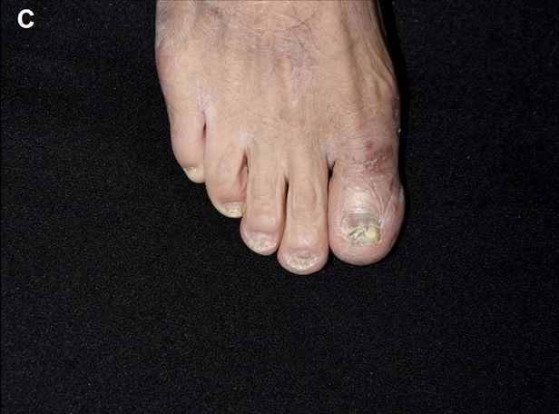
Brief Report

                                                                                        Fig. 1. (A, B) Multiple erythematous
                                                                                        to black nodules on the right foot
                                                                                        at initial presentation. (C, D) Four
                                                                                        months after treatment with oral
                                                                                        terbinafine.

                                                                                        Fig. 2. (A) Parakeratosis, pseudo-
                                                                                        epitheliomatous hyperplasia of epi-
                                                                                        dermis and mixed cell infiltration
                                                                                        in dermis (H&E, ×40). (B, C) Mul-
                                                                                        tiple small yeasts with narrow based
                                                                                        budding in dermis (B: GMS stain,
                                                                                        ×200; C: PAS stain, ×400). (D)
                                                                                        White and smooth glabrous yeast-
                                                                                        like colonies on Sabouraud agar.
                                                                                        (E) Result of polymerase chain re-
                                                                                        action amplification of the internal
                                                                                        transcribed spacer (ITS) region of
                                                                                        the fungus using ITS1 and ITS4 pri-
                                                                                        mers.

tous nodules mimicking Kaposi sarcoma are found in low-        this information.
er extremities of immunocompromised patients, a deep der-
matomycosis by Candida species should also be consi-           CONFLICTS OF INTEREST
dered. Tissue culture and PCR are helpful for diagnosis if
the infection is caused by a rare fungus species, such as C.   The authors have nothing to disclose.
guilliermondii.
                                                               FUNDING SOURCE
ACKNOWLEDGMENT
                                                               This research was supported by Basic Science Research
We thank the patient for granting permission to publish        Program through the National Research Foundation of Korea

92 Ann D erm atol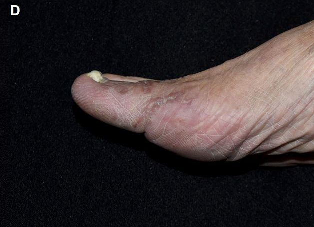
Brief Report

(NRF) funded by Ministry of Education, Science and Tech-               ten-year survey. J Mycol Med 2017;27:506-513.
nology (NRF-2019R1A2C1004114).                                      2. Nakazawa H, Nishina S, Senoo Y, Sakai H, Ito T, Kikuchi K,
                                                                       et al. Breakthrough Candida guilliermondii (Meyerozyma
DATA SHARING STATEMENT                                                 guilliermondii) fungemia after cord blood transplantation for
                                                                       extranodal NK-cell lymphoma with azole prophylaxis. Transpl
Research data are not shared.                                          Infect Dis 2018;20:e12922.
                                                                    3. Pfaller MA, Diekema DJ, Mendez M, Kibbler C, Erzsebet P,
ORCID                                                                  Chang SC, et al. Candida guilliermondii, an opportunistic
                                                                       fungal pathogen with decreased susceptibility to fluconazole:
Soo-Jung Kim, https://orcid.org/0000-0002-5140-1432                    geographic and temporal trends from the ARTEMIS DISK anti-
Jung-Min Shin, https://orcid.org/0000-0001-7465-5243                   fungal surveillance program. J Clin Microbiol 2006;44:3551-
Kang Wook Lee, https://orcid.org/0000-0003-3407-1205                   3556.
Yeon-Sook Kim, https://orcid.org/0000-0003-1142-5488                4. Brod C, Benedix F, Röcken M, Schaller M. Trichophytic
Babar Rao, https://orcid.org/0000-0002-9638-1279                       Majocchi granuloma mimicking Kaposi sarcoma. J Dtsch
Young Lee, https://orcid.org/0000-0001-9205-1785                       Dermatol Ges 2007;5:591-593.
                                                                    5. Okata-Karigane U, Hata Y, Watanabe-Okada E, Miyakawa
REFERENCES                                                             S, Ota M, Uzawa Y, et al. Subcutaneous abscesses caused
                                                                       by Trichophyton rubrum in the unilateral groin of an immuno-
 1. Cebeci Güler N, Tosun I, Aydin F. The identification of            compromised patient: a case report. Med Mycol Case Rep
    Meyerozyma guilliermondii from blood cultures and surveil-         2018;21:16-19.
    lance samples in a university hospital in Northeast Turkey: a

                                                                                                        V ol. 33, N o. 1, 2021   93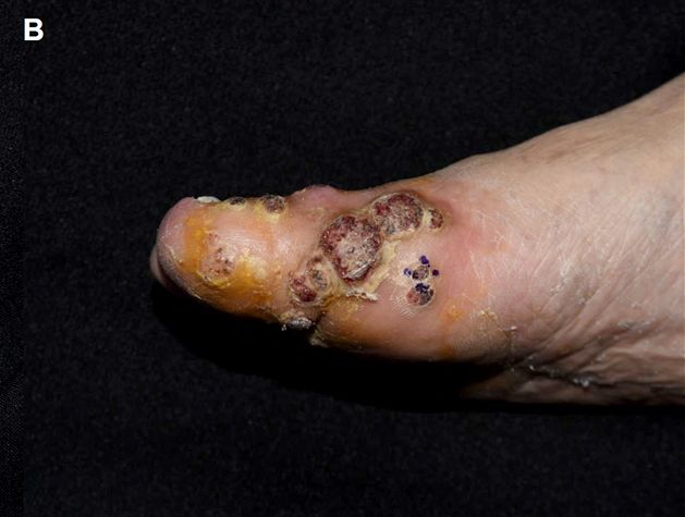
You can also read