KEYNOTE-SPEAKERS THOMAS BLANPIED, PHD | UNIVERSITY OF MARYLAND SCHOOL OF MEDICINE, USA - FENS
←
→
Page content transcription
If your browser does not render page correctly, please read the page content below
CAJAL Neuroscience Training Course
Advanced imaging techniques for cellular and systems in neuroscience
Bordeaux School of Neuroscience – March 23 to April 10, 2020

                               Keynote-speakers
    Thomas BLANPIED, PhD | University of Maryland School of Medicine, USA
                               I graduated from Yale University with a bachelor's degree in
                               Psychology. My long-standing interest in cognition and
                               learning has lead to my current work to understand the
                               cellular processes that underlie mental health and psychiatric
                               disorder. At the University of Pittsburgh, I obtained a Ph.D. in
                               the Department of Neuroscience with Jon Johnson, Ph.D.,
                               where I used single-channel recordings to study the
                               mechanisms by which the anti-Parkinsonian and anti-
                               Alzheimer's drugs amantadine and memantine act on NMDA
                               receptors. I then undertook postdoctoral training with George
Augustine, Ph.D. and Michael Ehlers, M.D. Ph.D. at Duke University in the Department of
Neurobiology. I joined the Department as an Assistant Professor in 2005, and was promoted
to Associate Professor with tenure in 2012.

Selected publications:
- Divakaruni SS, Van Dyke AM, Chandra R, LeGates TA, Contreras M, Dharmasri PA, Higgs HN,
    Lobo MK, Thompson SM,Blanpied TA. (2018) Long-Term Potentiation Requires a Rapid Burst of
    Dendritic Mitochondrial Fission during Induction. Neuron in press.
- Biederer T, Kaeser PS, Blanpied TA. (2017) Transcellular Nanoalignment of Synaptic Function.
    Neuron. 96:680-696.
- Francis TC, Chandra R, Gaynor A, Konkalmatt P, Metzbower SR, Evans B, Engeln M, Blanpied
    TA, Lobo MK. (2017) Molecular basis of dendritic atrophy and activity in stress susceptibility. Mol
    Psychiatry. 22:1512-1519.
- Tang AH*, Chen H*, Li TP, Metzbower SR, MacGillavry HD, Blanpied TA. (2016) A trans-synaptic
    nanocolumn aligns neurotransmitter release to receptors. Nature 535 (7215) August 11. (*These
    authors contributed equally.)
- Li TP and Blanpied TA. (2016) Control of Transmembrane Protein Diffusion within the
    Postsynaptic Density Assessed by Simultaneous Single-Molecule Tracking and Localization
    Microscopy. Frontiers in Synaptic Neuroscience 8: 8-22-16.
- Perez de Arce K, Schrod N, Metzbower SR, Kong G K-W, Tang A, Krupp AJ, Stein V, Liu X,
    Blanpied TA, Lucic V and Biederer T. (2015) Topographic Mapping of the Synaptic Cleft into
    Adhesive Nanodomains. Neuron 88(6): 1165–1172.
- Lu HE, MacGillavry HD, Frost NA, Blanpied TA (2014) Multiple spatial and kinetic
    subpopulations of CaMKII in spines and dendrites as resolved by single-molecule tracking PALM.
    J Neurosci. 2014 May 28;34(22):7600-10

-     MacGillavry, HD, Song, Y, Raghavachari, S, and Blanpied TA. (2013) Nanoscale scaffolding
      domains within the postsynaptic density concentrate synaptic AMPA receptors. Neuron. 78: 615-
      22.

                             The CAJAL Advanced Neuroscience Training Programme                      1
                                             www.cajal-training.org
CAJAL Neuroscience Training Course
Advanced imaging techniques for cellular and systems in neuroscience
Bordeaux School of Neuroscience – March 23 to April 10, 2020

 Daniel CHOQUET, PhD | University of Bordeaux (Bordeaux Neurocampus), France
                                 Daniel Choquet obtained an engineering degree from Ecole
                                Centrale (Paris, France) in 1984. He then got attracted to
                                neuroscience and completed his PhD in the lab of Henri Korn
                                at the Pasteur Institute (Paris), studying ion channels in
                                lymphocytes. He got appointed tenure Research officer at the
                                CNRS in 1988. He then performed a post-doctoral/sabbatical
                                at the Duke University (North Carolina, USA) in the laboratory
                                of Michael Sheetz where he studied the regulation of integrin-
                                cytoskeletal linkage by force, and demonstrated that cells can
                                sense and respond to extracellular traction. He then setup his
group in Bordeaux (France) at the Institute for Neuroscience where he got a directorship
position at the CNRS. He launched an interdisciplinary program on the combination of
physiology, cell and chemical biology and high resolution imaging to study the functional role
of the dynamic organization and trafficking of neurotransmitter receptors in synaptic
transmission. He is now heading the Institute for Interdisciplinary Neuroscience and the
Bordeaux Imaging Center core facility. He is also the director of the center of excellence
BRAIN, Bordeaux Region Aquitaine Initiative for Neuroscience. He has been the recipient of
several awards including the 1990 Bronze Medal from the CNRS, the Research prize from the
Fondation pour la Recherche Médicale (FRM), 1997, the Grand Prix from the French Academy
of Sciences, Prix du CEA and the 2009 Silver Medal from the CNRS. He is a Member of the
Institut de France, the French Science Academy since November 2010 and Officier de la Légion
d’honneur. He has been awarded three ERC advanced grants in 2008, 2013 and
2018. His team develops several research topics, combining neuroscience, physics and
chemistry in order to unravel the dynamics and nanoscale organization of multimolecular
receptor complexes and their functional role in glutamatergic synaptic
transmission. Recently, the team has engaged in a major program to analyse and understand
the interplay between AMPA type glutamate receptor nanoscale dynamics, synaptic plasticity
and memory formation in the healthy and diseased brain.

Selected publications:
- Linking Nanoscale Dynamics of AMPA Receptor Organization to Plasticity of Excitatory Synapses
    and Learning. Choquet D. J Neurosci. 2018 Oct 31;38(44):9318-9329.
- Modulation of AMPA receptor surface diffusion restores hippocampal plasticity and memory in
    Huntington's disease models. Zhang H, Zhang C, Vincent J, Zala D, Benstaali C, Sainlos M, Grillo-
    Bosch D, Daburon S, Coussen F, Cho Y, David DJ, Saudou F, Humeau Y, Choquet D.
- Nat Commun. 2018 Oct 15;9(1):4272
- CaMKII Metaplasticity Drives Aβ Oligomer-Mediated Synaptotoxicity. Opazo P, Viana da Silva S,
    Carta M, Breillat C, Coultrap SJ, Grillo-Bosch D, Sainlos M, Coussen F, Bayer KU, Mulle C, Choquet
    D. Cell Rep. 2018 Jun 12;23(11):3137-3145
- Lengthening of the Stargazin Cytoplasmic Tail Increases Synaptic Transmission by Promoting
    Interaction to Deeper Domains of PSD-95. Hafner AS, Penn AC, Grillo-Bosch D, Retailleau N, Poujol
    C, Philippat A, Coussen F, Sainlos M, Opazo P, Choquet D. Neuron. 2015 Apr 22;86(2):475-89.

 Rosa COSSART, PhD | Aix-Marseille University, France
More to come…

                             The CAJAL Advanced Neuroscience Training Programme                     2
                                             www.cajal-training.org
CAJAL Neuroscience Training Course
Advanced imaging techniques for cellular and systems in neuroscience
Bordeaux School of Neuroscience – March 23 to April 10, 2020

 Olga GARASCHUK, PhD | University of Tübingen, Germany
                                 Olga Garaschuk holds the Chair for Neurophysiology (W3) at
                                 the Eberhard Karls University of Tübingen since 2008. She is
                                 an elected member of Academia Europaea, a Member of the
                                 scientific advisory board of the Center of Advanced European
                                 Studies and Research (Bonn), the scientific advisory board of
                                 the Bernstein Centre for Computational Neuroscience and
                                 Bernstein Fokus Neurotechnology (Göttingen University) and
                                 a Mentor at the "Helmholtz Management Academy". Her
                                 research (supported by e.g. DFG, VolkswagenStiftung,
                                 Alexander von Humboldt-Stiftung and Horizon 2020) focuses
on (i) functional analysis of neuronal networks in vivo, especially in context of perception of
sensory stimuli; (ii) interactions between the nervous and the immune systems of the brain
as well (iii) aging and neurodegeneration (in particular, Alzheimer disease). Among her
scientific achievements is development of several techniques for functional in vivo imaging of
neural networks, pioneering research on in vivo Ca2+-signaling in microglia as well as adult
neural stem cells. Olga Garaschuk graduated in physico-chemical biology from the Moscow
Institute for Physics & Technology (MFTI) with top honors. She gained her PhD at the
Bogomoletz Institute for Physiology in Kyiv in 1992, and moved first to the Max Planck
Institute for Biophysical Chemistry in Gottingen and thereafter to the Institute of Physiology
at the University of Saarland, where she was awarded the Irène Curie habilitation Scholarship.
She habilitated in Physiology at the Ludwig-Maximilians University, Munich in 2003. Before
moving to Tübingen Olga Garaschuk was appointed as Professor for Neuroimaging (W2) at
the Institute of Neuroscience of the Technical University in Munich (2006). Since 2016 Olga
Garaschuk is a president of the German-Ukrainian Academic Society. She was born in Kyiv,
Ukraine, is married and has three daughters.

Selected publications (86 in total, Times cited: 6117 (Scopus) h-index 32, cumulative IF: 501.54)
- Lerdkrai C., Asavapanumas N., Brawek B., Kovalchuk Y., Mojtahedi N., Olmedillas del Moral M.,
    Garaschuk O. 2018 Intracellular Ca2+ stores control in vivo neuronal hyperactivity in a mouse
    model of Alzheimer’s disease. PNAS USA 115: E1279-E1288.
- Liang Y, Li K., Riecken K, Maslyukov A, Gomez-Nicola D, Kovalchuk Y, Fehse B, Garaschuk O. 2016
    Long-term in vivo single cell tracking reveals the switch of migration patterns in adult-born
    juxtaglomerular cells of the mouse olfactory bulb. Cell Research 26: 805-821.
- Kovalchuk Y, Homma R, Liang Y, Maslyukov A, Hermes M, Thestrup T, Griesbeck O, Ninkovic J,
    Cohen LB, Garaschuk O. 2015 In vivo odorant response properties of migrating adult-born neurons
    in the mouse olfactory bulb. Nature Communications 6, 6349.
- Brawek B, Schwendele B, Riester K, Kohsaka S, Lerdkrai C, Liang Y, Garaschuk O. 2014 Impairment
    of in vivo calcium signaling in amyloid plaque-associated microglia. Acta Neuropathol. 127, 495-
    505.
- Busche MA, Eichhoff G, Adelsberger H, Abramowski D, Wiederhold KH, Haass C, Staufenbiel M,
    Konnerth A, Garaschuk O. 2008 Clusters of hyperactive neurons near amyloid plaques in a mouse
    model of Alzheimer’s disease. Science 321: 1686-1689.

                             The CAJAL Advanced Neuroscience Training Programme                   3
                                             www.cajal-training.org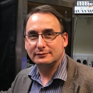
CAJAL Neuroscience Training Course
Advanced imaging techniques for cellular and systems in neuroscience
Bordeaux School of Neuroscience – March 23 to April 10, 2020

 Laurent GROC, PhD | University of Bordeaux (Bordeaux Neurocampus), France
                                       Laurent Groc identified a form of glutamate receptor plasticity
                                       in immature synapses as a post-doc with Eric Hanse
                                       (Goteborg, Sweden). Laurent then explored glutamate
                                       receptor trafficking in developing neurons using single
                                       molecule tracking and electro-physiology. He currently heads
                                       a lab at the Interdisciplinary Institute for Neuroscience in
                                       Bordeaux1,2, focusing on receptor dynamics in developing
                                       networks. He will instruct single nanoparticle tracking of
                                       membrane receptors in developing neurons.

Selected publications :
- Differential Nanoscale Topography and Functional Role of GluN2-NMDA Receptor Subtypes at
    Glutamatergic Synapses. Kellermayer B, Ferreira JS, Dupuis J, Levet F, Grillo-Bosch D, Bard L,
    Linarès-Loyez J, Bouchet D, Choquet D, Rusakov DA, Bon P, Sibarita JB, Cognet L, Sainlos M,
    Carvalho AL, Groc L. Neuron 2018. doi: 10.1016/j.neuron.2018.09.012.
- Pathogenicity of Antibodies against NMDA Receptor: Molecular Insights into Autoimmune
    Psychosis. Jézéquel J, Johansson EM, Leboyer M, Groc L. Trends Neurosci. 2018. doi:
    10.1016/j.tins.2018.05.002.
- Dynamic disorganization of synaptic NMDA receptors triggered by autoantibodies from psychotic
    patients. Jézéquel J, Johansson EM, Dupuis JP, Rogemond V, Gréa H, Kellermayer B, Hamdani N, Le
    Guen E, Rabu C, Lepleux M, Spatola M, Mathias E, Bouchet D, Ramsey AJ, Yolken RH, Tamouza R,
    Dalmau J, Honnorat J, Leboyer M, Groc L. Nat Commun. 2017. doi: 10.1016/j.tins.2018.05.002.
- Cell- and Single Molecule-Based Methods to Detect Anti-N-Methyl-D-Aspartate Receptor
    Autoantibodies in Patients With First-Episode Psychosis From the OPTiMiSE Project.
    Jézéquel J, Rogemond V, Pollak T, Lepleux M, Jacobson L, Gréa H, Iyegbe C, Kahn R, McGuire P,
    Vincent A, Honnorat J,

 Michael HÄUSSER, PhD | University College London, UK
                             Michael Häusser is Professor of Neuroscience at University
                             College London and a Principal Research Fellow of the
                             Wellcome Trust. He did his PhD work at Oxford University
                             under the supervision of Julian Jack. He subsequently worked
                             with Bert Sakmann at the Max-Planck-Institute for Medical
                             Research in Heidelberg and with Philippe Ascher at the Ecole
                             Normale Superieure in Paris. He established his own lab at UCL
                             in 1997, where his group aims to understand the cellular basis
                             of neural computation in the mammalian brain using a
                             combination of experiments and theory, with a special focus
on the role of dendrites. He is also the Facilitator of the International Brain Laboratory
(www.internationalbrainlab.com), a new global open-science collaboration which aims to
understand how the brain makes decisions. Lab website: www.dendrites.org

Selected publications:
- Smith, S., Smith, I.T., Branco, T., Häusser, M. (2013). Dendritic spikes enhance stimulus selectivity
    in cortical neurons in vivo. Nature 503(7474):115-20.
                             The CAJAL Advanced Neuroscience Training Programme                      4
                                             www.cajal-training.org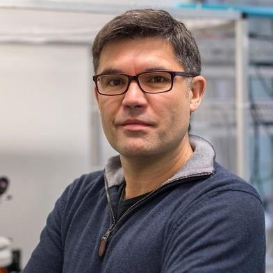
CAJAL Neuroscience Training Course
Advanced imaging techniques for cellular and systems in neuroscience
Bordeaux School of Neuroscience – March 23 to April 10, 2020

-     Packer A.M., Russell L.E., Dalgleish H.W., Häusser M. (2015). Simultaneous all-optical manipulation
      and recording of neural circuit activity with cellular resolution in vivo. Nature Methods 12(2):140-
      6.
-     Schmidt-Hieber C, Toleikyte G, Aitchison L, Roth A, Clark BA, Branco T, Häusser M (2017). Active
      dendritic integration as a mechanism for robust and precise grid cell firing. Nature Neuroscience
      20(8):1114-1121.
-     Zhang Z, Russell LE, Packer AM, Gauld OM, Häusser M (2018). Closed-loop all-optical interrogation
      of neural circuits in vivo. Nature Methods 15(12):1037-1040.
-     Kostadinov D, Beau M, Pozo MB, Häusser M (2019). Predictive and reactive reward signals
      conveyed by climbing fiber inputs to cerebellar Purkinje cells. Nature Neuroscience 22(6):950-962.

    Christian HENNEBERGER, PhD | University of Bonn, Germany
                                Christian Henneberger is a Professor for Neurophysiology at
                                the University of Bonn in the Institute of Cellular
                                Neurosciences. He was trained in Germany at the Humboldt
                                University Berlin and in the United Kingdom at the University
                                College London in the labs of Rosemarie Grantyn and Dmitri
                                Rusakov. His main scientific interest are the mechanisms of
                                synaptic transmission and plasticity and how they are shaped
                                by signal exchange with glial cells and by the geometry of the
                                tripartite synapse. Recent work of his lab investigates the
                                mechanisms by which neurons and astrocytes supply the
NMDA receptor (NMDAR) co-agonists D-serine and glycine and thus govern NMDAR-
dependent synaptic plasticity in the hippocampus. In collaboration with Colin Jackson’s lab
(ANU, Canberra) his lab is developing novel optical sensor for these co-agonists and uses them
to investigate the spatiotemporal dynamics of co-agonist signalling. In the course, Christian
will introduce in situ imaging of extracellular neurotransmitter dynamics.
Selected publications:
- Zhang WH, Herde MK, Mitchell JA, Whitfield JH, Wulff AB, Vongsouthi V, Sanchez-Romero I,
    Gulakova PE, Minge D, Breithausen B, Schoch S, Janovjak H, Jackson CJ, Henneberger C (2018)
    Monitoring hippocampal glycine with the computationally designed optical sensor GlyFS. Nat.
    Chem. Biol. 14(9):861-869.
- Minge D, Senkov O, Kaushik R, Herde MK, Tikhobrazova O, Wulff AB, Mironov A, van Kuppevelt TH,
    Oosterhof A, Kochlamazashvili G, Dityatev A, Henneberger C (2017) Heparan sulfates support
    pyramidal cell excitability, synaptic plasticity and context discrimination. Cereb. Cortex 27(2):903-
    918.
- Anders S, Minge D, Griemsmann S, Herde MK, Steinhäuser C, Henneberger C (2014) Spatial
    properties of astrocyte gap junction coupling in the rat hippocampus. Phil. Trans. R. Soc. B
    369(1654):20130600.
- Henneberger C, Papouin T, Oliet SHR, Rusakov DA (2010). Long-term potentiation depends on
    release of d-serine from astrocytes. Nature 463: 232-236.
- Kochlamazashvili G*, Henneberger C*, Bukalo O*, Dvoretskova E, Senkov O, Lievens PM,
    Westenbroek R, Engel AK, Catterall WA, Rusakov DA, Schachner M, Dityatev A. (2010) The
    Extracellular Matrix Molecule Hyaluronic Acid Regulates Hippocampal Synaptic Plasticity by
    Modulating Postsynaptic L-Type Ca2+ Channels. Neuron 67: 116-128. (* equal contribution)

                             The CAJAL Advanced Neuroscience Training Programme                         5
                                             www.cajal-training.org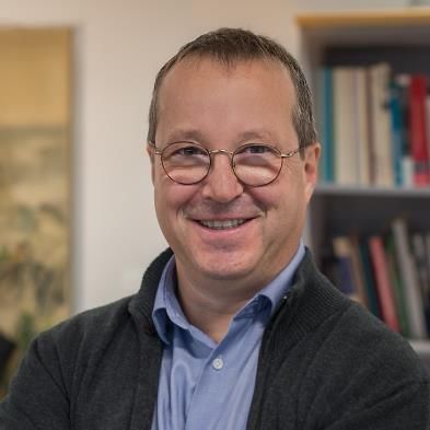
CAJAL Neuroscience Training Course
Advanced imaging techniques for cellular and systems in neuroscience
Bordeaux School of Neuroscience – March 23 to April 10, 2020

 Christophe LETERRIER, PhD | Aix-Marseille University, Germany
                             Christophe Leterrier has been working on the organization of
                             the axon since his PhD with Zsolt Lenkei in Paris, where he
                             studied the axonal targeting of the CB1 cannabinoid receptor.
                             For his postdoc in Bénédicte Dargent's lab in Marseille, he
                             worked on revealing new cytoskeletal components of the
                             axon initial segment, as well as their nanoscale organization.
                             He started the NeuroCyto lab in 2017, with the aim of
                             deciphering the axonal cytoskeleton architecture using
                             advanced microscopy techniques. The team currently focuses
                             the organization of axonal actin and its partners in order to
understand the function of newly discovered axonal actin structures: rings, hotspots and
trails.

Selected publications:
- The functional architecture of axonal actin. Papandréou MJ, Leterrier C. Mol Cell Neurosci. 2018
    Sep;91:151-159.
- Quantitative mapping and minimization of super-resolution optical imaging artifacts. Culley S,
    Albrecht D, Jacobs C, Pereira PM, Leterrier C, Mercer J, Henriques R. Nat Methods. 2018
    Apr;15(4):263-266.
- The nano-architecture of the axonal cytoskeleton. Leterrier C, Dubey P, Roy S. Nat Rev Neurosci.
    2017 Dec;18(12):713-726.
- A dynamic formin-dependent deep F-actin network in axons. Ganguly A, Tang Y, Wang L, Ladt K,
    Loi J, Dargent B, Leterrier C, Roy S., J Cell Biol. 2015 Aug 3;210(3):401-17.

 Valentin NÄGERL, PhD | University of Bordeaux (Bordeaux Neurocampus), France
                                       Valentin Nägerl was trained in Germany, working with Tobias
                                       Bonhoeffer, Arthur Konnerth and the Nobel laureate Stefan
                                       Hell. He made several crucial observations on how structural
                                       changes in the postsynaptic spine contribute to synaptic
                                       plasticity. Valentin is now a full professor of neuroscience and
                                       bio-imaging at the University of Bordeaux and continues to
                                       study the nanoscale structural mechanisms of neural plasticity
                                       using super-resolution imaging. In the course, Valentin will
                                       instruct spine motility in living neurons using STED
                                       microscopy.

Selected publications:
- Chéreau et al. (2017) PNAS Feb 7;114(6):1401-1406.
- Tønnesen et al. (2014) Nat Neurosci. 17:678-85.
- Becker et al. (2008) Neuron 60:590-7.
- Nägerl et al. (2004) Neuron 44:759-67.

                             The CAJAL Advanced Neuroscience Training Programme                       6
                                             www.cajal-training.org
CAJAL Neuroscience Training Course
Advanced imaging techniques for cellular and systems in neuroscience
Bordeaux School of Neuroscience – March 23 to April 10, 2020

    Timothy A. RYAN, PhD | Weill Cornell Medical College, USA
                              Timothy Ryan is a Rockefeller/Sloan-Kettering/Cornell Tri-
                              Institutional Professor in the department of Biochemistry at
                              Weill Cornell Medical College and a Senior fellow at the
                              Howard Hughes Medical Institute Janelia Research Campus.
                              He received his BSc with honors in Physics at McGill University
                              and his PhD in Physics at Cornell University. He carried out
                              postdoctoral work at Stanford University in the department of
                              Molecular and Cellular Physiology. Dr. Ryan’s lab has
                              pioneered the development and use of quantitative optical
                              tools to interrogate nerve terminal function. He was an Alfred
P. Sloan Fellow, a two-time recipient of the McKnight Technological Innovations in
Neuroscience Award, a recipient of the NINDS Javits Award and a recipient of the Siegel Family
Award for Outstanding Biomedical Research.
Selected publications:
-     V. Rangaraju, N. Calloway & T. A. Ryan. Activity-driven local ATP synthesis is required for
      synaptic function. Cell (2014) 156(4):825-35.
-     M. Hoppa, G. Gouzer, M. Armbruster & T. A. Ryan. Control and plasticity of the presynaptic
      action potential waveform at small CNS nerve terminals. Neuron (2014) 84(4):778-789
-     G. Ashrafi, Z. Wu, R.J. Farrell & T. A. Ryan. Glut4 mobilization supports energetic demands
      of active synapses. Neuron (2017) 93(3):606-615.e3. doi: 10.1016.
-     J. de Juan Sanz, G. T. Holt, F. de Juan, D. S. Kim & T. A. Ryan. Axonal endoplasmic reticulum
      Ca2+ content controls release probability in CNS nerve terminals. Neuron (2017)
      93(4):867-881.e6. doi: 10.1016/j.neuron.2017.01.010
-     G. Ashrafi, J. de Juan Sanz, R. Farrell & T. A. Ryan. Molecular tuning of the axonal
      mitochondrial Ca2+ uniporter ensures metabolic flexibility of neurotransmission. Neuron
      (2019) in press

    Ilaria TESTA, PhD | KTH Royal Institute of Technology, Sweden
                                I performed my PhD between 2006 and 2009 at the University
                                of Genoa (Italy) in the group of Professor Alberto Diaspro
                                working on the use of Photoswitchable Fluorescent Proteins
                                in two photon microscopy for functional analysis in living cells.
                                Between 2009-2014 I worked as a Postdoc Researcher at the
                                Department of NanoBiophotonics directed by Professor
                                Stefan Hell at the Max Planck Institute for Biophysical
                                Chemistry in Göttingen (Germany). During this time I actively
                                designed and developed several nanoscopes that implement
                                two different approaches for achieving super-resolution
imaging: (1) the first set-up was based on stochastic switching of single molecule between a
fluorescent and a non-fluorescent state (GSDIM/PALM/STORM) with multicolour ability
based on ratiometric detection (2) the second set-up showed the practical accomplishment
of RESOLFT, a target switching technique based on reversible long lived molecular transition.
Within an interdisciplinary team of biologists and physicists we succeeded to demonstrate
                             The CAJAL Advanced Neuroscience Training Programme                   7
                                             www.cajal-training.org
CAJAL Neuroscience Training Course
Advanced imaging techniques for cellular and systems in neuroscience
Bordeaux School of Neuroscience – March 23 to April 10, 2020

RESOLFT in living cells and even tissues. Since 2015 I’m conducting my independent research
at the Science for Life Laboratory as Faculty at the Royal Institute of Technology (KTH,
Stockholm, Sweden). The goal of my group is to develop the novel paradigms made available
by super-resolution microscopy to address contemporary challenges in biophysics and
neuroscience. To achieve this goal we push forward the quantitative aspect of live cell imaging
by setting-up and applying different concepts of super-resolution microscopy based on target
switching such as automated STED microscopy. We developed two new nanoscopes named
MoNaLISA and smart RESOLFT which allow the precise identification of populations of
neuronal proteins and organelles depending on their localization, abundance and dynamics
inside their native environment. We also use our technology to investigate neuronal proteins,
especially in synapses, where trafficking organelles and protein complexes are so tight in
space that resolving them requires high resolution in space and time.

Selected publications:
- Jes Dreier, Marco Castello, Giovanna Coceano, Rodrigo Cáceres, Julie Plastino, Giuseppe Vicidomini
    and Ilaria Testa, “Smart scanning for low-illumination and fast RESOLFT nanoscopy in vivo” Nature
    Communications, volume 10, Article number: 3281.
- Luciano A. Masullo*, Andreas Bodén*, Francesca Pennacchietti*, Giovanna Coceano, Michael Ratz
    and Ilaria Testa (2018) Enhanced photon collection enables four dimensional fluorescence
    nanoscopy of living systems. Nature Communications, volume 9, Article number: 3281
- Francesca Pennacchietti*, Ekaterina O. Serebrovskaya*, Aline R. Faro*, Irina I. Shemyakina*, Nina
    G. Bozhanova, Alexey A. Kotlobay, Nadya G. Gurskaya, Andreas Bodén, Jes Dreier, Dmitry M.
    Chudakov, Konstantin A. Lukyanov, Vladislav V. Verkhusha, Alexander S. Mishin and Ilaria Testa.
    (2018) Fast reversibly photoswitching red fluorescent proteins for live-cell RESOLFT nanoscopy
    Nature Methods 15, 601–604
- Ilaria Testa, Nicolai T. Urban, Stefan Jakobs, Christian Eggeling, Katrin I. Willig, Stefan W. Hell
    (2012) Nanoscopy of living brain slice with low light levels, Neuron 75: 992–1000 doi:
    10.1016/j.neuron.2012.07.028

 Andrea VOLTERRA, PhD | University of Lausanne, Switzerland
                                Andrea Volterra, Italian, 62 years-old, is Full Professor at the
                                Department of Fundamental Neurosciences, University of
                                Lausanne, Switzerland since 2001, which he directed from
                                2004 to 2012. He obtained a PhD in Pharmacology from Univ
                                Milan, Italy (1985), joined the labs of Steven Siegelbaum and
                                the Nobel Prize Eric Kandel at Columbia University, New York,
                                as Research Scientist (1986-89) and then started his
                                independent career as Assistant and then Associate Professor
                                at the Dept. Pharmacology, Univ Milan (19902000). In 2001 he
                                moved to Switzerland. He is member of Academia Europea
and Swiss Academy of Medical Sciences (SAMS), and of Swiss, European and American
Societies of Neuroscience, and won several prices, including the Theordore Ott price 2017
from SAMS. Prof. Volterra obtained >30 grants as PI, including currently, the prestigious ERC
Advanced, Swiss National Science Foundation individual grant and participates in two of the
Swiss National Centers of Excellence (NCCRs), “Transcure” and “Synapsy”. He is author of 125
publications with >10000 citations and h-index of 42 (Thomas Reuters’ Web of Science). His
research field is astrocyte-neuron communication and its contribution to synaptic integration
                             The CAJAL Advanced Neuroscience Training Programme                    8
                                             www.cajal-training.org
CAJAL Neuroscience Training Course
Advanced imaging techniques for cellular and systems in neuroscience
Bordeaux School of Neuroscience – March 23 to April 10, 2020

and the pathogenesis of neuropsychiatric disorders, to which he contributed some of the
seminal work in the last twenty years. Initially he worked in the field of biochemical
neuropsychopharmacology; at Columbia University studied memory paradigms in Aplysia
Californica with electrophysiology techniques (Piomelli et al., Nature, 1987; Buttner et al.,
Nature, 1989; Sweatt et al., Nature, 1989). Between 1990 and 2000 focused on the role of
glutamate transport in physiology and disease. In 1997-2001 he discovered a Ca2+-dependent
pathway of transmitter release from astrocytes and demonstrated its role in synaptic function
and pathology (Bezzi et al., Nature, 1998; Bezzi et al. Nature Neurosci, 2001). In the last 18
years, his research has widened up from the initial discoveries to establish his group as one
of the pioneers in deciphering the modulatory role of astrocytes in synaptic function via the
release of neuroactive messengers and in showing that perturbation of this astrocyte input
to synapses causes neuronal damage, supporting the idea of non-cell autonomous
mechanisms of neuronal degeneration, possibly one of the key advances in neurology in
recent years (Bezzi et al., Nature Neurosci, 2004; Jourdain et al. Nature Neurosci., 2007; Di
Castro et al., Nature Neurosci., 2011; Santello et al., Neuron, 2011; Habbas et al., Cell 2015;
Bindocci et al., Science, 2017; reviewed in Volterra and Meldolesi, Nature Rev Neurosci., 2005
and recently in Araque et al., Neuron, 2014 and Volterra et al., Nature Review Neurosci., 2014;
Santello et al., Nature Neurosci 2019). His most recent work, using a multidisciplinary
approach and novel imaging technologies, is engaged in decoding the language of astrocytes
in cognitive function and dysfunction.

Selected publications:
- Savtchouk I, Carriero G, Volterra A. Studying Axon-Astrocyte Functional Interactions by 3D Two-
    Photon Ca(2+) Imaging: A Practical Guide to Experiments and "Big Data" Analysis. Front Cell
    Neurosci. 2018 Apr 13;12:98. doi: 10.3389/fncel.2018.00098. eCollection 2018. PubMed PMID:
    29706870; PubMed Central PMCID: PMC5908897.
- Bindocci E, Savtchouk I, Liaudet N, Becker D, Carriero G, Volterra A. Three-dimensional Ca(2+)
    imaging advances understanding of astrocyte biology. Science. 2017 May 19;356(6339). pii:
    eaai8185. doi: 10.1126/ science.aai8185. PubMed PMID: 28522470.
- Volterra A, Liaudet N, Savtchouk I. Astrocyte Ca²⁺ signalling: an unexpected complexity. Nat Rev
    Neurosci. 2014 May;15(5):327-35. doi: 10.1038/ nrn3725. Review. PubMed PMID: 24739787.
- Di Castro MA, Chuquet J, Liaudet N, Bhaukaurally K, Santello M, Bouvier D, Tiret P, Volterra A. Local
    Ca2+ detection and modulation of synaptic release by astrocytes. Nat Neurosci. 2011 Sep
    11;14(10):1276-84. doi: 10.1038/ nn.2929. PubMed PMID: 21909085.
- Habbas S, Santello M, Becker D, Stubbe H, Zappia G, Liaudet N, Klaus FR, Kollias G, Fontana A,
    Pryce CR, Suter T, Volterra A. Neuroinflammatory TNFα Impairs Memory via Astrocyte Signaling.
    Cell. 2015 Dec 17;163(7): 1730-41. doi: 10.1016/j.cell.2015.11.023. Epub 2015 Dec 10. PubMed
    PMID:26686654

                             The CAJAL Advanced Neuroscience Training Programme                      9
                                             www.cajal-training.org
CAJAL Neuroscience Training Course
Advanced imaging techniques for cellular and systems in neuroscience
Bordeaux School of Neuroscience – March 23 to April 10, 2020

 Kirill VOLYNSKI, PhD | University College London, UK
                               Kirill Volynski is a Professor of Neuroscience at UCL Queen
                               Square Institute of Neurology. His research is focused on
                               understanding the mechanisms of Ca2+-dependent regulation
                               of transmitter release at the level of individual presynaptic
                               boutons. His group established a powerful suite of
                               quantitative fluorescence microscopy methods in neuronal
                               cultures (Ermolyuk et al., 2013;Ermolyuk et al., 2012;Novak et
                               al., 2013). This resulted in the discovery that two equally
                               important mechanisms contribute to the basal heterogeneity
                               of neurotransmitter release probability: the size of the readily
releasable pool of vesicles, which scales with bouton size, and the fusion probability of
individual release-ready vesicles, which in turn is determined by the size of the AP-evoked
Ca2+ influx (Ermolyuk et al., 2012). Using combination of electrophysiology and
computational modelling Volynski lab showed that stochastic opening of individual Ca2+
channels contributes to both evoked and spontaneous glutamate release (Ermolyuk et al.,
2013). In collaboration with Y. Korchev (Imperial College London) he has obtained the first
nanoscale-targeted patch-clamp recordings from small presynaptic boutons and applied this
unique methodology to understand the presynaptic mechanisms of episodic ataxia type 1
which is caused by mutations in Kv1.1 potassium channel (Novak et al., 2013;Vivekananda et
al., 2017).

Selected publications:
- Ermolyuk,Y.S., Alder,F.G., Surges,R., Pavlov,I.Y., Timofeeva,Y., Kullmann,D.M., and Volynski,K.E.
    (2013). Differential triggering of spontaneous glutamate release by P/Q-, N- and R-type Ca(2+)
    channels. Nat. Neurosci. 16, 1754-1763.
- Ermolyuk,Y.S., Alder,F.G., Henneberger,C., Rusakov,D.A., Kullmann,D.M., and Volynski,K.E. (2012).
    Independent Regulation of Basal Neurotransmitter Release Efficacy by Variable Ca2+
    Influx and Bouton Size at Small Central Synapses. PLoS Biol 10, e1001396.
- Novak,P., Gorelik,J., Vivekananda,U., Shevchuk,A.I., Ermolyuk,Y.S., Bailey,R.J., Bushby,A.J.,
    Moss,G.W., Rusakov,D.A., Klenerman,D., Kullmann,D.M., Volynski,K.E., and Korchev,Y.E. (2013).
    Nanoscale-targeted patch-clamp recordings of functional presynaptic ion channels. Neuron 79,
    1067-1077.
- Vivekananda,U., Novak,P., Bello,O.D., Korchev,Y.E., Krishnakumar,S.S., Volynski,K.E., and
    Kullmann,D.M. (2017). Kv1.1 channelopathy abolishes presynaptic spike width modulation by
    subthreshold somatic depolarization. Proc. Natl. Acad. Sci. U. S. A. 114, 2395-2400.

                             The CAJAL Advanced Neuroscience Training Programme                 10
                                             www.cajal-training.org
You can also read