Lemur Loris Digestive Tract Pathologies (Dissection Study)
←
→
Page content transcription
If your browser does not render page correctly, please read the page content below
E3S Web of Conferences 285, 04014 (2021)                                 https://doi.org/10.1051/e3sconf/202128504014
ABR 2021

       Lemur Loris Digestive                                            Tract             Pathologies
       (Dissection Study)
       L. V. Tkachenko *
       Altai State Agrarian University, Barnaul, Russia

                     Abstract. A necropsy study of the Lemur Lori was carried out.
                     Physiologically, Lemurs have a very sensitive gastrointestinal tract.
                     Therefore, these animals almost keep the lead in the incidence of
                     pathologies of the oral cavity, stomach, and intestines. The study was
                     carried out in the dissection room of the Veterinary Medicine Faculty of
                     the Altai State Agrarian University. The object was the animal corpse of a
                     Lemur Loris: animal species: monkey; gender: female; age: 1.5 years;
                     breed: Loris; the owner is a private zoo; provisional diagnosis: not known.
                     The following methods were used: history taking; postmortem examination
                     according to Shor's method with a description of the results obtained;
                     photographing; analysis of the data obtained with the help of fact-finding.
                     As a result of the postmortem examination, the following changes were
                     recorded: pulmonary edema, acute congestive hyperemia and emphysema;
                     acute expansion of the right atrium and ventricle of the heart; acute
                     catarrhal-hemorrhagic esophagitis and gastroenteritis; acute hemorrhagic
                     pancreatitis; acute serous-hemorrhagic lymphadenitis of the mesenteric
                     lymph nodes; liver and kidneys fatty degeneration; cyanosis of the
                     conjunctiva, tunica mucosa of the mouth; obesity. Thus, death occurred as
                     a result of pulmonary arrest (pulmonary edema), which was the result of
                     autointoxication (inflammatory processes in the stomach, intestines,
                     pancreas). The cause of autointoxication is a long-term and major violation
                     of the norms of feeding and maintenance, stress, and presumably a drug
                     that was administered to the animal, namely, an incorrect dose or
                     individual intolerance of its constituent components.

       1 Introduction
       In the modern world, exotic animals are increasingly purchased for home keeping, for
       private animal collections or ethnic parks. At the same time, it is not always possible and
       sometimes even impossible to reproduce fully the way of life, hierarchical ties, and pair
       formation.
           As a result, animals suffer from various pathologies that lead to their death.
           That is why each case of exotic animals death should be studied, and the results of
       necropsy studies should be accurately described. This will allow veterinary specialists,
       biologists, zoologists to understand better all the mechanisms of pathology development,
       and therefore give competent advice to the owners of exotic animals.

       *
           Corresponding author : rabota36@bk.ru

© The Authors, published by EDP Sciences. This is an open access article distributed under the terms of the Creative Commons
Attribution License 4.0 (http://creativecommons.org/licenses/by/4.0/).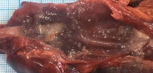
E3S Web of Conferences 285, 04014 (2021)                      https://doi.org/10.1051/e3sconf/202128504014
ABR 2021

          Lemur Loris is a representative of the Strepsirrhini suborder, the Prosimii suborder, the
      Lemuridae family, the Lemur genus. Naturalist G. Buffon ranked Lemur Loris among
      Lemurs [3].
          The name of the animal Loris (Stenops) comes from "Loeris", translated from Dutch as
      "a clown". What is more, the Romans called Lemurs the souls of the dead, who guarded the
      family and home [16].
          They are the arboreal animals, whose historical homeland is Madagascar and the
      Comoros. It is impossible to take lemurs out of their natural habitats, so they can only be
      purchased from breeders [2, 3]. Lemurs are listed as endangered species, which are
      protected in accordance with the Law of the Russian Federation and by different
      international environmental organizations. Lemur trade is prohibited in Russia and
      throughout the world [12, 13].
          Lemurs Loris are sometimes compared to cats. This explains their great popularity
      among private breeders in any part of the world. It is difficult to reproduce Loris’ living
      conditions at home. In addition to society, cubicles and other things, it is necessary to keep
      these animals to the diet, on which the quality of Loris’ life directly depends [6, 10].
          In captivity, these animals get used to almost any food [7, 14]. Despite being
      omnivorous, they have a very sensitive gastrointestinal tract. Consequently, these animals
      almost keep the lead in the incidence of pathologies of the oral cavity, stomach, and
      intestines, such as inflammatory processes, bezoars, intestinal obstructions, etc. [14].
          Trichobezoars are stones formed due to the accumulation of wool in the stomach of an
      animal. For their painless removal, some drugs are prescribed, and some vegetables are
      used as preventive measures [5, 15].
          In general, Loris’ diet in captivity should contain vegetables, seedless fruits, berries,
      eggs and meat, as well as insects, larvae, plus vitamin complexes. Everything should be
      pre-treated with hot water. It is strictly forbidden to feed them with any dishes from the
      table [3].
          The average age of lemurs is 8-13 years [3]. But in nature, mortality in the first year of
      life is 50%, and in zoos this indicator is minimal [9, 6]. The source [4, 8] mentions a high
      level of stress due to improper maintenance, care and lack of veterinary care, which leads to
      a rapid death coming.
          There are no data available in any literature concerning animal mortality and description
      of pathological changes in the gastrointestinal tract of Lemurs Loris kept at home.
          The aim of our study was to establish the cause of death of the Lemur Loris.
          Research objectives are as follows: 1. To determine the pathological changes of the
      Lemur Loris.
          2. To establish the cause of death of the Lemur Lori.
          3. To establish fact-findings between the cause of death and the conditions of
      maintenance and feeding.

      2 Methodology
      The study was carried out in the dissection room of the Veterinary Medicine Faculty of the
      Altai State Agrarian University. The internal organs of a deceased Lemur served as the
      object for the study.
         Animal species: monkey
         gender: female;
         age: 1.5 years;
         breed: Loris;
         the owner is a private zoo;
         provisional diagnosis: not known.

                                                   2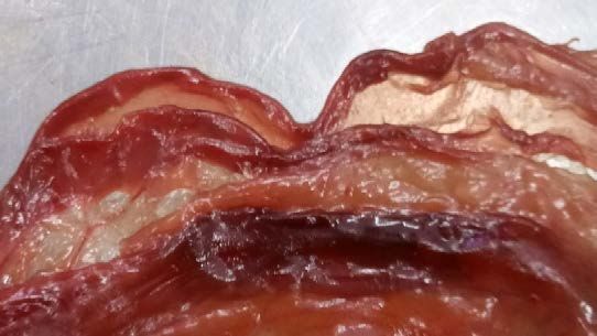
E3S Web of Conferences 285, 04014 (2021)                         https://doi.org/10.1051/e3sconf/202128504014
ABR 2021

         Methods: 1. History taking [1]. 2. Postmortem examination according to Shor's method
      with a description of the results obtained [11, 14]. 3. Macrophotography performed with a
      Sony camera. 4. Analysis of the data obtained with the help of fact-finding [2].

      3 Results and Discussion
      The history taking showed the following. The animal was kept at home, then, due to its
      excess weight, it was transferred to a private zoo. After strict diet crossover (vegetables,
      fruits), with the loss of weight, the Lemur felt unwell sharply. As a result, it was suggested
      that the stomach was clogged with hair (trichobezoars), and a drug used to remove hair
      from the cats’ stomach was prescribed to solve this problem. Duration of admission made
      14 days, after which the animal died. The name of the drug, dosage, frequency rate was not
      indicated by the owner.
          As a result of the necropsy study, the following changes were recorded – an animal of
      strong proportional constitution, fatty fill (Fig. 1).

      Fig. 1. Lemur Loris. Age – 1.5 years. Gender - female. General form. Strong proportional
      constitution, fat.
          Esophagus: the position is not disturbed, adventitia without overlaps and enlargements,
      blood vessels highly filled. A small amount of content, consisting of white particles of 0.2-
      0.3 mm, soft consistency (pieces of bread) (Fig. 2.1.), permeability is not impaired. The
      mucosa, submucosa, muscular membranes are thickened, rugosity is well expressed,
      cherry-colored, moist, abundantly covered with mucoid masses of a reddish color (Fig.
      2.2.).

                                                       3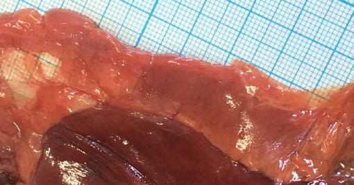
E3S Web of Conferences 285, 04014 (2021)                          https://doi.org/10.1051/e3sconf/202128504014
ABR 2021

                             2

                                                                          1

      Fig. 2. Lemur Loris. Age – 1.5 years. Gender - female. Esophagus (dissected). 1. A small amount of
      content: white particles of 0.2-0.3 mm, soft consistency (pieces of bread). 2. Section of acute
      catarrhal-hemorrhagic esophagitis.
         Stomach: the position is not disturbed, the serous membrane without overlaps and
      enlargements, blood vessels highly filled. No content. Permeability is not impaired. The
      mucosa, submucosa, muscular membranes are thickened, rugosity is well expressed, red-
      cherry, moist. The surface is abundantly covered with a red mucus mass (Fig. 3).

                                                           1

      Fig. 3. Lemur Loris. Age – 1.5 years. Gender - female. Stomach (dissected). 1. Section of acute
      catarrhal-hemorrhagic esophagitis.
          Pancreas: the volume is increased, the shape is specific, the surface is slightly bumpy,
      red-cherry in color with sections of white and yellow color, the consistency is doughy, the
      structure in the dissection – the pattern is erased (Fig. 4).

                                                       4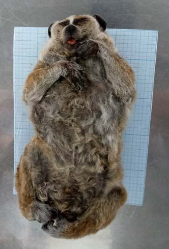
E3S Web of Conferences 285, 04014 (2021) https://doi.org/10.1051/e3sconf/202128504014
ABR 2021
1
2
Fig. 4. Lemur Loris. Age – 1.5 years. Gender - female. Stomach (dissected). 1. Section of acute
hemorrhagic pancreatitis. 2. Stomach.
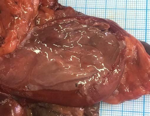
Intestines: on the mesentery, there is a large amount of fatty deposits of gray and pink
color (Fig. 5.1.), the blood vessels are highly filled with blood. The mesenteric lymph nodes
(Fig. 5.3.) are sharply enlarged in volume and elongated, the surface is smooth, red-cherry,
of doughy consistency, the structure in the dissection – the pattern is smoothed, red mucus-
like mass flows down in the dissection (Fig. 5.2.).
2
1
3
Fig. 5. Lemur Loris. Age – 1.5 years. Gender - female. Mesenteric lymph node (dissected) 1. A large
amount of fat deposits. 2. A section of acute serous-hemorrhagic lymphadenitis of the mesenteric
lymph nodes. 3. Mesenteric lymph node.
Duodenum: the position is not disturbed, the serous membrane without overlaps and
enlargements, the blood vessels highly filled. No content. Permeability is not impaired. The
mucosa, submucosa, muscular membranes are thickened, the rugosity is moderately
expressed, red-cherry, moist, abundantly covered with red mucus-like mass.
The jejunum: the position is not disturbed, the serous membrane without overlaps and
enlargements, the blood vessels highly filled. No content. Permeability is not impaired. The
mucosa, submucosa, muscular membranes are thickened, the rugosity is moderately
expressed, red-cherry, moist, abundantly covered with red mucus-like mass.
5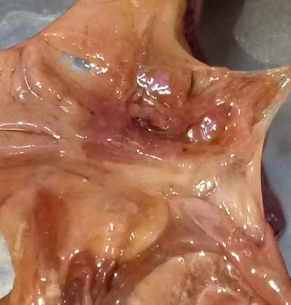
E3S Web of Conferences 285, 04014 (2021)                           https://doi.org/10.1051/e3sconf/202128504014
ABR 2021

         The ileum: the position is not disturbed, the serous membrane without overlaps and
      enlargements, the blood vessels highly filled. No content. Permeability is not impaired. The
      mucosa, submucosa, muscular membranes are thickened, the rugosity is moderately
      expressed, red-cherry, moist, abundantly covered with red mucus-like mass (Fig. 6).

                                                        1

      Fig. 6. Lemur Loris. Age – 1.5 years. Gender - female. Small intestine (dissected). 1. Section of acute
      catarrhal-hemorrhagic enteritis.
          Blind intestine: the position is not disturbed, the serous membrane without overlaps and
      enlargements, the blood vessels highly filled. No content. Permeability is not impaired. The
      mucosa, submucosa, muscular membranes are thickened, the rugosity is moderately
      expressed, red-cherry, moist, abundantly covered with red grout.
          Segmented intestine: the position is not disturbed, the serous membrane without
      overlaps and enlargements, the blood vessels highly filled. No content. Permeability is not
      impaired. The mucosa, submucosa, muscular membranes are thickened, the rugosity is
      moderately expressed, red-cherry, moist, abundantly covered with red grout.
          Straight intestine: the position is not disturbed, the serous membrane without overlaps
      and enlargements, the blood vessels highly filled. No content. Permeability is not impaired.
      The mucosa, submucosa, muscular membranes are thickened, the rugosity is moderately
      expressed, red-cherry, moist, abundantly covered with red grout.
          Liver: the volume is increased, of a specific shape, smooth surface, red-brown, sloppy
      texture, the structure in the dissection is slightly smoothed. Scraping contains ample drops
      of fat
          Gallbladder: the position is not disturbed, the serous membrane without overlaps and
      enlargements, the blood vessels highly filled. Content: homogeneous, moderate amount,
      liquid consistency, green color, specific odor. Permeability is not impaired. Mucous,
      fibromuscular, sub-serous base, serous membrane are not thickened, the rugosity is not
      pronounced, greenish-yellowish in color, moist, without overlaps.
          Kidneys: the volume is increased, of a specific shape, smooth surface, red-brown,
      sloppy texture, the structure in the dissection bulges out, slightly smoothed. Scraping
      contains ample drops.
          In general, the following diagnoses were established based on the results of the
      necropsy study:
          1. Pulmonary edema, acute congestive hyperemia and emphysema.
          2. Acute expansion of the right atrium and ventricle of the heart.
          3. Acute catarrhal-hemorrhagic esophagitis, gastroenteritis.
          4. Acute hemorrhagic pancreatitis.
          5. Acute serous-hemorrhagic lymphadenitis of the mesenteric lymph nodes.
          6. Liver and kidneys fatty degeneration.

                                                        6
E3S Web of Conferences 285, 04014 (2021) https://doi.org/10.1051/e3sconf/202128504014
ABR 2021
7. Cyanosis of the conjunctiva, tunica mucosa of the mouth.
8. Obesity.
4 Conclusion
Thus, having analyzed the information obtained, we conclude that the death of the animal
occurred as a result of pulmonary arrest (pulmonary edema), which was the result of
autointoxication (inflammatory processes in the stomach, intestines, pancreas).
In our opinion, the cause of autointoxication is a long-term and major violation of the
norms of feeding and maintenance, stress, and presumably a drug that was administered to
the animal, namely, an incorrect dose or individual intolerance of its constituent
components.
References
1. Anamnesis. - URL: https://big_medicine.academic.ru/1583/ ANAMNESIS (Accessed
on 2.04.2021).
2. Bolshakov O.P. Didactic and ethical aspects of research on biomodels and laboratory
animals. O.P. Bolshakov, N.G. Neznanov, R.V. Babakhanyan. High-quality clinical
practice. - 2002. - No. 1. - pp. 58-61.
3. Bram A.E. Life of animals. M.: Terra, 1992. pp. 135-143.
4. Types of Lemur Lori. – URL:
http://zhivotnue.ru/index_ru.php?cat=zhivotnue_krasnoi_knigi&obl=lyamyr_lori&ind=
60 (Accessed on 2.04.2021).
5. Davenport G.M. Method and composition for regulation of wool excretion with feces
and prevention of trichobezoar formation. G.M. Davenport, G.D. Sunwold, G.A.
Reinhart, M.G. Hake. Patent for invention - RU 2250776 C2, 27.04.2005. - Application
No. 2002120921/15 dated 05.01.2001.
6. Ten unexpected facts about lemurs. – URL: https://fishki.net/2619609-10-
neozhidannyh-faktov-o-Lemurah.html (Accessed on 2.04.2021).
7. Animal life. V.7. Mammals. Ed. by V.E. Sokolov. - 2nd ed., Rev. - M.: Education,
1989. - pp. 122-129.
8. Lemur Lori. - URL: https://simple-fauna.ru/wild-animals/Lemur-lori (Accessed on
2.04.2021).
9. Small and large fat Lori. - - URL:https://chins.ru/showthread.php/203-Лемуры-
Лори/page19 (accessed on 2.04.2021).
10. Meshik V.A. Formation of pairs and groups of primates in the zoo .V.A. Meshik.
Research in zoological parks. - 2014. - No. 30. - pp. 161-205.
11. Pathological autopsy of animals. Training for private veterinarians, 2013. - URL:
http://vsb.kg/uploads/Old_Modul/Руководство%20для%20участника_Патан_Роксан
а.pdf (Accessed on 2.04.2021).
12. Prokhorov V.G. Wildlife samples smuggling: Contemporary history .V.G. Prokhorov.
Eurasian Law Journal. - 2009. - No. 12 (19). - pp. 95-99.
13. Rare animals from the Red Book of Russia and the whole world. – URL: https://simple-
fauna.ru/animals-life/redkie-zhivotnye-iz-krasnoj-knigi (Accessed on 2.04.2021).
7E3S Web of Conferences 285, 04014 (2021) https://doi.org/10.1051/e3sconf/202128504014
ABR 2021
14. Tkacheva E.Yu. Possibilities of non-contact monitoring of the reproductive state and
stress level of zoo animals by methods of hormonal analysis. E.Yu. Tkacheva, O.B.
Lifanova. Scientific research in zoological parks. - 1998. - No. 10 (10). - pp. 236-245.
15. Trichobezoars - wool in the stomach. - URL: http://lorises.ru/zdorovie.html (Accessed
on 2.04.2021).
16. R. Mittermeier, J. Ganzhorn, W. Konstant, K. Glander, I. Tattersall, C. Groves, A.
Rylands, A. Hapke, J. Ratsimbazafy, M. Mayor, E. Louis jr., Y. Rumpler, C. Schwitzer
und R. Rasoloarison: Lemur Diversity in Madagascar. In: International Journal of
Primatology 29 (2008), pp. 1607–1656.
8You can also read