Lens regeneration in axolotl: new evidence of developmental plasticity
←
→
Page content transcription
If your browser does not render page correctly, please read the page content below
Suetsugu-Maki et al. BMC Biology 2012, 10:103
http://www.biomedcentral.com/1741-7007/10/103

 RESEARCH ARTICLE                                                                                                                             Open Access

Lens regeneration in axolotl: new evidence of
developmental plasticity
Rinako Suetsugu-Maki1,2, Nobuyasu Maki1,2, Kenta Nakamura1,3, Saulius Sumanas4, Jie Zhu5, Katia Del Rio-Tsonis5
and Panagiotis A Tsonis1*

  Abstract
  Background: Among vertebrates lens regeneration is most pronounced in newts, which have the ability to
  regenerate the entire lens throughout their lives. Regeneration occurs from the dorsal iris by transdifferentiation of
  the pigment epithelial cells. Interestingly, the ventral iris never contributes to regeneration. Frogs have limited lens
  regeneration capacity elicited from the cornea during pre-metamorphic stages. The axolotl is another salamander
  which, like the newt, regenerates its limbs or its tail with the spinal cord, but up until now all reports have shown
  that it does not regenerate the lens.
  Results: Here we present a detailed analysis during different stages of axolotl development, and we show that
  despite previous beliefs the axolotl does regenerate the lens, however, only during a limited time after hatching.
  We have found that starting at stage 44 (forelimb bud stage) lens regeneration is possible for nearly two weeks.
  Regeneration occurs from the iris but, in contrast to the newt, regeneration can be elicited from either the dorsal
  or the ventral iris and, occasionally, even from both in the same eye. Similar studies in the zebra fish concluded
  that lens regeneration is not possible.
  Conclusions: Regeneration of the lens is possible in the axolotl, but differs from both frogs and newts. Thus the
  axolotl iris provides a novel and more plastic strategy for lens regeneration.
  Keywords: Lens, Regeneration, Axolotl, Plasticity

Background                                                                          lens, normally it never contributes to the process [4]. This
Salamanders, especially newts, are capable of regenerating                          restriction can be very instructive when it comes to inves-
tissues, organs and even body parts, such as limbs and                              tigating the mechanisms underlying the process.
tails throughout their adult lives [1,2]. Among all tissues                            The only other vertebrates that have been shown to
that can be regenerated, the lens is a special case. In con-                        regenerate the lens are frogs and some fish. In frogs, the
trast to removal of a part of limb or tail (total removal dis-                      process has been studied well and it has been established
allows regeneration), the lens can be removed in its                                that regeneration occurs only in pre-metamorphic stages
entirety. It has also been shown that newts can regenerate                          and that the lens is derived from the cornea by transdiffer-
the lens very faithfully no matter how many repeated len-                           entiation [5]. After metamorphosis, the ability for lens
tectomies are performed or the age of the animal [3].                               regeneration ceases. Another salamander, the neonate
Notably, the lens regenerates by another tissue, the pig-                           axolotl, is also capable of regenerating limbs and tails as is
ment epithelial cells of the iris by the process of transdif-                       the newt. However, in a paper by Stone in 1967 it was
ferentiation. There is, however, a restriction: The lens can                        concluded that this species is not able to regenerate the
only be regenerated from the dorsal iris. Even though the                           lens, even though no staging details were provided [6]. We
ventral iris can be induced experimentally to regenerate a                          have decided to revisit this issue and have undertaken a
                                                                                    detailed study starting with larvae at stage 44. At this stage
* Correspondence: ptsonis1@udayton.edu                                              the eye tissues including the lens have been well differen-
1
 Department of Biology and Center for Tissue Regeneration and                       tiated, while other body parts (such as limbs) are begin-
Engineering, University of Dayton, 300 College Park, Dayton, OH 45469-2320,
USA                                                                                 ning to form [7]. We find that starting at this stage
Full list of author information is available at the end of the article

                                       © 2012 Suetsugu-Maki et al; licensee BioMed Central Ltd. This is an Open Access article distributed under the terms of the Creative
                                       Commons Attribution License (http://creativecommons.org/licenses/by/2.0), which permits unrestricted use, distribution, and
                                       reproduction in any medium, provided the original work is properly cited.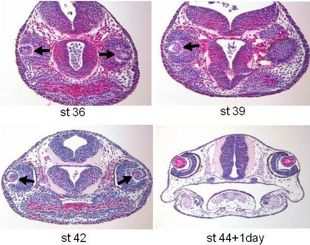
Suetsugu-Maki et al. BMC Biology 2012, 10:103                                                                     Page 2 of 8
http://www.biomedcentral.com/1741-7007/10/103

axolotls, similarly to newts, can regenerate a perfect lens     anti-rabbit Cy3 conjugated antibody (1/100 dilution,
from the iris and that this ability persists for about two to   Millipore). Immunohistochemistry images were taken
three weeks beyond that stage. After that, the ability for      using a BX51 microscope (Olympus, Tokyo, Japan) with a
lens regeneration is lost. Surprisingly, however, we find       CCD camera (Cool SNAP cf2; Photometrics, Tucson, AZ,
that the lens can be regenerated by either the dorsal or the    USA) and imaging software (Metamorph, Molecular
ventral iris. In some cases regeneration occurred from          Devices, Eugene, OR, USA), or by confocal imaging
both irises in the same eye. A similar series of experiments    (Olympus FV500 confocal microscope).
employing zebrafish failed to show any evidence that this
species can regenerate their lens underscoring the impor-       Results and discussion
tance of urodeles in the study of lens regeneration.            Based on the embryonic stages of axolotl outlined by
                                                                Armstrong and Malacinski [7], we histologically evaluated
Methods                                                         lens development in embryos as early as stage 36. Our eva-
Lentectomy                                                      luation showed that by stage 44, the lenses had developed
Axolotl larvae (st35 and st43, pre-hatched) were supplied       well with full differentiation of the globe having a clear
from the Ambystoma Genetic Stock Center (Department             lens epithelium and lens fibers (Figure 1). Since this is the
of Biology, Univ. of Kentucky, KY, USA) and kept at 27°C.       stage at which hatching occurs (and no further staging is
The hatched larvae were fed brine shrimp. Larvae were           available), we decided to remove the lens at different time
anesthetized in 0.1% ethyl 3-aminobenzoate (#E10521;            intervals after stage 44 starting with day 1 to day 27. Each
Sigma, St. Louis, MO, USA) at different stages. Using a         group was examined at different times after lentectomy to
sharp-edged blade, an incision was made in the cornea,          evaluate if animals were able to regenerate their lenses.
and then the lens was removed in its entirety. At different     We found that lens regeneration was possible within a
time intervals after lentectomy, animals were fixed in          particular time window after lens removal in animals up
methanol acetic acid solution (methanol: acetic acid = 3:1)     to 14 days after hatching (stage 4; Table 1, Figure 2). After
at 4°C overnight and processed for paraffin embedding.          that window of time, axolotls were found incompetent of
Similar series were also performed using zebrafish              regenerating their lenses. The regeneration process was
embryos (wild-type Ekkwill strain) at different stages. Ani-    very fast. Within one to two days after lentectomy, a well
mal care adhered to the guide lines of the Institutional        differentiated lens was present. The frequency of lens
Animal Care and Use Committee (IACUC), University of            regeneration was highest when lentectomy was performed
Dayton (axolotl) and of the IACUC, Cincinnati Children’s        3 or 7 days after stage 44; there were 10/16 (62.5%) and
Hospital Medical Center (zebrafish).                            8/10 (80%) regenerated lenses, respectively. We believe
                                                                that the failed cases were most likely the result of trauma
Hematoxylin and Eosin staining and                              to the eye due to lentectomy. The eye of the axolotl at
immunohistochemistry                                            these stages is very small and no matter how carefully len-
Paraffin sections of 15 μm were deparaffinized and used         tectomy is performed some injury is unavoidable. When
for H & E staining and immunohistochemistry. For H & E          we examined later stages, such as 27 days past stage 44,
staining, hematoxylin (#26754-01; Electron Microscope           axolotls were no longer able to regenerate the lens. In
Sciences, Hatfield, PA, USA) and eosin (#26762-01; Elec-        Table 2 in parentheses we include staging beyond stage
tron Microscope Sciences) were used. To examine cell            44, according to limb development [8].
proliferation in the iris after lentectomy, 5-bromo-2’-           Histological sections at day 1 after lentectomy hinted
deoxyuridine (BrdU; #B5002; SIGMA, 75 ug/g body                 that the lens regenerates from the iris; however, it could
weight) was injected into larvae 3 hours before fixation.       be formed from either the dorsal or the ventral iris.
Sections from BrdU injected larvae were treated with 1N         Prompted by these findings we decided to analyze lens
HCl for 5 minutes at room temperature, blocked in TNB           regeneration in more detail. For this, we removed the lens
buffer (0.1 M Tris-HCl, pH7.5; 0.15 M NaCl; and 0.5%            at stage 44 + 8 days and analyzed the process of regenera-
blocking reagent) supplied in the TSA kit (Perkin Elmer,        tion 3, 6, 12, 24 and 48 hours post-lentectomy. Three
Waltham, MA, USA) and incubated with mouse anti-                hours before collection, animals were injected with BrdU
BrdU antibody (1/100 dilution, #MAB3510; Millipore,             to examine cell proliferation. The collected animals were
Billerica, MA, USA) overnight at 4°C, and subsequently          embedded, sectioned and incubated with BrdU antibody
washed and incubated with Alexa 488 conjugated anti-            as well as with g-crystallin antibody that marks lens fibers.
immunoglobulin G (IgG) (1/100 dilution, Invitrogen,             The histological series are presented in Figure 3. In the
Grand Island, NY, USA) for 90 minutes at room tempera-          top panel, sections at 0 hour, just after removing the lens,
ture. To monitor lens regeneration, sections were incu-         are shown. BrdU staining was only observed at the ciliary
bated with anti g-crystallin rabbit antibody (1/300 dilution,   margin, where proliferating retinal stem/progenitor cells
source bovine crystallin) and then detected with an             contribute to the growing retina. As expected, g-crystallin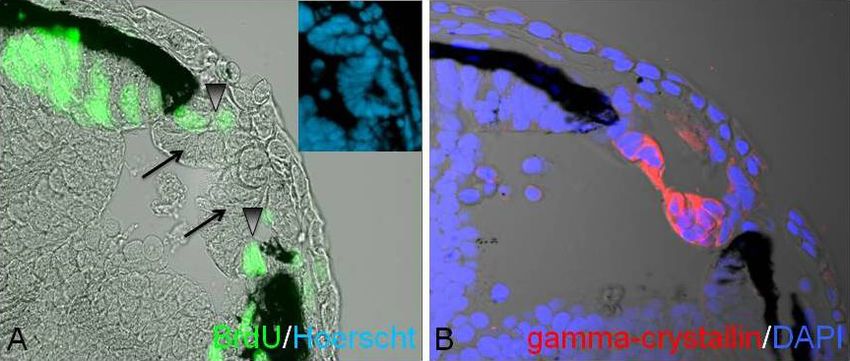
Suetsugu-Maki et al. BMC Biology 2012, 10:103                                                                                                     Page 3 of 8
http://www.biomedcentral.com/1741-7007/10/103

  Figure 1 Histological sections through the head of developing axolotls at different stages, to show the degree of differentiation of
  the eye and especially the lens. At stage 36, there is only a lens vesicle (arrows) with no clear differentiation of lens fibers yet. At stage 39,
  cells at the posterior part of the lens vesicle start elongating (arrow) and differentiating to lens. This is the beginning of lens fibers differentiation.
  At stage 42, differentiation of lens fibers covering most of the lens is evident (red color, arrow). At stage 44 +1 the lens is fully differentiated
  with a lens epithelium in the anterior region and differentiated lens fibers at the posterior region. H & E staining.

expression is absent. At 3 hours post-lentectomy no crys-                        post-lentectomy a lens vesicle positive for g-crystallin is
tallin synthesis was documented (not shown). At 6 hours                          present. The vesicle becomes more organized and polar-
                                                                                 ized at 12, 24 and 48 hours with an anterior region having
Table 1 Lens regeneration in axolotl evaluated at                                proliferating lens epithelial cells and a posterior part with
different stages starting with 1 day post-hatch after                            differentiating lens fibers. This represents the normal pro-
stage 44.                                                                        cess of lens development as well. Interestingly, the lens
       Stage at lentectomy                     Regenerated lenses                was elicited from the dorsal iris (9/14, 64.3%) or the ven-
          44+1day (44-45)                                4/8                     tral iris (3/14, 21.5%). Most surprisingly, however, is that
          44+2days (46-48)                               4/8                     in 2/14 (14.2%) cases lenses were regenerated from both
          44+3days (48-49)                              10/16                    the dorsal and the ventral iris. The regenerating lens at 6
          44+7days (49-50)                              8/10                     hours originated from the dorsal iris, while the regenerat-
         44+10days (50-51)                              4/16                     ing lens at 48 hours originated from the opposite, the ven-
         44+13days (51-52)                               2/4                     tral, iris (Figure 3). In a different eye, a regenerating lens
          44+27days (>54)                               0/10                     24 hours after lentectomy was produced from the ventral
                                                                                 iris (Figure 4). The lens was attached to the tip of the iris,
In parentheses equivalent stages as described by Nye et al. according to
forelimb and hindlimb development [8].                                           which was also positive for BrdU. The lens epithelium was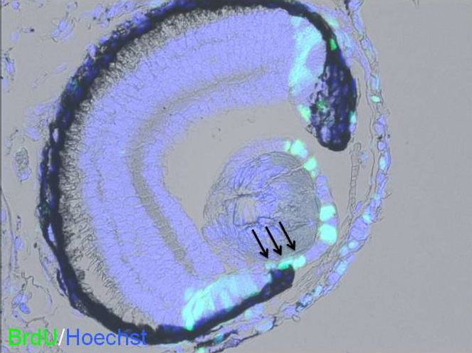
Suetsugu-Maki et al. BMC Biology 2012, 10:103                                                                                              Page 4 of 8
http://www.biomedcentral.com/1741-7007/10/103

  Figure 2 Examples of lens regeneration in axolotl larvae. A: A regenerating lens 2 days after lentectomy (lentectomy was performed at
  stage 44 + 2 days). Note that the lens is attached to the iris and shows the characteristic structure of the lens epithelium at the anterior (arrow)
  and differentiated lens fibers at the posterior region (arrowhead). B: A section through the head of an un-operated animal at the same stage as
  in A (in this case stage 44 + 4 days) to compare the degree of lens differentiation. C: A regenerated lens 7 days after lentectomy (lentectomy
  was performed at stage 44 + 3 days). Note an almost complete differentiation of lens. D: A section through the head of an un-operated animal
  at the same stage as in C (in this case stage 44 + 10 days) to compare the degree of lens differentiation. H & E staining.

also positive as expected. The two cases of the double                        and in Figure 5B two crystallin-positive lenses can be
lenses can be seen in Figure 5. In Figure 5A two lens vesi-                   observed.
cles derived from both dorsal and ventral iris are depicted                     Our results, thus, convincingly show that lens regen-
                                                                              eration is possible in the axolotl larvae. In this respect,
                                                                              there are differences and similarities when axolotl lens
Table 2 Lens regeneration in zebrafish at different stages
of development.
                                                                              regeneration is compared with the same process in frogs
                                                                              and in newts. Similarly to frogs, regeneration is possible
Stage                           Regenerated lenses at Day 4 post-
                                          lentectomy                          during a limited time window, but in contrast, the iris is
Prim 22 (35 hpf)                                 0/9
                                                                              the source of regeneration in the axolotl and not the
High or Long Pec (42-48                          0/5
                                                                              cornea. Similarly to newts, regeneration takes place
hpf)                                                                          from the iris pigment epithelium, but in contrast to the
Long Pec + FGF2 (48 hpf)                         0/17                         newt, it is not restricted to only the dorsal iris, and this
Protruding mouth (72                             0/2                          capacity is not present at later stages or as an adult.
hpf)                                                                            It is interesting to also note here that the competent
Hatching is around 42 hpf.                                                    stages for lens regeneration coincide with early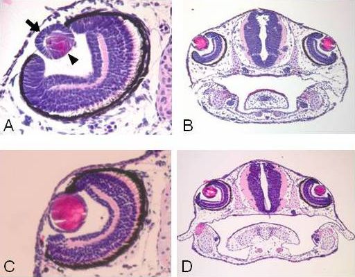
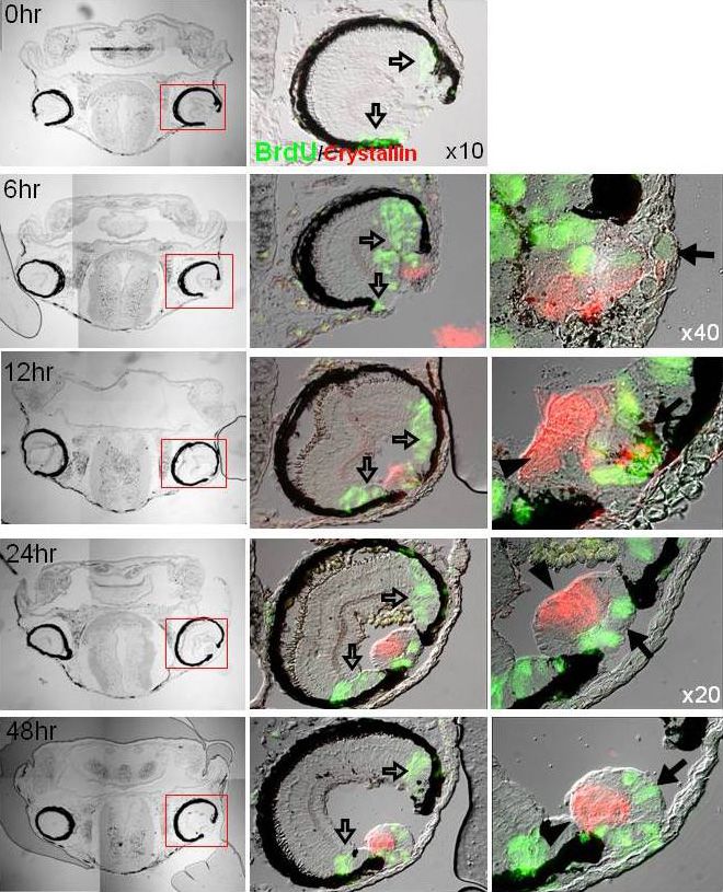
Suetsugu-Maki et al. BMC Biology 2012, 10:103                                                                                             Page 5 of 8
http://www.biomedcentral.com/1741-7007/10/103

 Figure 3 A detailed analysis of lens regeneration, showing lens proliferation and crystalline synthesis. All lentectomies were performed
 at stage 44 + 8 days. The first column shows bright field sections through the heads and the eyes. The second column shows bright field
 sections with BrdU staining (green) and g-crystallin staining (red) X10. The third column shows higher magnification images to depict better the
 regenerating lenses. The different rows represent samples at different time points after lentectomy. Note that the regenerating lens comes from
 the dorsal iris (the case at 6 hours) or the ventral iris (case at 48 hours). The regenerating lens is obvious at 6 hours post-lentectomy and starts
 organizing well with proliferating lens epithelium at the anterior (arrows) and differentiating lens fibers at the posterior (arrowheads) as soon as
 12 hours post-lentectomy. The proliferating cells in the ciliary margin (open arrows) are retina stem/progenitor cells that contribute to the
 growing retina.
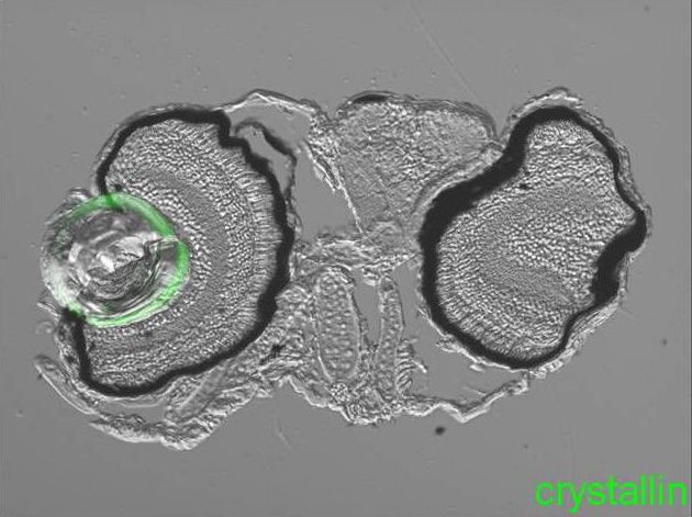
Suetsugu-Maki et al. BMC Biology 2012, 10:103                                                                                              Page 6 of 8
http://www.biomedcentral.com/1741-7007/10/103

 Figure 4 A different case of regenerating lens 24 hours after lentectomy (lentectomy was performed at stage 44 + 8 days. The
 regenerated lens is clearly attached to the iris (ventral) whose cells at the tip are actively dividing (arrows). As expected many other cells of the
 anterior lens epithelium are dividing as well. X40.

development of forelimbs and hindlimbs. Stage 44 is                          beginning of hindlimb bud differenrtiation. While a
morphologically marked by the development of the fore-                       direct correlation between the two events cannot be
limb bud [7,8]. The capacity for regeneration is termi-                      made at present, one idea could be that crucial factors
nated around stage 54 which is marked by the                                 for both events act during that time window.

 Figure 5 Two cases of lenses regenerated from both dorsal and ventral iris seen at 6 hours after lentectomy (lentectomy was
 performed at stage 44 + 8 days). A: note two early lens vesicles (arrows) with proliferating cells (arrowheads) at the tip of both irises. In the
 insert the same vesicles are shown stained with Hoerstch. B: Another case with two regenerating lenses. Blue is 4’,6-diamidino-2-phenylindole
 (DAPI) and red is g-crystallin. X40.
Suetsugu-Maki et al. BMC Biology 2012, 10:103                                                                                            Page 7 of 8
http://www.biomedcentral.com/1741-7007/10/103

 Figure 6 An example of lack of lens regeneration in zebrafish embryo. Lentectomy was performed at Long Pec Stage (48 hpf). Only the
 lens from the right eye was removed. The lens on the left eye was not touched. There is no sign of lens regeneration or crystallin synthesis
 (green) in the lentectomized eye when the animal was examined three days after lentectomy. Note that there was no severe trauma in the
 lentectomized eye.

   Prompted by the axolotl results, we also examined                     ventral iris. Such analysis can pinpoint crucial regulation
lens regeneration in zebrafish. Lens formation in zebra-                 underlying induction of lens regeneration.
fish with well differentiated anterior lens epithelium and
posterior lens fibers occurs within 28 hours post fertili-               Conclusions
zation [9]. We removed lenses at different stages starting               In this study, we have shown that lens regeneration is
at prim 22 (35 hpf), high pec (42 hpf) or long pec (48                   possible in the axolotl for a limited period of time. Lens
hpf) and protruding mouth (72 hpf). Hatching begins at                   can be regenerated between stage 44 (hatching) and
high pec stage. In none of the cases was a regenerated                   stage 52 (2 weeks post hatching). Our results show that
lens obtained (Table 2, Figure 6). Given the role of                     contrary to the newt that regenerates the lens only from
fibroblast growth factor (FGF) in zebrafish fin regenera-                the dorsal iris, the lens can be regenerated from either
tion [10] and in newt lens regeneration [11,12], we also                 the dorsal or the ventral iris or even from both sites.
added this growth factor to zebrafish embryos or to axo-                 Thus, the axolotl lens regeneration provides evidence
lotls at non-permissive stages post-lentectomy. FGF                      for a new developmental plasticity.
failed to induce any regeneration (not shown).
   Taken together our results underscore the importance of
                                                                         Abbreviations
newts in the study of lens regeneration. However, the fact               BrdU: 5-bromo-2’-deoxyuridine; FGF: fibroblast growth factor; H & E:
that there is a time window in which the axolotl can actu-               hematoxylin and eosin.
ally regenerate the lens provides an excellent comparative
                                                                         Acknowledgements
addition to the study of lens regeneration. Recently, the                This study was supported by NIH grant EY10540 to PAT and EY 17319 to
transcriptome of newt lens regeneration has been com-                    KDRT.
pleted (unpublished) as well as similar studies during axo-
                                                                         Author details
lotl limb regeneration [13]. Thus, this technology could be              1
                                                                          Department of Biology and Center for Tissue Regeneration and
of great value to regeneration studies. It will be of great              Engineering, University of Dayton, 300 College Park, Dayton, OH 45469-2320,
interest to compare, in the future, the transcriptome of                 USA. 2Current address: Institute of Protein Research, Osaka University, 3-2
                                                                         Yamadaoka, Suita-Shi, Osaka 565-0871, Japan. 3Current address: Faculty of
lens regeneration in axolotl (permissive and non-permis-                 Life and Environmental Sciences, University of Tsukuba, Tennoudai 1-1-1,
sive stages) with the newt transcriptome of the dorsal and               Tsukuba, Ibaraki 305-8572 Japan. 4Division of Developmental Biology,
Suetsugu-Maki et al. BMC Biology 2012, 10:103 Page 8 of 8
http://www.biomedcentral.com/1741-7007/10/103
Cincinnati Children’s Hospital Medical Center, 3333 Burnet Ave., Cincinnati,
OH 45229, USA. 5Department of Zoology, Miami University, 700 High Street,
Oxford, OH 45056, USA.
Authors’ contributions
RS-M and NM designed the research, performed the experiments and
analyzed data. KN, SS, JZ and KDRT performed experiments and analyzed
data. PAT designed the research, analyzed data and wrote the paper. All
authors have read and approved the manuscript for publication.
Competing interests
The authors declare that they have no competing interests.
Received: 7 November 2012 Accepted: 17 December 2012
Published: 17 December 2012
References
1. Tsonis PA: Limb Regeneration New York, NY, USA: Cambridge University
Press; 1996.
2. Sánchez Alvarado A, Tsonis PA: Bridging the regeneration gap: genetic
insights from diverse animal models. Nat Rev Genet 2006, 7:873-884.
3. Eguchi G, Eguchi Y, Nakamura K, Yadav MC, Millán JL, Tsonis PA:
Regenerative capacity in newts is not altered by repeated regeneration
and ageing. Nat Commun 2011, 2:384.
4. Tsonis PA, Del Rio-Tsonis K: Lens and retina regeneration:
transdifferentiation, stem cells and clinical applications. Exp Eye Res 2004,
78:161-172.
5. Henry JJ, Tsonis PA: Molecular and cellular aspects of amphibian lens
regeneration. Prog Retin Eye Res 2010, 29:543-555.
6. Stone LS: An investigation recording all salamanders which can and
cannot regenerate a lens from the dorsal iris. J Exp Zool 1967, 164:87-104.
7. Armstrong JB, Malacinski GM, Eds: Developmental Biology of the Axolotl
Oxford, UK: Oxford University Press; 1989.
8. Nye HLD, Cameron JA, Chernoff EAG, Stocum D: Extending the table of
stages of normal development of the axolotl: limb development. Dev
Dyn 2003, 226:555-560.
9. Greiling TM, Clark JI: Early lens development in the zebrafish: a three-
dimensional time-lapse analysis. Dev Dyn 2009, 238:2254-2265.
10. Wills AA, Kidd AR, Lepilina A, Poss KD: Fgfs control homeostatic
regeneration in adult zebrafish fins. Development 2008, 135:3063-3070.
11. Del Rio-Tsonis K, Jung JC, Chiu IM, Tsonis PA: Conservation of fibroblast
growth factor function in lens regeneration. Proc Natl Acad Sci USA 1997,
94:13701-13706.
12. Hayashi T, Mizuno N, Ueda Y, Okamoto M, Kondoh H: FGF2 triggers iris-
derived lens regeneration in newt eye. Mech Dev 2004, 121:519-526.
13. Monaghan JR, Epp LG, Putta S, Page RB, Walker JA, Beachy CK, Zhu W,
Pao GM, Verma IM, Hunter T, Bryant SV, Gardiner DM, Harkins TT, Voss SR:
Microarray and cDNA sequence analysis of transcription during nerve-
dependent limb regeneration. BMC Biol 2009, 7:1.
doi:10.1186/1741-7007-10-103
Cite this article as: Suetsugu-Maki et al.: Lens regeneration in axolotl:
new evidence of developmental plasticity. BMC Biology 2012 10:103.
Submit your next manuscript to BioMed Central
and take full advantage of:
• Convenient online submission
• Thorough peer review
• No space constraints or color figure charges
• Immediate publication on acceptance
• Inclusion in PubMed, CAS, Scopus and Google Scholar
• Research which is freely available for redistribution
Submit your manuscript at
www.biomedcentral.com/submitYou can also read