Metastatic Ovarian Tumor of Vulvar Malignant Melanoma in a 43-Year-Old Woman: A Case Report and Literature Review
←
→
Page content transcription
If your browser does not render page correctly, please read the page content below

Open Journal of Obstetrics and Gynecology, 2021, 11, 1-11
                                                                                             https://www.scirp.org/journal/ojog
                                                                                                         ISSN Online: 2160-8806
                                                                                                           ISSN Print: 2160-8792

Metastatic Ovarian Tumor of Vulvar Malignant
Melanoma in a 43-Year-Old Woman: A Case
Report and Literature Review

Yasuyuki Kawagoe1*, Midori Fujisaki1, Tomoko Goto1, Noriko Ueno1, Junji Onishi1,
Kosuke Mochida2, Yuichiro Sato3, Hiroshi Sameshima1
1
 Department of Obstetrics and Gynecology, Faculty of Medicine, University of Miyazaki, Kihara, Kiyotake, Miyazaki,
Japan
2
 Department of Dermatology, University of Miyazaki Hospital, Faculty of Medicine, University of Miyazaki, Kihara, Kiyotake,
Miyazaki, Japan
3
 Department of Diagnostic Pathology, University of Miyazaki Hospital, Faculty of Medicine, University of Miyazaki, Kihara,
Kiyotake, Miyazaki, Japan

How to cite this paper: Kawagoe, Y., Fu-      Abstract
jisaki, M., Goto, T., Ueno, N., Onishi, J.,
Mochida, K., Sato, Y. and Sameshima, H.       An ovarian malignant melanoma sometimes occurs from ovarian teratoma.
(2021) Metastatic Ovarian Tumor of Vul-       Ovarian metastatic malignant melanoma is extremely rare. We describe a pa-
var Malignant Melanoma in a 43-Year-Old       tient in whom vulvar melanoma (previously resected) metastasized to the
Woman: A Case Report and Literature
Review. Open Journal of Obstetrics and
                                              ovary, making ovarian metastatic malignant melanoma. A 43-year-old Japa-
Gynecology, 11, 1-11.                         nese woman was referred to us because of left ovarian tumor. She had un-
https://doi.org/10.4236/ojog.2021.111001      dergone resection for malignant melanoma on the right labia minora with
                                              inguinal lymph node metastasis (pT1bN1aM0, stage IIIA, FIGO 2008). Eigh-
Received: December 10, 2020
                                              teen months after this surgery, CT scans revealed left ovarian tumor and
Accepted: January 11, 2021
Published: January 14, 2021                   swelled pelvic lymph nodes, with a pelvic examination disclosing a left ad-
                                              nexal solid mass, with normal serum CA125 level (21.7 U/mL). Laparotomy
Copyright © 2021 by author(s) and             revealed a left solid ovarian tumor measuring 4 cm, which was covered with a
Scientific Research Publishing Inc.
                                              smooth grayish capsule. The right ovary, uterus, and pelvic cavity appeared
This work is licensed under the Creative
Commons Attribution International             normal. Upon sectioning during the surgery, the cross-sectional surface of the
License (CC BY 4.0).                          left ovary revealed a dark brown solid tumor. Following an intra-operative
http://creativecommons.org/licenses/by/4.0/   frozen-section diagnosis as metastatic melanoma, total hysterectomy with bi-
                Open Access                   lateral salpingo-oophorectomy and pelvic lymph dissection was performed.
                                              Histological examination confirmed the diagnosis as malignant melanoma
                                              metastasis to the left ovary and the obturator lymph node: the same laterality
                                              (left) as the primary site. The tumor was entirely composed of malignant me-
                                              lanoma cells with no evidence of teratoma. Combined chemotherapy with
                                              dabrafenib mesylate and trametinib was planned based on the positive BRAF
                                              mutations. This case highlights the importance that physicians should have

DOI: 10.4236/ojog.2021.111001 Jan. 14, 2021                      1                    Open Journal of Obstetrics and Gynecology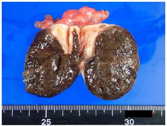
Y. Kawagoe et al.

                                  high index of suspicion for the occurrence of ovarian melanoma metastasis
                                  after melanoma surgery. We also made extensive literature review on this is-
                                  sue, of which description may contribute to better understanding of this con-
                                  dition.

                                  Keywords
                                  Vulva, Malignant Melanoma, Ovarian Metastasis

                                1. Introduction
                                Primary ovarian melanoma is sporadic, and most cases originate within a ma-
                                ture cystic teratoma, while metastatic malignant melanoma of the ovary is a rare
                                condition. Most commonly originates from cutaneous tumors, and sometimes
                                from ocular melanoma. The vulva is the most common site of melanoma in the
                                female genital tract, while only one case of vulvar melanoma metastasis to the
                                ovary has been reported [1]. The median age at diagnosis of cutaneous melano-
                                ma in sun-exposed skin is 56 years, while that of the vulva is 68 years, but it
                                rarely occurs in women of a reproductive age [2] [3]. We describe here an un-
                                usual case of a woman who underwent surgical removal of melanoma of the
                                vulva at 41 years of age, and subsequently developed left ovarian metastasis 18
                                months after the initial surgery.

                                2. Case Presentation
                                A 43-year-old Japanese gravida 2 para 2 premenopausal woman was referred to
                                our gynecological department because of significant enlargement of the left
                                ovary. At 36 years of age, pigmentation of the external genitalia was detected at
                                the time of her second delivery, although it was left untreated. Her past and fam-
                                ily medical histories were uneventful. Five years after the delivery, when she was
                                41 years old, she noticed a slight bump on the surface of the pigmented skin ac-
                                companied by scant bleeding. She visited a local dermatological hospital, whe-
                                reupon a gray-white irregular lesion was detected on the left labia minora, which
                                was diagnosed as a malignant melanoma by biopsy. The patient then consulted
                                the Department of Dermatology at the University Hospital. A darkly pigmented
                                lesion was confirmed on the left labia minora measuring approximately 10 mm
                                in size. The patient subsequently underwent radical local excision plus sentinel
                                lymph node sampling. Pathologically, the nodular lesion was composed of poly-
                                gonal cell proliferation with lesional melanin deposition (Figure 1(a)). The tu-
                                mor cells were diffusely positive for HMB-45 by immunohistochemical analysis.
                                The thickness of the tumor was 3.5 mm, and the width was 8 mm, with no ulcer
                                formation. With a positive sentinel lymph node result, the entire left inguinal
                                lymph node was dissected a few weeks after the initial surgery. The final histo-
                                logical examination revealed a vulvar melanoma with inguinal lymph node

DOI: 10.4236/ojog.2021.111001                         2                   Open Journal of Obstetrics and Gynecology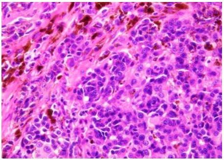
Y. Kawagoe et al.

                                                     (a)                                         (b)

                                Figure 1. (a) Vulva: Polygonal tumor cells proliferate in nests or diffusely. Melanin pig-
                                ments are also present; (b) Left ovary: Polygonal tumor cells with round to oval-shaped
                                hyperchromatic nuclei and prominent nucleoli proliferatein sheets. Melanin deposition is
                                also observed (Hematoxylin and eosin, ×400).

                                metastasis (pT1bN1aM0, stage IIIA, FIGO 2008). No adjuvant therapy was in-
                                itiated, and the patient continued to be regularly followed up.
                                  Eighteen months after the surgery, CT scans taken at a routine medical ex-
                                amination revealed significant swelling of the left ovary and pelvic lymph nodes,
                                compared with previous CT scans taken just before the surgery. After the refer-
                                ence to the gynecological department, pelvic examination disclosed a left adnex-
                                al solid mass, a normal-sized uterus and a right ovary. She had no symptoms or
                                complaints on her first visit. Pelvic MRI (T-weighted image) also showed a
                                low-intensity solid tumor of the left ovary measuring 35 × 40 mm and significant
                                enlargement of a pelvic lymph node (Figure 2). Serum CA125 level was 21.7
                                U/mL, which was within the normal range. At laparotomy, a left solid ovarian
                                tumor covered with a smooth grayish and intact capsule was confirmed without
                                adhesions to its surroundings (Figure 3). The right ovary, uterus, and pelvic
                                cavity appeared grossly normal. The left obturator and external iliac lymph
                                nodes were swollen. Infracolic-omentum and peritoneal surfaces were smooth
                                with no evidence of dissemination. Upon sectioning, the cross-sectional surface
                                of the left ovary showed characteristic findings of a homogenous dark brown solid
                                tumor (Figure 4). The diagnosis of the intra-operative frozen section was metastat-
                                ic malignant melanoma. Total hysterectomy with bilateral salpingo-oophorectomy
                                and pelvic lymph dissection was performed. As a final diagnosis, metastases to
                                both the left ovary and the obturator lymph node were determined histological-
                                ly. The tumor was entirely composed of malignant melanoma cells with no evi-
                                dence of teratoma, and peritoneal cytology was negative for atypical cells (Figure
                                1(b)). These metastatic lesions were evident only on the left side, which was on
                                the same side as the primary lesion on the vulva. Her postoperative course was
                                uneventful. Two months after surgery, combined chemotherapy with dabrafenib
                                mesylate and trametinib was planned based on the positive results of molecular
                                testing for BRAF mutations.

                                3. Discussion
                                Incidence rates of malignant melanoma differ according to ethnic background,

DOI: 10.4236/ojog.2021.111001                           3                     Open Journal of Obstetrics and Gynecology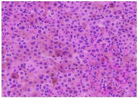
Y. Kawagoe et al.

                                Figure 2. Pelvic MRI study (T2-weighted image) showed a low-intensity solidtumor of
                                left ovary (arrow A) and significant enlargement of an obturator lymphnode (arrow B).

                                Figure 3. The left ovary was palpable about 40 × 35 mm (arrows), covered with the
                                grayish surface, while the right ovary and uterus were grossly normal appearance.

                                Figure 4. The cross-sectional surface of the left ovary showed a solid tumor colored in
                                dark brown.

                                and the incidence rate was 24.9 in White, 1.3 in Asian/Pacific Islander, and 1.0 in
                                black individuals per 100,000 between 2012 and 2016 [4]. Melanoma commonly
                                arises from cutaneous sites in regions exposed to ultraviolet radiation. Extracuta-
                                neous sites lined with mucosal membranes can also include an original site of
                                melanoma such as the respiratory, gastrointestinal, and urogenital tracts. Vulvar
                                melanoma is a mucosal type as well, which is rare and is more aggressive. It
                                usually results in less favorable prognoses compared with cutaneous melanoma.
                                  Malignant melanoma in the ovary can involve either a primary or metastatic
                                tumor, but most cases are metastatic [5]. Primary ovarian melanoma is sporadic,
                                and most cases originate within a mature cystic teratoma. Metastases of mela-
                                noma to the ovary occur from the skin, and choroid of the eye, but rarely from
                                primary vulva tumors, the prognosis of which is poor with a 5-year survival rate
                                of only 5% [6]. In postmortem cases, approximately 18% of women with disse-

DOI: 10.4236/ojog.2021.111001                          4                     Open Journal of Obstetrics and Gynecology
Y. Kawagoe et al.
minated melanoma have ovarian implants [7]. Taken together, it is conceivable
that our case was vulvar melanoma in a woman of reproductive age, resulting in
metastasis to the ovary. Both findings are extremely unusual phenomena.
We searched PubMed and Google Scholar, from January 1980 to December
2019, using the terms “ovary/ovarian”; “melanoma”; and “metastatic/metastasis/
metastases”. The literature was limited to English-language case reports and case
series. They comprised four case series [5] [8] [9] [10] and 22 case reports [1] [6]
[11]-[30], totaling 61 cases. We excluded cases that met any of the following cri-
teria: no medical information available; metastases confirmed at autopsy; and
unknown original site of melanoma. Forty-one out of the 61 cases plus our case,
totaling 42 cases, were available for this review and are listed in Table 1 [1] [5]
[6] [8] [10]-[27] [29] [30]. Based on this search, this is the second report of ova-
rian metastasis of vulvar melanoma. The most frequent primary site was the skin
(83.3%, 35/42) followed by the choroid (11.9%, 5/42), while two cases were from
the vulva (4.8%, 2/42). The median age at ovarian metastasis was 38.5 years
(range, 8 - 65 years), and 85.7% (36/42) of patients were less than 50 years old.
The median interval for relapse was 66 months (range, 8 - 300). The median
tumor size was 100 mm (range, 40 - 190 mm), and bilateral ovaries were affected
in 26.8% (11/41) of cases. In deceased patients (n = 27), the median survival pe-
riod after the confirmation of ovarian metastasis was 10 months (range, 1 - 96).
In an analysis of 1521 cases of melanoma, which included male patients, the
most frequent site of first metastasis was the lung, followed by the skin, and
lymph nodes, and 14% of cases had multiple metastatic sites [31]. Our review
showed that 81.0% (34/42) of the cases involved multiple lesions when ovarian
implants were identified, while this was rare for solitary metastasis. Once ova-
rian metastasis had established, extensive multisystemic spread of the disease
occurred in most cases.
In patients in which an ovarian tumor is detected long after the initial treat-
ment, it is a diagnostic challenge. Therefore, for patients with such a history, a
precise medical interview is essential, and systemic screening is mandatory. In
our case, routine CT scans allowed for the discovery of a unilateral ovarian tu-
mor of a relatively small size. We were able to classify metastatic sites without
difficulty because of the short interval since the last surgery. As such, regular
screening could contribute to the early detection of metastases. However, this
does not always lead to a preferred prognosis because most cases are accompa-
nied by multiple metastatic lesions, resulting in a survival period of less than 1
year after relapse. It is interesting to note that the interval for ovarian metastasis
was long, at 5 years or more in 64.2% (27/42) of cases, and 10 years or more in
23.8% (10/42) of cases. Especially, metastases from choroidal melanoma tend to
have a latent period than metastasis from cutaneous melanoma. In most cases, it
was associated with multiple metastases. At the initial therapy for melanoma, it
is crucial to inform patients and their families about the possibility of relapse af-
ter a long period.
DOI: 10.4236/ojog.2021.111001 5 Open Journal of Obstetrics and GynecologyY. Kawagoe et al.
Table 1. Published cases of metastatic malignant melanoma to the ovary (n = 42).
Laterality Size of Interval
Extent Other Outcome
Author (year) Case Age Primary of the ovarian Clinical from
disease sites of (time after
[Reference] No. (years) site affected tumor** symptoms primary
adimitted metastasis surgery)
ovary (mm) (months)
Fitzgibbons PL
1 38 skin (scalp) Bil 80 NA Yes axillary LN 48 NA
(1987) [8]
2 45 skin (thumb) Uni NA NA No 24 Died (12 Mo)
3 32 skin (leg) Uni 180 NA Yes chest wall 60 NA
lung,
4 37 skin (leg) Bil NA NA Yes 60 NA
omentum
5 29 skin (arm) Bil 120 NA Yes axillary LN 24 NA
Moselhi M
6 36 skin (leg) Uni 100 pelvic pain No 96 Died (NA)
(1998) [10]
regular large bowel
7 25 skin (leg) Bil 80 Yes 24 Died (NA)
screening mesentery
abdominal pelvic bone,
8 45 skin (arm) Uni* 80 Yes 24 Died (NA)
bloating lung
Gupta D skin
9 29 Uni 110 regular screening Yes multiple 15 Died (10 Mo)
(2004) [5] (shoulder)
10 39 skin (arm) Bil 70 regular screening Yes multiple 120 Died (26 Mo)
dysuria, para-aortic
11 25 skin (back) Uni 135 Yes 96 Died (32 Mo)
abdominal pain LN
12 29 skin (back) Uni 150 regular screening Yes multiple 34 Died (40 Mo)
13 43 skin (arm) Uni 100 pelvic mass Yes multiple 132 Died (7 Mo)
lungs,
14 45 skin (arm) Uni 180 regular screening Yes abdominal 228 Died (4 Mo)
wall
lungs,
15 49 skin (arm) Uni 55 pelvic mass Yes perirectal 20 Died (8 Mo)
mass
16 40 skin (arm) Uni 60 regular screening Yes multiple 30 Died (30 Mo)
skin
17 41 Uni 100 regular screening Yes multiple 83 Died (76 Mo)
(shoulder)
18 38 skin (pubis) Uni 120 regular screening Yes lungs 74 Alive (96 Mo)
skin
19 27 Uni 80 regular screening Yes axillary LN 46 Alive (6 Mo)
(shoulder)
20 52 skin (scalp) Uni 50 regular screening Yes multiple 54 Died (10 Mo)
axillary LN,
Murphy DJ
21 25 skin (scalp) Bil 120 regular screening Yes subcutaneous 26 NA
(1994) [11]
tissue
Remadi S
22 33 skin (chest) Uni 100 abdominal pain No 60 Died (24 Mo)
(1997) [12]
pelvic pain,
Nakano J multiple
23 36 skin (arm) Bil 125 Yes subcutaneus 132 Died (13 Mo)
(1998) [13] subcutaneous
nodules
DOI: 10.4236/ojog.2021.111001 6 Open Journal of Obstetrics and GynecologyY. Kawagoe et al.
Continued
Piura B abdominal pain, subcutaneus
24 45 skin (back) Uni 100 Yes 84 Alive (7 Mo)
(1998) [14] pelvic mass tissue, brain
Fuller PN
25 34 skin (arm) Bil 190 abdominal pain Yes brain 180 Died (1 Mo)
(1999) [15]
Bilgin T abdominal omentum,
26 35 skin (scalp) Bil 94 Yes 96 Died (5 Mo)
(2000) [16] bloating liver
Oliver R
27 58 skin (arm) Uni 120 asymptomatic Yes pelvic LN 120 Died (12 Mo)
(2005) [17]
Boutis A abdominal peritnium,
28 43 skin (arm) Uni* NA Yes 108 Died (8 Mo)
(2008) [18] distension omentum
abdominal
Abe Y
29 39 skin (leg) Uni 65 fullness, Yes iliac bone 8 Died (7 Mo)
(2009) [19]
appetite loss
Sbitti Y
30 45 skin (leg) Uni 150 pelvic pain No 38 Died (13 Mo)
(2011) [20]
acute abdomen,
Fenzl V
31 35 skin (back) Bil 90 fever, Yes bone 60 Died (15 Mo)
(2011) [21]
appetite loss
Habek D
32 35 skin (back) Uni 110 abdominal pain Yes brain 84 Died (3 Mo)
(2012) [22]
Berisavac M
33 48 skin (arm) Uni 125 abdominal pain No 60 Died (48 Mo)
(2013) [23]
Araki Y skin
34 8 Uni 60 regular screening Yes liver, bone 8 Died (14 Mo)
(2014) [24] (buttock)
liver, spleen,
Mendel A
35 65 skin (leg) Uni 76 regular screening Yes esphagus, 84 NA
(2017) [6]
lung
lung,
omentum,
Ben David M abdominal pain, serosal
36 62 choroid Uni 150 Yes 300 NA
(1984) [25] vaginal bleeding surface of
small and
large bowel
Santeusanio G
37 47 choroid Uni 170 pelvic pain No 168 NA
(2000) [26]
Rey-Caballero
38 38 choroid Uni 61 abdominal pain No 108 Alive (7 Mo)
VE (2004) [27]
Bloch-Marcotte
39 50 choroid Uni 40 abdominal pain Yes liver 240 Alive (5 Mo)
C (2008) [29]
pancreas,
Mandato VD hypogastric pain,
40 57 choroid Bil 110 Yes para-aortic 72 Died (7 Mo)
(2010) [30] weight loss
LN
Spatz A vulva
41 28 Uni 65 regular screening No 180 Alive (12 Mo)
(1999) [1] (labia majora)
Kawagoe vulva
42 43 Uni 50 regular screening Yes obturator LN 18 Alive (4 Mo)
Y (2020) (labia minora)
*Laterality was determined by CT imaging without operation. **Tumor sizes are expressed by the maximum dimension. LN: lymph node, NA: Not available,
Mo: month, Uni: Unilateral, Bil; Bilateral.
DOI: 10.4236/ojog.2021.111001 7 Open Journal of Obstetrics and GynecologyY. Kawagoe et al.
The ovary is a small organ in the pelvis and is a relatively common site of me-
tastases. This has been speculated to be as a result of its hypervascularity [32]
[33]. Ovarian metastases are thought to mainly occur in reproductive-age wom-
en, and less frequently in postmenopausal women. Our review showed that the
median age of diagnosis was 38.5 years and 85.7% of patients were less than 50
years old, even though melanoma is common in older adults.
Ovarian metastases are typically from the stomach, colon, and breast. These
metastatic lesions in the ovary are known to show bilateral and multicystic le-
sions, namely mucinous carcinoma, compared with primary lesions. Our patient
had a solid homogenous unilateral tumor, the cross-section of which was dark
brown. This was the first case in which we had observed such characteristics of
metastatic melanoma. Its entire capsule was also a grayish color with a smooth
surface. Young et al. reported in a review of ovarian metastases that 55% of the
tumors were solid, while 45% were solid and cystic with the solid component
predominating. Thirty percent of the tumors were either entirely black or had
discernible black or brown foci [9]. Gupta et al. reported that 35% of the lesions
were grossly pigmented [5]. Regarding laterality, bilateral involvement was
commonly seen in cutaneous melanoma in 45% of cases [9]. In our review,
which included choroidal and vulvar melanoma, its incidence was less frequent
at 26.8%.
4. Conclusion
We here described the second reported case of ovarian metastasis of vulvar me-
lanoma in a 43-year-old woman. In patients who have a history of melanoma
and a significant enlargement of the ovary, metastasis should be ruled out. Me-
tastasis usually occurs after a long period and accompanies disseminated disease,
resulting in a poor prognosis of generally less than 1-year survival.
Consent
Informed consent was obtained.
Conflicts of Interest
The authors have no conflicts of interest relevant to this article.
References
[1] Spatz, A., Zimmermann, U., Bachollet, B., Pautier, P., Michel, G. and Duvillard, P.
(1998) Malignant Blue Nevus of the Vulva with Late Ovarian Metastasis. The
American Journal of Dermatopathology, 20, 408-412.
https://doi.org/10.1097/00000372-199808000-00016
[2] Ragnarsson-Olding, B., Johansson, H., Rutqvist, L. and Ringborg, U. (1993) Malig-
nant Melanoma of the Vulva and Vagina. Trends in Incidence, Age Distribution,
and Long-Term Survival among 245 Consecutive Cases in Sweden 1960-1984. Can-
cer, 71, 1893-1897.
https://doi.org/10.1002/1097-0142(19930301)71:53.
DOI: 10.4236/ojog.2021.111001 8 Open Journal of Obstetrics and GynecologyY. Kawagoe et al.
0.CO;2-7
[3] Sugiyama, V.E., Chan, J.K., Shin, J.Y., Berek, J.S., Osann, K. and Kapp, D.S. (2007)
Vulvar Melanoma: A Multivariable Analysis of 644 Patients. Obstetrics & Gynecol-
ogy, 110, 296-301. https://doi.org/10.1097/01.AOG.0000271209.67461.91
[4] Centers for Disease Control and Prevention (2019) Melanoma Incidence and Mor-
tality, United States 2012-2016. USCS Data Brief, No. 9. Centers for Disease Control
and Prevention, US Department of Health and Human Services, Atlanta.
https://www.cdc.gov/cancer/uscs/about/data-briefs/no9-melanoma-incidence-mort
ality-UnitedStates-2012-2016.htm
[5] Gupta, D., Deavers, M.T., Silva, E.G. and Malpica, A. (2004) Malignant Melanoma
involving the Ovary: A Clinicopathologic and Immunohistochemical Study of 23
Cases. The American Journal of Surgical Pathology, 28, 771-780.
https://doi.org/10.1097/01.pas.0000126786.69232.55
[6] Mendel, A., Terzibachian, J.J., Aubin, F. and Malicenco, L. (2017) Ovarian Metasta-
sis of a Malignant Melanoma: A Case Report. Journal of Gynecology Obstetrics and
Human Reproduction, 46, 461-462. https://doi.org/10.1016/j.jogoh.2017.02.016
[7] Das Gupta, T.K. and Brasfield, R.D. (1969) Primary Melanomas in Unusual Sites.
Surgery, Gynecology and Obstetrics, 128, 841-848.
[8] Fitzgibbons, P.L., Martin, S.E. and Simmons, T.J. (1987) Malignant Melanoma Me-
tastatic to the Ovary. The American Journal of Surgical Pathology, 11, 959-964.
https://doi.org/10.1097/00000478-198712000-00006
[9] Young, R.H. and Scully, R.E. (1991) Malignant Melanoma Metastatic to the Ovary.
A Clinicopathologic Analysis of 20 Cases. The American Journal of Surgical Pa-
thology, 15, 849-860. https://doi.org/10.1097/00000478-199109000-00005
[10] Moselhi, M., Spencer, J. and Lane, G. (1998) Malignant Melanoma Metastatic to the
Ovary: Presentation and Radiological Characteristics. Gynecologic Oncology, 69,
165-168. https://doi.org/10.1006/gyno.1998.4992
[11] Murphy, D.J., Hickling, D.J. and Pathak, U.N. (1994) Metastatic Malignant Mela-
noma of the Ovary. Case Report. European Journal of Gynaecological Oncology, 2,
119-120.
[12] Remadi, S., McGee, W., Egger, J.F. and Ismail, A. (1997) Ovarian Metastatic Mela-
noma. A Diagnostic Pitfall in Histopathologic Examination. Archives D’anatomie et
de Cytologie Pathologiques, 45, 43-46.
[13] Nakano, J., Shimizu, T., Hirota, T. and Muto, M. (1998) An Unusual Female Mela-
noma Patient with Late Metastases to Both Skin and Ovaries. The Journal of Der-
matology, 25, 126-128. https://doi.org/10.1111/j.1346-8138.1998.tb02363.x
[14] Piura, B., Kedar, I., Ariad, S., Meirovitz, M. and Yanai-Inbar, I. (1998) Malignant
Melanoma Metastatic to the Ovary. Gynecologic Oncology, 68, 201-205.
https://doi.org/10.1006/gyno.1997.4919
[15] Fuller, P.N. (1999) Malignant Melanoma of the Ovary and Exposure to Clomiphene
Citrate: A Case Report and Review of the Literature. American Journal of Obstetrics
and Gynecology, 180, 1499-1503. https://doi.org/10.1016/S0002-9378(99)70045-1
[16] Bilgin, T. (2000) Late Ovarian Metastases of a Cutaneous Malignant Melanoma.
Journal of Obstetrics and Gynaecology, 20, 323.
https://doi.org/10.1080/01443610050009791
[17] Oliver, R., Dasgupta, C., Coker, A., Al-Okati, D. and Weekes, A.R.L. (2005) Ovarian
Malignant Melanoma: Unusual Presentation of a Solitary Metastasis. Gynecologic
Oncology, 99, 412-414. https://doi.org/10.1016/j.ygyno.2005.06.066
DOI: 10.4236/ojog.2021.111001 9 Open Journal of Obstetrics and GynecologyY. Kawagoe et al.
[18] Boutis, A., Valeri, R., Korantzis, I., Valoukas, D., Andronikidis, I. and Andreadis, C.
(2008) Delayed Malignant Melanoma Recurrence Simulating Primary Ovarian
Cancer: Case Report. World Journal of Surgical Oncology, 6, 124.
https://doi.org/10.1186/1477-7819-6-124
[19] Abe, Y., Takeuchi, M., Matsuzaki, K., Uehara, H., Furumoto, H. and Nishitani, H.
(2009) A Case of Metastatic Malignant Melanoma of the Ovary with a Multilocular
Cystic Appearance on MR Imaging.Japanese Journal of Radiology, 27, 458-461.
https://doi.org/10.1007/s11604-009-0368-6
[20] Sbitti, Y., Fadoukhair, Z., Kadiri, H., Oukabli, M., Essaidi, I., Kharmoum, S.,
M’rabti, H., Albouzidi, A., Ichou, M. and Errihani, H. (2011) Diagnostic Challenge
for Ovarian Malignant Melanoma in Premenopausal Women: Primary or Metastat-
ic? World Journal of Surgical Oncology, 9, Article No. 65.
https://doi.org/10.1186/1477-7819-9-65
[21] Fenzl, V., Prkacin, I. and Skrtic, A. (2011) Malignant Melanoma Ovarian Metastasis
Mimicking Acute Tuboovarian Abscess. European Journal of Gynaecological On-
cology, 32, 582-584. https://doi.org/10.2478/s11536-011-0133-y
[22] Habek, D., Marton, I., Bauman, R., Prka, M. and Bobic, M.V. (2012) Ruptured Ova-
rian Metastatic Malignant Melanoma Caused Acute Abdomen. Central European
Journal of Medicine, 7, 241-244.
[23] Berisavac, M., Kotlica, B.K., Pilic, I. and Atanackovic, J. (2013) Metastatic Malignant
Ovarian Melanoma—A Case Report. Vojnosanitetski Pregled, 70, 229-232.
https://doi.org/10.2298/VSP110719030B
[24] Araki, Y., Kaneda, H., Oashi, K., Okada, S. and Tsutsumida, A. (2014) Ovarian Me-
tastasis of Malignant Melanoma: The First Pediatric Case. Journal of Pediatric Sur-
gery Case Reports, 2, 473-475. https://doi.org/10.1016/j.epsc.2014.09.010
[25] Ben David, M., Feldberg, D., Dicker, D., Kessler, H. and Goldman, J.A. (1984) Ova-
rian Melanoma: An Interesting Case. International Journal of Gynecology & Obste-
trics, 22, 77-79. https://doi.org/10.1016/0020-7292(84)90107-3
[26] Santeusanio, G., Ventura, L., Mauricello, A., Carosi, M., Spagnoli, L.G., Maturo, P.,
Terranova, L. and Romanini, C. (2000) Isolated Ovarian Metastasis from a Spindle
Cell Malignant Melanoma of the Choroid 14 Years after Enucleation: Prognostic
Implication of the Keratin Immunophenotype. Applied Immunohistochemistry &
Molecular Morphology, 8, 329-333.
https://doi.org/10.1097/00022744-200012000-00011
[27] Rey-Caballero, V.E., Lopez-Gonzalez, B., Garcia-Benitez, J.L., Boix-Fos, A. and Di-
az-Lagama, A.M. (2004) Solitary Ovarian Metastasis from Ocular Melanoma.
American Journal of Obstetrics and Gynecology, 191, 368-369.
https://doi.org/10.1016/j.ajog.2003.10.713
[28] Coutts, M.A., Borthwick, N.J., Hungerford, J.L. and Cree, I.A. (2006) Post-Menopausal
Bleeding: A Rare Presentation of Metastatic Uveal Melanoma. Pathology Oncology
Research, 12, 184-187. https://doi.org/10.1007/BF02893367
[29] Bloch-Marcotte, C., Ambrosetti, D., Novellas, S., Caramella, T., Dahman, M., Thyss,
A. and Chevallier, P. (2008) Ovarian Metastasis from Choroidal Melanoma. Clinical
Imaging, 32, 318-320. https://doi.org/10.1016/j.clinimag.2008.04.001
[30] Mandato, V.D., Kobal, B., Di Stefano, A., Sinkovec, J., Levicnik, A., La Sala, G.B.
and Rakar, S. (2010) Choroidal Melanoma Metastasized to the Ovary: Case Report
and Review of the Literature. European Journal of Gynaecological Oncology, 31,
109-113.
[31] Barth, A., Wanek, L.A. and Morton, D.L. (1995) Prognostic Factors in 1,521 Mela-
DOI: 10.4236/ojog.2021.111001 10 Open Journal of Obstetrics and GynecologyY. Kawagoe et al.
noma Patients with Distant Metastases. Journal of the American College of Surge-
ons, 181, 193-201.
[32] Young, R.H. and Scully, R.E. (1991) Metastatic Tumors in the Ovary: A Prob-
lem-Oriented Approach and Review of the Recent Literature. Seminars in Diagnos-
tic Pathology, 8, 250-276.
[33] Singh, N., (2004) The Pathology of Metastasis in the Ovary. Journal of Gynecologic
Oncology, 9, 96-102.
DOI: 10.4236/ojog.2021.111001 11 Open Journal of Obstetrics and GynecologyYou can also read