PCOS - An Updated Overview and Current Trends in Ultrasound Imaging - journal of evidence based medicine and healthcare
←
→
Page content transcription
If your browser does not render page correctly, please read the page content below
Jebmh.com                                                                                                           Review Article

PCOS - An Updated Overview and Current Trends in Ultrasound Imaging
                                        Dev Prakash Singh Rathour1, Shubham Singh2

              1
               Associate Professor, Department of Radiology, Era’s Lucknow Medical College, Lucknow,
                           Uttar Pradesh, India. 2Physician, Lucknow, Uttar Pradesh, India.

                                                            A BS T R A C T

BACKGROUND
Poly-Cystic Ovary Syndrome (PCOS) is a widespread complex endocrine disorder                      Corresponding Author:
of women in the reproductive age group. It may present as mild menstrual                          Dr. Dev Prakash Singh Rathour,
disorder which affects metabolic functions severely. PCOS results in chronic                      C-24, H-Park,
                                                                                                  Mahanagar Extension,
anovulation. There is abnormal production of oestrogen and androgens due to
                                                                                                  Lucknow- 226006,
imbalance of LH and FSH. LH/FSH ratio is elevated. Women with PCOS are prone                      Uttar Pradesh, India.
to insulin resistance, type II diabetes mellitus, obesity and infertility, psychological          E-mail: drdpsrathour@gmail.com
disorder like depression, cardiovascular diseases, and endometrial and ovarian
cancer. Presenting symptoms may be acne and hirsutism. To define PCOS, there                      DOI: 10.18410/jebmh/2020/267
has to be two of the three following features- menstrual irregularity, clinical and
                                                                                                  Financial or Other Competing Interests:
biochemical evidence of androgen excess and multiple cysts in the ovary.
                                                                                                  None.
            PCOS is manifestation of various interrelated mechanisms; it may not be
known which if any, is primary. Probably PCOS is common end result of different                   How to Cite This Article:
mechanisms and pathologies. There may be pituitary dysfunction resulting in high                  Rathour DPS, Singh S. PCOS- An
serum LH and high serum prolactin. Menstrual cycles may be anovulatory                            updated overview and current trends in
                                                                                                  ultrasound imaging. J. Evid. Based Med.
presenting as oligomenorrhea, secondary amenorrhea, cystic ovaries and
                                                                                                  Healthc. 2020; 7(26), 1255-1260. DOI:
infertility. Patients are prone to obesity which leads hyperglycaemia and elevated
                                                                                                  10.18410/jebmh/2020/267
oestrogen and sometimes insulin resistance leading to type II diabetes mellitus,
dyslipidaemia and hypertension.                                                                   Submission 26-04-2020,
            However, despite significant progress in understanding the                            Peer Review 29-04-2020,
                                                                                                  Acceptance 30-05-2020,
pathophysiology and diagnosis of the disorder, over the past 20 years, the disorder
                                                                                                  Published 29-06-2020.
remains underdiagnosed and misunderstood. The diagnostic criteria are indefinite
with numerous intricacies, PCOS remains a challenging area of research. The aim
of this article is to review the present status and formulate an interesting clinically
relevant research direction with emphasis on ultrasound imaging in diagnosis of
polycystic ovary with stress on various aspects of 3D, colour & power Doppler
study that is essential to move the field of PCOS forward.

KEYWORDS
PCOS, Poly Cystic Ovary, Ultrasound Imaging

J. Evid. Based Med. Healthc., pISSN- 2349-2562, eISSN- 2349-2570/ Vol. 7/Issue 26/June 29, 2020                               Page 1255
Jebmh.com Review Article
INTRODUCTION hyperandrogenaemia and oligo-ovulation, or (c) hirsutism
and oligo-ovulation. Alternatively, fasting insulin levels were
PCOS is a heterogeneous disorder of functional androgen highest in patients with hirsutism, hyperandrogenaemia, and
excess, detected clinically or by laboratory testing, with oligo-ovulation, and lowest in those women with oligo-
ovulatory dysfunction and polycystic ovarian morphology ovulation and hirsutism only.
also affecting a large proportion of these patients. PCOS is a
diagnosis of exclusion, with other androgen excess or
related disorders to be ruled out. The diagnosis of PCOS is ESHRE/ASRM (Rotterdam) Criteria
based on well-defined criteria, at present for utilization in Another expert conference was organized in Rotterdam in
clinical practice there are three major sets of diagnostic May of 2003 in part sponsored by ESHRE and ASRM. The
criteria available. Regional prevalence of PCOS depends on proceedings of the conference noted that PCOS could be
diagnostic criteria utilized and ethnicity. Targeted screening diagnosed, after the exclusion of related disorders, by two
in women with isolated symptoms of acne, hirsutism, and of three features:6
irregular menstrual cycles should be practiced. 1. Oligo and/or Anovulation (up to 90% patients) -
Oligomenorrhea with less than eight periods per year or
amenorrhea with no periods for more than three
REVIEW OF LITERATURE months.
2. Hyperandrogenism (seen in approx. 60% patients) -
Assessed clinically by: hirsutism, acne, alopecia or
Diagnostic Criteria
biochemically: raised circulating androgens.
Stein and Leventhal originally described the combination of
3. Polycystic ovaries on Ultrasound (US): ovarian volume
oligo-ovulation and hyperandrogenism1 in the year 1935.
10 cc or more and/or has >10 follicles of 2-9 mm in
Description of the syndrome was based upon case reports in
diameter per ovary. The prevalence of Polycystic ovary
the literature. Abnormal uterine bleeding was the most
in PCOS patients is estimated to be 17-33%.
common symptom associated with the condition. Over time,
multiple efforts have been made to better characterize this
As for the NIH 1990 criteria, other disorders should be
syndrome to allow for better appreciation of this complex
excluded. It should be noted that these recommendations
entity. For clinicians there are three major sets of diagnostic
did not replace the NIH 1990 criteria; rather they expanded
criteria in diagnosing PCOS.
the definition of PCOS.
The first set of criteria was proposed by National
Institutes of Health (NIH) in Bethesda, Maryland, in 1990,
but has been replaced in clinical practice by the relatively
AES 2006 Criteria
recently proposed Rotterdam criteria.
Because of the continuing controversy regarding the
To date, three major criteria have been proposed, with
definition of the PCOS, the AES, an international
other investigators proposing modifications of these. We will
organization dedicated to promoting knowledge and original
review the criteria arrived at a NIH/NICHD expert conference
clinical and basic research in every aspect of androgen
sponsored in 1990,2 that proposed by an expert conference
excess disorders, charged a Task Force to recommend an
of the European Society for Human Reproduction and
evidence-based definition for PCOS, whether already in use
Embryology (ESHRE) and the American Society for
or not, to guide clinical diagnosis and future research. The
Reproductive Medicine (ASRM) in 2003,3 and that proposed
Task Force, after review of all available published data,
by the Androgen Excess Society in 2006.4
proposed that PCOS should be diagnosed by the presence of
three features: (a) androgen excess (clinical and/or
biochemical hyperandrogenism), (b) ovarian dysfunction
National Institute of Health Criteria
(oligo-anovulation and/or polycystic ovarian morphology),
The first useful definition of PCOS arose from the
and (c) exclusion of other androgen excess or ovulatory
proceedings of an expert conference sponsored by the US
disorders.6
National Institutes of Health (NIH) in April 1990. Participants
were surveyed, and tabulation of the results indicated the
features of PCOS were:
Diagnosis of PCOS
a) Hyperandrogenism and/or hyperandrogenaemia,
The diagnosis of PCOS needs: (a) to assess features
b) Chronic anovulation and
suggestive of PCOS are present and (b) to exclude related
c) Exclusion of related disorders such as
androgen excess or ovulatory disorders. It will depend on
hyperprolactinemia, thyroid disorders, and congenital
chosen diagnostic criteria.
adrenal hyperplasia.5 Polycystic ovaries were
suggestive, not diagnostic, of the syndrome.
Assess whether Features of PCOS are Present
Three principal phenotypes of PCOS are recognized
Features suggesting PCOS are: (a) long-term menstrual
using the NIH 1990 criteria, including women with: (a)
dysfunction or irregularity, suggestive of chronic ovulatory
hirsutism, hyperandrogenaemia, and oligo-ovulation, (b)
J. Evid. Based Med. Healthc., pISSN- 2349-2562, eISSN- 2349-2570/ Vol. 7/Issue 26/June 29, 2020 Page 1256Jebmh.com Review Article
dysfunction, (b) dermatologic signs suggestive of congenital adrenal hyperplasia. These disorders account for
hyperandrogenism, like hirsutism, acne or alopecia, and (c) 5-10% of all women with androgen excess.10
polycystic ovarian morphology on ultrasonography.
All women with menstrual dysfunction should be
reviewed for hyperandrogenism presenting as acne, Normal Ovary
unwanted hair growth, scalp hair shedding or loss with a Normal ovary has a relatively homogeneous echotexture
more in-depth evaluation for PCOS. Clinical signs of insulin with a central, more echogenic medulla. Well-defined, small
resistance (e.g., acanthosis nigricans) needs more thorough anechoic or cystic follicles may be seen peripherally in the
evaluation for PCOS (and metabolic syndrome). Finally, in a cortex (Figure 1). The appearance of the ovary changes with
patient with menstrual dysfunction, assessment of polycystic age and with the phase of the menstrual cycle. During the
ovaries on Ultrasonography is needed to exclude PCOS. early proliferative phase, many follicles that are stimulated
by both follicle-stimulating hormone (FSH) and luteinizing
hormone (LH) develop and increase in size until about day 8
Exclusion of Other Androgen Excess or Ovulatory or 9 of the menstrual cycle. At that time one follicle becomes
Disorders dominant, destined for ovulation, and increases in size,
Diagnosis of PCOS needs exclusion of other disorders like reaching up to 2.0 to 2.5 cm at the time of ovulation. The
21-hydroxylase deficient NCAH (by a basal and/or stimulated other follicles become atretic. A follicular cyst develops if the
17-hydroxyprogesterone), androgen-secreting neoplasms fluid in one of these non-dominant follicles is not resorbed.
(by history and clinical exam and appropriate studies in Following ovulation, the corpus luteum develops and may be
selected patients), adreno cortical hyperactivity (by clinical identified sonographically as a small hypoechoic or isoechoic
exam and appropriate testing), and drug-induced structure peripherally within the ovary. The corpus luteum
hyperandrogenism. involutes before menstruation.
Laboratory and Radiological/Sonographic Evaluation
Women should undergo measurement of circulating TSH,
prolactin, 17-HP levels. Androgen levels: measurement of
circulating androgen levels (generally total and free
testosterone, and DHEAS), assessment of TSH, prolactin,
and 17-HP levels required. Ovarian Ultrasonography in
assessing PCO is essential.
Figure 1. Normal Ovaries on Transvaginal Scan
DIFFERENTIAL DIAGNOSIS
PCOS remains a diagnosis of exclusion, and it is useful to
exclude other potential aetiologies that can present with the
triad of polycystic ovaries, hyperandrogenism, and chronic
anovulation. For instance, chronic anovulation alone may be
due to failure or dysfunction of the hypothalamic-pituitary
axis or to frank ovarian failure, states of steroid deficiency
without androgen excess. In series of adult women
presenting with amenorrhea alone, PCOS is present in about
one-third of these patients,7 but rises to 70% or more when
other symptoms such as hirsutism are considered.8 Other
than PCOS, other potentially serious causes of
hyperandrogenism include such disorders as Cushing’s
syndrome and an androgen-secreting tumour.9 These
disorders are acquired and are often preceded by a period
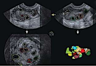
Figure 2. PCO with Cysts Arranged at the Periphery
of normal menses without symptoms of hyperandrogenism.
In contrast, PCOS presents in the post menarche and tends
Because of the variability in shape, ovarian volume has
to affect women throughout much of their reproductive life.
been considered the best method for determining ovarian
Androgen secreting tumours are rare in this age group, are
size. The volume measurement is based on the formula for
usually ovarian in origin, tend to have markedly elevated
a prolate ellipse (0.523 x length x width x height). Studies
circulating androgen levels above the usual disorder that can
have shown that ovarian volumes are larger than previously
present peripubertally in a similar indolent fashion as PCOS
thought. In the first 2 years of life. The mean ovarian volume
is 21-hydroxylase (21-OH) deficient non classic congenital
is slightly greater than 1 cc in the first year and 0.7 cc in the
adrenal hyperplasia (NCAH), also known late-onset
J. Evid. Based Med. Healthc., pISSN- 2349-2562, eISSN- 2349-2570/ Vol. 7/Issue 26/June 29, 2020 Page 1257Jebmh.com Review Article
second year. The upper limit of normal has been reported eyeballing. But AFC is more, as typically seen in polycystic
as 3.6 cc in the first 3 months, 2.7 cc from 4 to 12 months, ovary 3D US & advanced calculation software Sono AVC is
and 1.7 cc in the second year. Ovarian volume remains required. To use Sono AVC, region of interest is selected to
relatively stable up to 5 years of age and then gradually include the whole ovary in all three orthogonal planes on
increases up to menarche when the mean volume is 4.2±2.3 acquired ovarian volume. The count is done automatically
cc, with an upper limit of 8.0 cc.11 In 87% pre menarchal & but post processing may be required. Automated 3D
neonatal ovaries small follicles or cysts are present.12 In measures do provide reliable information on follicle number
adult menstruating female a normal ovary has a volume as and size and appear to be more reflective of ovarian reserve
large as 22 cc. In one study by transvaginal scan reported a and response.16
mean volume of 6.8 cc with upper limit of 18 cc.13
Following menopause, the ovary atrophies and follicles
disappear over the subsequent few years with ovary
decreasing in size.14
Ultrasound Technique
This scan is done on day 2-3 of menstrual cycle. It was done Figure 3. Ovarian
by transvaginal route using a transducer with 8 MHz Area Calculation on
B-Mode US Image
frequency. If trans-abdominal ultrasound scan is used, AFC
(Antral Follicle Count) is not to be used as criteria to
diagnose PCO (Polycystic ovary). Doppler is used to assess
ovarian stromal vascularity. Spectral Doppler is used for
quantitative assessment of the flows by measuring intra-
ovarian resistance index (RI) and peak systolic velocity
(PSV). For colour doppler, pulse repetition frequency (PRF)
is set at 0.3, wall filters are lowest, with optimum gains and
balance settings. For pulse Doppler PRF around 0.9-1.3 is
set, and wall filters are set at 30 HZ as stromal flows on Figure 4. Stromal
Volume Calculation
baseline scan are usually low-velocity flows. on B-Mode US Image
Ovarian Volume is calculated by formula for ellipse
(0.523 x length x width x height). Measure the largest
longitudinal, transverse, and AP diameter of the ovary in
centimeters (cm). 3D US provides a new method for
objective quantitative assessment of follicle count, ovarian
volume, and blood flow in the ovary.15
Volume histogram gives values of 3D power Doppler
indices, VI (Vascularization Index), FI (Flow Index) and VFI
(Vascularization-Flow Index). VI indicates abundance of flow
in the selected volume, FI is an index for average intensity
of flow in a selected volume, and VFI is a perfusion index.
Stromal Volume
Applying threshold volume on the same VOCAL (Virtual
Organ Computer Aided Analysis) calculated volume will Figure 5. Colour Coded Antral Follicle on Sono AVC
define stromal volume when threshold is set to differentiate
follicles from rest of the ovarian tissues. When AFC is much
more, as typically seen in polycystic ovaries, 3D US and Stromal Echogenicity and Polycystic Ovary
advanced calculation software. Sono 3D volume acquisition Morphology (PCOM)
of the ovary and volume calculation by VOCAL (virtual organ Stromal echogenicity is assessed against echogenicity of
computer - aided analysis for volume) is a more reliable myometrium on B-mode US. Polycystic ovary morphology
technique especially when the ovarian shape is not round or (PCOM) is more accurate for PCOS diagnosis for women
oval. between 30 and 39 years of age.17 It has also been
suggested that the threshold for PCOM should be revised
regularly with advancing ultrasound technology and age-
Antral Follicle Count (AFC) specific cut off values for PCOM should be defined.
Antral follicles are counted in the whole ovary by taking a 2D
sweep across whole ovary and counting the follicles by
J. Evid. Based Med. Healthc., pISSN- 2349-2562, eISSN- 2349-2570/ Vol. 7/Issue 26/June 29, 2020 Page 1258Jebmh.com Review Article
Ultrasound Features and Review of Literature (Sono AVC & VOCAL) have provided tools for more accurate
Volume: Though 10 cc is the cut off defined in Rotterdam’s assessment of number of follicles, ovarian volume, ovarian
criteria, ovarian volume 6.6 cc has shown 91% sensitivity morphology & stromal volume. US remains a good tool for
and 91% specificity for polycystic ovarian' syndrome.18 assessment of normal physiological changes and variations
Polycystic ovary morphology is therefore a better in volume and size of ovaries during puberty. Transvaginal
discriminator than ovarian volume between women with sonography in adolescent has definite role in diagnosis of
polycystic ovary syndrome and control women. PCOS.
The best sensitivity and specificity for diagnosis of PCOS
were obtained using different threshold volume and AFC at
different ages. RE F E R E N C E S
This study also quoted that the polycystic ovary
morphology (PCOM) is more accurate for PCOS diagnosis for [1] Stein IF, Leventhal ML. Amenorrhea associated with
women between 30 and 39 years of age. It has also been bilateral polycystic ovaries. Am J Obstet Gynecol
suggested that the threshold for PCOM should be revised 1935;29(2):181-191.
regularly with advancing ultrasound technology. [2] Rotterdam ESHRE/ASRM-Sponsored PCOS Consensus
Therefore, now it is decided that ultrasound should not Workshop Group. Revised 2003 consensus on
be used as one of two features to diagnose PCOS in females diagnostic criteria and long-term health risks related to
up to 8 years of gynaecological age. For diagnosis of PCOS polycystic ovary syndrome. Fertil Steril 2004;81(1):19-
in young adolescent female all the three features of PCOS 25.
according to Rotterdam criteria should be present. [3] Azziz R, Carmina E, Dewailly D, et al. The androgen
The antral and atretic follicles may be peripherally excess and PCOS society criteria for the polycystic ovary
arranged or are dispersed in the stroma and are named syndrome: the complete task force report. Fertil Steril
peripheral and general cystic pattern of PCO (polycystic 2009;91(2):456-488.
ovary).19 Lam et al. have concluded in their study that the [4] Rosenfield RL, Wroblewski K, Padmanabhan V, et al.
current criteria will fail to identify milder forms of PCOS and Antimüllerian hormone levels are independently related
further information, about the ovarian stroma and the to ovarian hyperandrogenism and polycystic ovaries.
degree of vascularization, is required. Hyperdense stroma Fertil Steril 2012;98(1):242-249.
and stromal abundance have been described with polycystic [5] Zawadzki JK, Dunaif A. Diagnostic criteria for polycystic
ovaries since the first definition of the syndrome by Stein- ovary syndrome: towards arational approach. In: Dunaif
Leventhal. US to diagnose PCOS, a cardinal feature has been A, Givens JR, Haseltine FP, et al, eds. Polycystic ovary
shown to be the presence of a bright, highly echogenic syndrome. Boston: Blackwell Scientific Publications
stroma and stromal echogenicity/total ovarian echogenicity 1992:377-384.
was significantly higher in PCOS than controls.20 Increased [6] Rotterdam ESHRE/ASRM-Sponsored PCOS consensus
stromal echogenicity for diagnosis of PCOS has a sensitivity workshop group. Revised 2003 consensus on diagnostic
of 94% and specificity of 90%.21 Stromal area of 4.6 cm2 has criteria and long-term health risks related to polycystic
91% sensitivity and 86% specificity for diagnosis of ovary syndrome (PCOS). Hum Reprod 2004;19(1):41-
polycystic ovarian syndrome. Ovarian area of 5.3 cm² has 47.
93% sensitivity and 91% specificity for diagnosis of [7] Azziz R, Carmina E, Dewailly D, et al. Positions
polycystic ovarian syndrome. Mean stromal area/mean statement: criteria for defining polycystic ovary
ovarian area ratio of 0.34 and above has a specificity of syndrome as a predominantly hyperandrogenic
100% and this parameter may be used in routine clinical syndrome: an androgen excess society guideline. J Clin
practice for improving US diagnosis of PCOS.22 In a study by Endocrinol Metab 2006;91(11):4237-4245.
Franks et al., it has been well derived that PCOM in normal [8] van Santbrink EJ, Hop WC, Fauser BC. Classification of
women is not a morphological variant of normal ovaries, but normogonadotropic infertility: polycystic ovaries
rather represents a functional entity - a silent form of diagnosed by ultrasound versus endocrine
PCOS.23 Along with evaluation of ovaries on US, endometrial characteristics of polycystic ovary syndrome. Fertil Steril
morphology and ovarian pathologies (if any) should be 1997;67(3):452-458.
evaluated. [9] Arroyo A, Laughlin GA, Morales AJ, et al. Inappropriate
gonadotropin secretion in polycystic ovary syndrome:
influence of adiposity. J Clin Endocrinol Metab
CONCLUSIONS 1997;82(11):3728-3733.
[10] Azziz R, Sanchez LA, Knochenhauer ES, et al. Androgen
excess in women: experience with over 1000
Assessment of anovulation and polycystic ovary (PCO) is
consecutive patients. J Clin Endocrinol Metab
very well possible on US. With advent of Transvaginal (TV)
2004;89(2):453-462.
sonography and improved image resolution, better
[11] Balen AH, Laven JSE, Tan SL, et al. Ultrasound
assessment of ovarian morphology is possible. 3D, colour
assessment of the polycystic ovary: International
and Power Doppler imaging, advanced calculation software
J. Evid. Based Med. Healthc., pISSN- 2349-2562, eISSN- 2349-2570/ Vol. 7/Issue 26/June 29, 2020 Page 1259Jebmh.com Review Article
consensus definitions. Hum Reprod Update [18] Kim HJ, Adams JM, Gudmundsson JA, et al. Polycystic
2003;9(6):505-514. ovary morphologv: age-based ultrasound criteria. Fertil
[12] Orsini LF, Salardi S, Pilu G, et al. Pelvic organs in Steril 2017;108(3):548-553.
premenarcheal girls: real-time ultrasonography. [19] Wu MH, Tang HH, Hsu CC, et al. The role of three-
Radiology 1984;153(1):113-116. dimensional ultrasonographic images in ovarian
[13] Holm K, Laursen V, Brocks V, et al. Pubertal maturation measurement. Fertil Steril 1998;69(6):1152-1155.
of the internal genitalia: an ultrasound evaluation of 166 [20] Matsunaga l, Hata T, Kitao M. Ultrasonographic
healthy girls. Ultrasound Obstet Gynecol identification of polycystic ovary. Asia Oceania J Obstet
1995;6(3):175-181. Gynecol 1985;11(2):227-232.
[14] Goswmy RK, Campbell S, Roysten JP, et al. Ovarian size [21] Buckett WM, Bouzayen R, Watkin KL, et al. Ovarian
in postmenopausal women. Br J Obstet Gynaecol stromal echogenicity in women with normal and
1988;95(8):795-801. polycystic ovaries. Hum Reprod 1999;14(3):618-621.
[15] Merz E, Miric-Tesanic D, Bahlmann F, et al. Sonographic [22] Pache TD, Wladimiroff JW, Hop WC, et al. How to
size of uterus and ovaries in pre and post-menopausal discriminate between normal and polycystic ovaries:
women. Ultrasound Obstet Gynecol 1996;7(1):38-42. Transvaginal US Study. Radiology 1992;183(2):421-
[16] Lam PM, Raine-Fenning N. The role of three- 423.
dimensional ultrasonography in polycystic ovary [23] Fulghesu AM, Angioni S, Frau E, et al. Ultrasound in
syndrome. Hum Reprod 2006;21(9):2209-2215. polycystic ovary syndrome--the measuring of ovarian
[17] Franks S, Webber LJ, Goh M, et al. Ovarian morphology stroma and relationship with circulating androgens:
is a marker of heritable biochemical traits in sisters with results of multicentric study. Hum Reprod
polycystic ovaries. J Clin Endocrinol Metab 2007;22(9):2501-2508.
2008;93(9):3396-3402.
J. Evid. Based Med. Healthc., pISSN- 2349-2562, eISSN- 2349-2570/ Vol. 7/Issue 26/June 29, 2020 Page 1260You can also read