Molecular Modeling 2021 - A course about proteins protein conformation protein shape Protein energy
←
→
Page content transcription
If your browser does not render page correctly, please read the page content below

Molecular Modeling 2021

     A course about proteins

       protein conformation
           protein shape
          Protein energy
        protein interactions
         protein dynamics
     protein drug interactions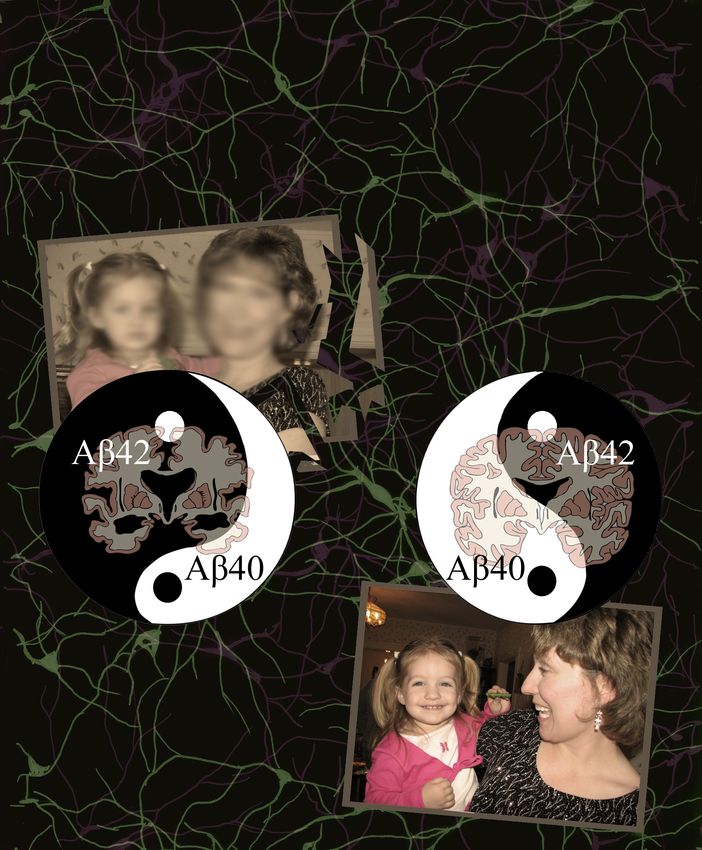
Administrative
• Course website: on Blackboard learn:
  https://lms.rpi.edu/
• Office hour: 4 – 5 PM Wednesdays, on webex
• Grading policy
  –   Assignment and quiz (30 % total)
  –   midterm (20%)
  –   final (20%)
  –   Presentation (20%)
  –   Class participation (answering questions in class) and
      attendance (10%)
• Questions ALWAYS welcome!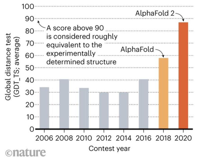
Syllabus

Installing MOE
• Download MOE to your personal computer using file
  on RPI box
   https://rpi.box.com/s/
   mdjcw69wffo5roe5p7nse4n16wznf97u
   !! If your RPI box account is not set up, use this link to
   set it up: https://itssc.rpi.edu/hc/en-us/articles/
   360004829931-Requesting-a-Box-RPI-Account
• Copy the license file (license.dat in the box directory)
  into the installed MOE local directory
• To run MOE, you need to access the license server
  located on RPI campus: got to have VPN running!
   – Install “cisco anyconnec”t: https://itssc.rpi.edu/hc/en-us/
     articles/360008783172-VPN-Installation-and-Connection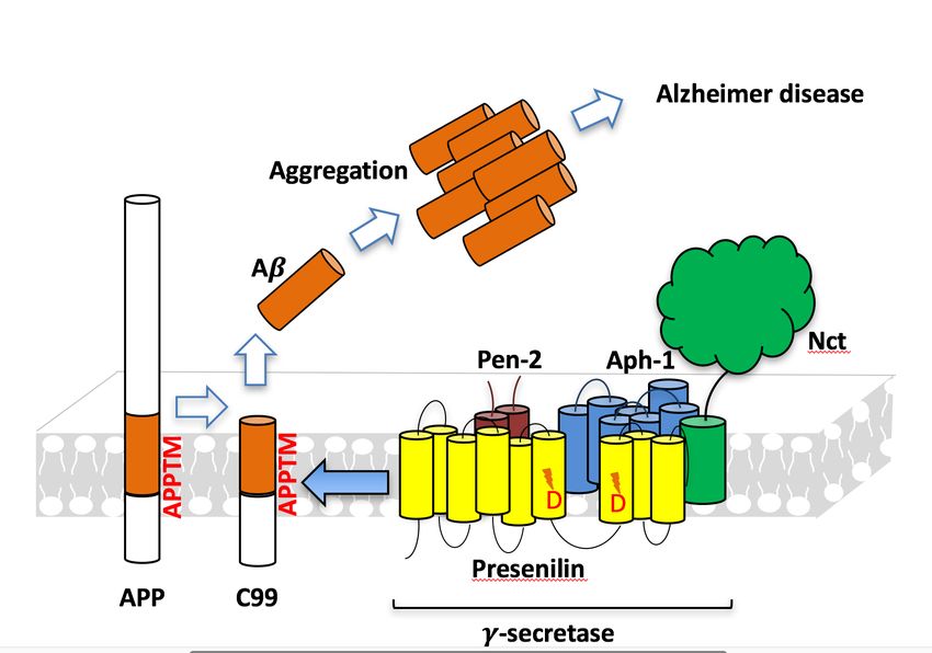
Instructor
• Chunyu Wang
• Associate Professor of Biological
  Sciences
• Expert in NMR, protein structure,
  Alzheimer's disease, Inteins and
  Hedgehog autoprocessing
• Hobbies: photography, astronomy,
  astrophotography, tennis, bridge,
  gardening, swimming etc.
NMR structure of Pab PolII intein: Hint fold

   Cover of Nov.4th
  2011 Issue of JBC

   Hint = Hedgehog
  Intein domain/fold

Du et.al. J. Biol. Chem.,
2011, 286, 38638-38648.
                                                         6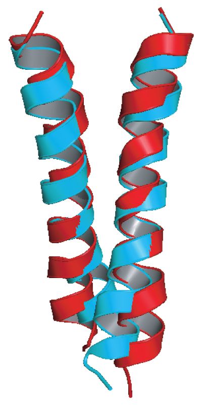
What are Aβ, Αβ40 and Αβ42 ?
      Aβ42

      Aβ40

Major component of the amyloid
   plaque

Aβ40 and Aβ42 are major species
   of Aβ

Aβ42 aggregates faster and is
   more toxic than Aβ40

In familial Alzheimer’s disease
    (FAD), Aβ42/Aβ40 ↑
                                           Aβ40                    Aβ42

.
                                  Sgourakis et al. (2007) JMB 368:1448-1457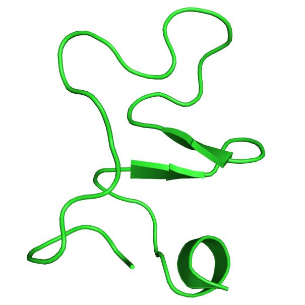
Yin and Yang of
                      Alzheimer’s

by Caitlin Piette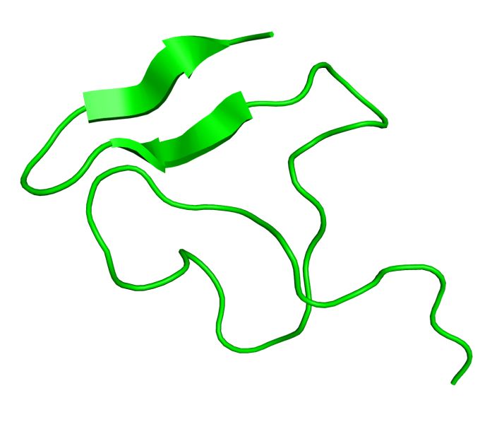
Aβ generation and AD

         Presenilin homolog
AD-Causing Mutations in APPTM
Increase Aβ42 by Opening T48
NMR structures of WT
APPTM (red) and V44M Wen Chen, Eric Gamache, David Rosenman, Jian Xie, Maria
Lopez, Yueming Li, and Chunyu Wang (2014). “Familial
(blue), an AD-causing Alzheimer's Mutations within APPTM Increase Abeta42
Production by Enhancing the Accessibility of the Epsilon-
mutant. Cleavage Site”. Nature Communications 4:3037 doi:
10.1038/ncomms4037.Intro to Molecular Modeling
and ProteinsWhat is molecular modeling ?
• Experiments performed on a computer
• Application of computational methods to
study the structure, dynamics, reaction, and
thermodynamics of molecules, and to
understand molecular processes
• The models and methods must be tailored to
the question at hand.
• This implies that molecular modeling must be
both multidisciplinary and multiscale
12Multidiscipline
• At the interface of chemistry, physics, mathematics,
computer science, molecular biology, material
science, nanotechnology
• Numerous names: computational chemistry,
molecular modeling, computational biochemistry,
computational biophysics, computational
nanoscience, computational structural biology,
computational material science ….
• Drawing increasing interest: academia, industry
(drug discovery)
• The field is developing with dazzling speed.
13Multiscale (temporal and spatial)
14Connection to Wet Lab
Experiments: Crucial
• Confirm, extrapolate and predict wet lab
experimental results --- representing one more
experimental tool, such as NMR
• Require information from wet-lab experiments
• Can help understand experimental results
• Can guide experiments (design)
• Can provide details not available in typical
experiments
15Some Important Applications
• Refine the X-ray and NMR structures
• Understand structure (dynamics) - function
relationship
• Understand chemistry of the biological
processes, including enzyme catalysis , DNA
damage ..
• Rational design of drugs, novel inhibitors,
new protein and materials
• Predict the biomolecular structure, property,
function
• ….
16Martin Karplus Michael Levitt Arieh
17 WarshelAlpha Fold 2: predicting protein structure w deep learning https://deepmind.com/blog/article/alphafold-a-solution-to-a-50-year-old-grand- challenge-in-biology https://www.nature.com/articles/d41586-020-03348-4
Today chemists experiment just as much on their computers as they do in their
labs. Theoretical results from computers are confirmed by real experiments that
  yield new clues to how the world of atoms works. Theory and practice cross-
                                fertilize each other.

                                                                   19
                                                          http://www.nobelprize.org/
Important Elements for Molecular Modeling
• Structure of the target
molecule
• Potential Energy
Surface
• Simulation. (sampling,
how to explore the
PES)
• analyze the results and
calculate the desired
properties
20Amino acids
21Amino acid states
Free amino acid, Zwitterionic form, Polymer repeating
solid phase aqueous, pH7 unit (“residue”)
R
R R
HO C O
H C C
C NH C H +
2 – N H H
C N
3
O H
O
O
The chemical nature of the AA backbone depends on the context.
Usually we are talking about AAs in the polymer (polypeptide)
context.peptide bond formation is dehydrating
R1 R2
-
O C
C H
H C H +
C +
N H3
N H
H O
O
R1 O
δ-
C +
C
H N H3 + H 2O
C H
N
C
H
O δ+
R2
Rx is catalyzed by the ribosome.the polypeptide backbone
peptide
bond
R2 O
O
H H
N C CH N C
C CH N
CH C
N
H
O R1 R3
O
Backbone atom names are “N, “C-alpha” and “C” (or N,CA,C).
Oxygen “O” is also considered a backbone atom. All atoms in all amino
acids have conventional atom names.the side chains
Which AA’s are
found on the
surface? In
turns? In the
core?
The shape and chemical nature of these 20 side chains account for the folding and function
of proteins.How and where to obtain a suitable initial
structure for your molecule ?
• The first and essential task for a molecular modeling
project.
• Building with softwares, such as MOE
• Crystal molecules of small molecules (Cambridge
structural database http://www.ccdc.cam.ac.uk/products/
csd )
• Large biomolecules: searching the protein data bank.
http://www.rcsb.org. The files can be downloaded from
the web
26What is a protein structure? • A collection of 3D coordinates (x, y, z) of all atoms in the protein • a pdb file in protein databank (pdb) • Visualized by many viewer programs, such as VMD, pymol etc.
PDB ATOM lines

          ATOM           1    N        VAL   A 101B           0.616      -1.613     20.826    1.00 68.81          1   8DFR 152
1-6 keyword ATOM
                                                                                                               67-80 footnotes and labels
     7-11 atom number
                                                                                                       61-66 B=Temperature factor‡
            13-16 atom name                                                                  55-60 Occupancy factor
                 17 altloc indicator
                                                                                  47-54 Z-coordinate
                   18-20 residue name
                      21 not used                                       39-46 Y-coordinate
                 22 chain identifier (optional)
                           23-26 residue number*
                        27 insertion code (optional)      31-38 X-coordinate**

                                                       28-30 not used

 * Usually, but not always, residues are numbered sequentially 1,2,3 etc. Often the numbering starts from a
 number other than 1.
 ** Coordinates are in orthogonal angstroms by convention. May be converted to crystallographic
 coordinates using CRYST lines.

 ‡Mean square displacement  is proportional to B:  = B/(8π2)
Sample pdb without header: specific atoms, x, y, z coordinates
pdb header (e.g. 2in0)
• A wealth of information
• Title: what protein
• What PSD methods; Resolution for X-
  ray
• Authors, citation
• Sequence (SEQRES)
• 2nd structure (HELIX, SHEET)
• Mutations (SEQADV)
Protein Data Bank (www.rcsb.org)

• The pdb archive is a repository for the
  processing and distribution of 3-D
  biological macromolecular structure data

• Tons of information, many useful tools but
  maybe challenging to explore …

• Understanding PDB Data: Looking at
                             31

  Structures
atom names: tryptophan
CZ3 CH2
CZ2
CE3
CE2
rotatable single CD2
bonds
NE1 H
CG
CD1
CB
CA
C N
H
PDB convention atom names O: polar H
follow the formula:
H-bond acceptor H
CA = Alpha Carbon, CB = Beta Carbon, OG = Gamma Oxygen, Delta.., Epsilon.., Zeta..,Hydrogen bonding holds it all together
δ-
donor Donor and acceptor both must
be electronegative, usually N
or O
H δ+
2.8Å H-bond angle (D-H...A) must be
linear (> 120 degrees)
:
Distance between Donor and
acceptor acceptor heavy atoms: 2.8 Å
δ- ideal, up to 3.1 Å
Hydrogen bonds are a linear arrangement of three atoms, two electronegative (O or N)
and an electropositive hydrogen in the middle. The atoms are closer together than
expected for a “non-bonded” interaction, but not close enough for a covalent interaction.
In that sense, they exist in the “limbo” world between covalent and non-covalent
interactions.H-bond stabilized 2nd Structure: helices,
strand and turns.
O δ-
These four atoms are
C C arranged in a line,
approximately.
CH N
H δ+ some orbital overlap
O δ- resonance
H double bonds
H
N C C
CH N δ+
H
H-bonds between backbone atoms define secondary structure like
buttons on a shirt, in the sense that donors match acceptors like
buttons to button-holes.Torsion angles
Protein flexibility 4 atoms define two planes
is due to rotations
around single Angle atom1 atom2 atom3 atom4
bonds, backbone
Φ Ci-1 Ni CAi Ci
and side chain.
Ψ Ni CAi Ci Ni+1
xDi Ω CAi Ci Ni+1 CAi+1
xGi χ1 Ci CAi CBi xGi
χ2 χ2 CAi CBi xGi xDi
χ = chi
CBi
χ1 4
Ni+1 CAi 2
Φ Ci-1 3
CAi+1 Ci
Ω Ψ Ni 1
H
O 35Measuring a torsion angle by eye
-90° = +270°
+90°
4
4
2
2
1 2
2 1
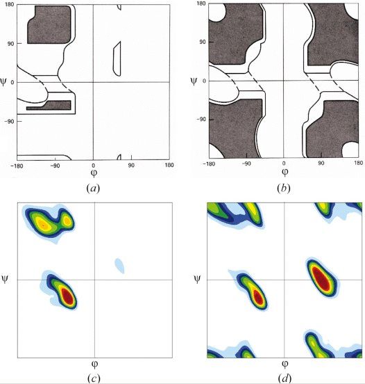
Positive direction: clockwiseRamachandran Plot maps allowable phi, psi regions
Ramachandran & Sasisekharan (1968)
Ramachandran used a physical
model of dipeptides to
determine the allowed (dark)
and disallowed (white)
combinations of phi and psi glycine
backbone angles. non-glycine, allowed
The observed frequencies non-proline regions
roughly agree with R’s allowed allowed
regions
regions.
glycine,
observed
CBi
non-glycine,
non-proline
Ni+1 CAi Φ observed
Ci-1
Ci
CAi+1 Ni
Ψ H
O 37MOE demo • Load 4Y6K, a membrane protease bound to an inhibitor (L679), rotate, translate, zoom in and out • open Sequence editor (SE) • delete unwanted chains • Synchronize selection in SE/Select • Select the drug: center, try different atom presentation • Select the catalytic aspartates D162, D200 • Measure distance to ligand: is there a hydrogen bond between the ligand and the catalytic aspartates? (add hydrogen, measure hbond length and angle)?
Assignments:
1. Complete MOE tutorial (help/tutorials/getting
                   started)
MOE Tour from "Building a Small Molecule" to
      "Introducing the Sequence Editor"
3. Review amino acid side chain structure: quiz
                 on Tuesday
Review questions
• What is a “residue”?
• What atoms make up the polypeptide backbone?
• What do we find in a PDB file?
• How are amino acid side chain atoms named?
• What is a Ramanchandran plot?
• What is a hydrogen bond made of?
• How do I measure a torsion angle?
• Is there any difference between a torsion angle and a bond angle?
40You can also read