Multi-center, Prospective, Randomized, Controlled Investigational Device Exemption Clinical Trial Comparing Mobi-C Cervical Artificial Disc to ...
←
→
Page content transcription
If your browser does not render page correctly, please read the page content below
Multi-center, Prospective, Randomized, Controlled
Investigational Device Exemption Clinical Trial Comparing
Mobi-C Cervical Artificial Disc to Anterior Discectomy and
Fusion in the Treatment of Symptomatic Degenerative Disc
Disease in the Cervical Spine
Michael S. Hisey, Hyun W. Bae, Reginald Davis, Steven Gaede, Greg Hoffman, Kee Kim, Pierce
D. Nunley, Daniel Peterson, Ralph Rashbaum and John Stokes

Int J Spine Surg 2014, 8 ()
doi: https://doi.org/10.14444/1007
http://ijssurgery.com/content/8/7
This information is current as of December 29, 2021.

   Email Alerts       Receive free email-alerts when new articles cite this article. Sign up at:
                      http://ijssurgery.com/alerts

The International Journal of Spine Surgery
2397 Waterbury Circle, Suite 1,
Aurora, IL 60504, Phone: +1-630-375-1432
© 2014 ISASS. All Rights  Reserved.
                      Downloaded from http://ijssurgery.com/ by guest on December 29, 2021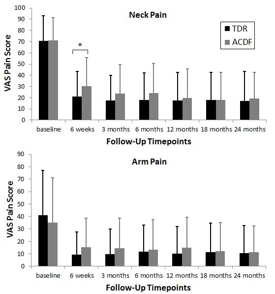
This article generously published free of charge by the International
           Society for the Advancement of Spine Surgery.

          Downloaded from http://ijssurgery.com/ by guest on December 29, 2021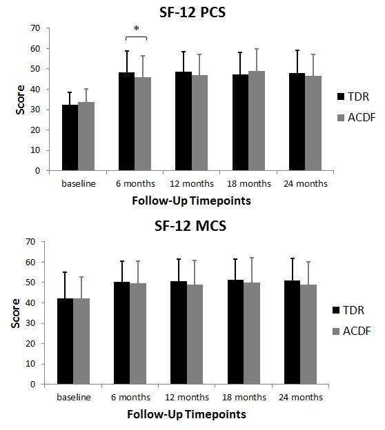
Multi-center, Prospective,
Randomized, Controlled
Investigational Device Exemption
Clinical Trial Comparing Mobi‑C
Cervical Artificial Disc to
Anterior Discectomy and Fusion
in the Treatment of Symptomatic
Degenerative Disc Disease in the
Cervical Spine
Michael S. Hisey, MD,1 Hyun W. Bae, MD,2 Reginald Davis, MD,3 Steven Gaede, MD,4 Greg
Hoffman, MD,5 Kee Kim, MD,6 Pierce D. Nunley, MD,7 Daniel Peterson, MD,8 Ralph Rashbaum,
MD,9 John Stokes, MD8
1
  Texas Back Institute, Denton, TX; 2The Spine Institute at St. John’s Health Center, Santa Monica,
CA; 3GBMC Healthcare Greater Baltimore Neurosurgical Associates, Baltimore MD; 4Oklahoma
Spine & Brain Institute, Tulsa, OK; 5Orthopedic North East, Fort Wayne, IN; 6University of
California Davis Medical Center, Sacramento, CA; 7Spine Institute of Louisiana, Shreveport, LA;
8
  Neurological Specialists of Austin, Austin, TX; 9Texas Back Institute, Plano, TX

Abstract
Background
Anterior cervical discectomy and fusion (ACDF) is the gold standard for treating
symptomatic cervical disc degeneration. Cervical total disc replacements (TDRs) have
emerged as an alternative for some patients. The purpose of this study was to evaluate the
safety and effectiveness of a new TDR device compared with ACDF for treating single-
level cervical disc degeneration.

Methods
This was a prospective, randomized, controlled, multicenter Food and Drug
Administration (FDA) regulated Investigational Device Exemption (IDE) study. A total
of 245 patients were treated (164 TDR: 81 ACDF). The primary outcome measure was
overall success based on improvement in Neck Disability Index (NDI), no subsequent
surgical interventions, and no adverse events (AEs) classified as major complications.

                    Downloaded from http://ijssurgery.com/ by guest on December 29, 2021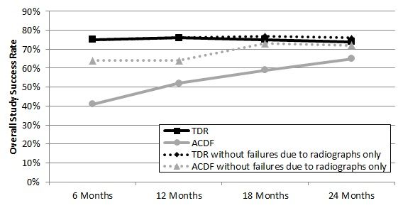
Secondary outcome measures included SF-12, visual analog scale (VAS) assessing neck
and arm pain, patient satisfaction, radiographic range of motion, and adjacent level
degeneration. Patients were evaluated preoperatively and postoperatively at 6 weeks, 3, 6,
12, 18, and 24 months. The hypothesis was that the TDR success rate was non-inferior to
ACDF at 24 months.

Results
Overall success rates were 73.6% for TDR and 65.3% for ACDF, confirming non-
inferiority (p < 0.0025). TDR demonstrated earlier improvements with significant
differences in NDI scores at 6 weeks and 3 months, and VAS neck pain and SF-12 PCS
scores at 6 weeks (p
Artificial cervical discs have been used since the 1990’s.12, 13 Total disc replacement
(TDR), as an alternative to ACDF in the treatment of symptomatic cervical DDD, is
becoming increasingly common and potentially provides some advantages over ACDF.
Not only does TDR provide the same anterior neural decompression and maintenance of
disc space as ACDF14, it can also preserve segmental mobility.15 Recently several Food
and Drug Administration (FDA) Investigational Device Exemption (IDE) trials have
demonstrated that TDR is at least as safe and effective as ACDF for of single-level
symptomatic cervical DDD.16, 17, 18 Furthermore, these studies found that TDR preserves
segmental mobility and these patients tended to have better clinical outcomes earlier after
surgery and maintained good quality clinical outcomes at 2 years postoperatively.
Additionally, longer term studies found that TDR provides excellent clinical outcomes 5
to 8 years post-operatively.19, 20, 21 Good clinical outcomes for cervical TDRs have also
been demonstrated in European studies.22, 23

One potential advantages of TDR over fusion was the reduction of adjacent segment
degeneration (ASD). Reports on the clinical impact of ASD have been mixed, with some
variation attributable to there being radiographic findings of ASD not associated with
clinical symptoms. Nunley et al. reported a rate of ASD in TDR patients of 3.1% per
year.24 Although not a primary part of their study, comparison to ACDF found no
significant difference in the occurrence rate. Other studies have found an increased rate of
ASD in fusion patients based on either radiographic findings25 or a trend toward a higher
rate of re-operation at the adjacent segment to treat new symptoms.26

The Mobi-C Artificial Cervical Disc (LDR Medical; Troyes, France) was introduced
outside the United States in November 2004. A European non-comparative, prospective,
multi-center study evaluated the safety and efficacy of this device in the treatment of
cervical DDD in 335 patients. This study found statistically significant improvements in
clinical outcomes and pain scores compared to baseline at 24 months post-operatively. 22,
23

The current study reports the 2-year follow-up results of a prospective, randomized,
concurrently controlled, multicenter clinical trial to determine the safety and efficacy of
TDR with the Mobi-C Artificial Cervical Disc versus ACDF at a single level for
treatment of symptomatic cervical DDD. The hypothesis was that clinical outcomes of
patients treated with TDR would not be inferior to outcomes of patients with the control
treatment at 24 months.

Material and Methods
Study design
A non-inferiority study design with a 2:1 randomization (TDR: control) was prospectively
planned for this FDA-regulated IDE study. A 5% advantage was hypothesized based on
pilot study data. A projected 75% success rate for control and 80% success rate for TDR
with respect to the primary endpoint was used along with the 2:1 randomization scheme.
A total sample size of 222 randomized cases (148 TDR vs. 74 control) was determined to
meet statistical power requirements to test for non-inferiority with the one-sided 95%
lower confidence bound on the difference exceeding -10%. An additional 10% was added

                  Downloaded from http://ijssurgery.com/ by guest on December 29, 2021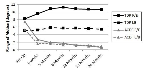
to the 222 to account for patients that may be lost to follow-up, bringing the total sample
size to 245. The population discussed in this report, with the exception of peri-operative
data and adverse events, is the Intent-To-Treat population (ITT), which consisted of all
subjects who were randomized and received study surgery. The ITT population consisted
of 164 TDR patients and 81 control patients. Peri-operative data is presented for the
Safety Population, which included all patients in the ITT population as well as the 15 non-
randomized training cases.

All principle investigators were trained on the surgical technique for the study device as
part of this IDE; which did consist of a separate two-level arm ran concurrently and using
the same investigational device.27 Each site was allotted one training case with the
investigational device; 15 sites performed training cases for a one level indication, all
others performed training with a two level case. This resulted in 15 additional TDR
patients (the non-randomized training cases) in the one level IDE.

The primary endpoint of the investigation was a composite measure assessed at 24
months. To be considered a success a patient had to be successful in each of the following
three measures: 1) NDI success, a 30 point improvement in score if baseline NDI score
was greater than or equal to 60, or a 50% of baseline score improvement in NDI score if
baseline NDI scores were less than 60, 2) No device related subsequent surgical
intervention at the index level (defined as removal, revision, supplemental fixation, or
reoperation), and 3) No study defined major complications which were defined as
radiographic failure, neurologic deterioration, and adverse events determined to be major
complications by an independent Clinical Events Committee (CEC). In the TDR group,
radiographic failure deemed to be a major complication was defined as: Spontaneous
fusion of the treatment level with radiographic evidence of bridging bone across the
treated disc space and less than 2° of angular motion from flexion to extension. In the
ACDF group, radiographic failure deemed to be a major complication was defined as:
Pseudoarthrosis at the treated level demonstrated by greater than or equal to 2° of angular
motion on flexion to extension, or radiolucent lines at greater than 50% of the graft
vertebral interfaces, or absence of bridging bone across the graft vertebral interfaces. This
composite measure of success was used to determine overall success rates for both
groups.

Study surgeries were performed for the 245 patients in the ITT population and the 15 non-
randomized training cases that received the investigational device between April 2006
and March 2008. The study was conducted by 25 principal investigators at 23 sites. All
patients were randomized to groups by an Interactive Voice Randomization System
(IVRS). The investigator or study coordinator called the IVRS after the pre-operative
inclusion/exclusion checklist confirmed eligibility and informed consent form was signed.
Patients were assigned to the TDR or control group by IVRS according to a stratified
randomization schedule (by baseline Neck Disability Index (NDI) score) with institutional
balancing. Due to the fact that the implant was evident to the surgeon, blinding the
physician to treatment was not possible. Patients remained blinded to the treatment group
assignment until after surgery had been performed to minimize the potential for
disproportionate patient dropouts.

                  Downloaded from http://ijssurgery.com/ by guest on December 29, 2021
The primary study hypothesis tested TDR non-inferiority against ACDF using a 10%
margin with respect to patient success at 24 months. Additional secondary endpoints
were: neck and arm pain (VAS), neck disability (NDI), patient satisfaction, quality of life
(SF-12), and radiographic outcomes (adjacent segment degeneration, range of motion
(ROM), significant radiolucency, and displacement or migration). Further measurements
of interest were analyzed and included operative time, blood loss, duration of
hospitalization, and time to return to work. Patients were evaluated pre-operatively and
post-operatively at 6 weeks, and 3, 6, 12, 18, and 24 months. Patients completed the
following questionnaires: NDI, VAS for neck and arm pain, SF-12 (Medical Outcomes
Study 12-Item Short Form Health Survey), Functional Outcome Swallowing Scale
(FOSS) for dysphagia, and patient satisfaction. Neurological examinations were
conducted and classified according to a pre-determined algorithm. Three neurological
tests were performed including motor, reflex, and sensory assessments. Diminished
neurological status was defined as a decrease in two points when compared to baseline
status in any of the treated level motor or reflex assessments or a decrease of one point
when compared to baseline of the treated level sensory tests. Any new or worsening signs
were reported as an adverse event (AE). A baseline for Nurick’s classification of cervical
spondylotic myelopathy (CSM) was established pre-operatively.
Patient selection criteria
The primary inclusion criteria were: diagnosis of DDD with radiculopathy or
myeloradiculopathy from C3 to C7 at one level without prior cervical fusion. DDD was
defined as discogenic neck and/or arm pain with degeneration of the disc confirmed by
patient history and by radiographic study. Patients were to have been unresponsive to
non-operative treatment for at least six weeks from symptom onset or had progressive
symptoms or signs of nerve root/spinal cord compression despite continued nonoperative
treatment (i.e. acute presentation). See Table 1 for detailed selection criteria.
Table 1. Study Inclusion and Exclusion Criteria
Inclusion criteria
Downloaded from http://ijssurgery.com/ by guest on December 29, 2021• Age 18-69 years
• Symptomatic cervical degenerative disc disease in only one level between
C3-C7 with:
◦ Neck and/or arm pain and/or
◦ Decreased muscle strength and/or
◦ Abnormal sensation and/or abnormal reflexes
• Deficit confirmed by imaging (CT, MRI, or X-ray)
• NDI score of ≥ 30
• Unresponsive to non-operative, conservative treatment for at least 6 weeks or
presence of progressive symptoms or signs of nerve root/spinal cord
compression despite continued non-operative treatment
• No prior surgery at the operative level and no prior cervical fusion procedure at
any level
• Physically and mentally able and willing to comply with the protocol
• Signed informed consent
• Willingness to discontinue all use of non-steroidal anti-inflammatory drugs
(NSAIDs) from one week before surgery until 3 months after surgery
Exclusion criteria
Downloaded from http://ijssurgery.com/ by guest on December 29, 2021• More than one vertebral level requiring treatment/immobile level between C1
and C7 from any cause
• Any prior spine surgery at operative level of any prior cervical fusion at any
level
• Disc height less than 3 mm
• T-score less than -1.5 (osteoporosis evaluation)
• Paget’s disease, osteomalacia, or any other metabolic bone disease other than
osteoporosis
• Active systemic infection of surgical site or history of or anticipated treatment
for systemic infection including HIV/Hepatitis C
• Active malignancy: a history of any invasive malignancy (except non-melanoma
skin cancer), unless treated with curative intent and there had been no clinical
signs or symptoms of the malignancy > 5 years
• Marked cervical instability on resting lateral or flexion-extension radiographs
• Known allergy to cobalt, chromium, molybdenum, or polyethylene
• Segmental angulation of greater than 11° at treatment or adjacent levels
• Rheumatoid arthritis, lupus, or other autoimmune disease
• Any diseases or conditions that would preclude accurate clinical evaluation
• Daily, high-dose oral and/or inhaled steroids or a history of chronic use of high
dose steroids
• BMI > 40
• Use of any other investigational drug or medical device within 30 days prior to
surgery
• Pending personal litigation relating to spinal injury (worker’s compensation not
included)
• Smoking more than one pack of cigarettes per day
• Reported to have mental illness or belonged to a vulnerable population
Device description
The TDR used in this study was the Mobi-C Cervical Artificial Disc (LDR Medical,
Troyes, France). This prosthesis is a three component, mobile-bearing device comprised
of two titanium plasma-sprayed and hydroxyapatite (HA) coated cobalt chromium alloy
endplates and an ultra-high molecular weight polyethylene (UHMWPE) mobile insert.
Downloaded from http://ijssurgery.com/ by guest on December 29, 2021The superior endplate incorporates a convex shape to match the natural cervical anatomy,
and both the superior and inferior endplates feature low-profile, inclined teeth along the
lateral edges to provide initial stability (Figure 1). The shape of the device and inclined
teeth were designed to facilitate a bone sparing surgical technique. The device is available
in several footprints and a range of heights to satisfy individual anatomical requirements.
The device allows five independent degrees of freedom, two translational and three
rotational (Figure 1).
Fig. 1. Mobi-C Artificial Cervical Disc (LDR Medical; Troyes, France).
Allows for five independent degrees of freedom.
Surgical approach
For both treatment groups, the patient was positioned physiologically neutral to avoid
hyperextension. This position was maintained throughout surgery and head rotation was
minimized. A standard Smith Robinson approach was used to access the anterior cervical
spine. A discectomy was performed in which the disc and the osteophytes were removed
and the involved neural structures were decompressed.
ACDF Procedure
For the control group, a standard ACDF technique was used. The disc space was filled
with corticocancellous allograft bone and an anterior cervical plate was placed over the
graft and secured with screws to the vertebrae according to the manufacturer’s published
surgical technique. Anterior cervical plate selection was limited to the SLIM-LOC™
Anterior Cervical Plate System (Depuy Spine; Raynham, MA) and the Sofamor Danek
ATLANTIS™ or ATLANTIS™ VISION Anterior Cervical Plate Systems (Medtronic;
Memphis, TN).
TDR Procedure
In the TDR group, following the discectomy, the disc space was distracted in parallel
using a vertebral distractor. With normal disc height restored, distraction was maintained
with a Caspar distractor. The cartilaginous endplates were removed to expose the
underlying bone but no endplate shaping was performed. Using fluoroscopy and the trial
Downloaded from http://ijssurgery.com/ by guest on December 29, 2021instruments provided, the appropriate Mobi-C footprint and height were selected. The
assembled prosthesis was then impacted into the prepared disc space using a self-retaining
inserter. An adjustable stop allowed precise modification of the implant’s anteroposterior
(AP) position. Once final position was obtained, the distraction was released and slight
compression was applied via the Caspar distractor to facilitate seating of the implant teeth
into the vertebrae. AP and lateral fluoroscopy was used to confirm final implant position
(Figure 2).
Fig. 2. TDR device in proper placement. Lateral flexion and extension
x-rays of the TDR at 24 months.
Post-operative care
Post-operative care was left to surgeon discretion. Antibiotic coverage and rehabilitation
were continued in accordance with the surgeon’s standard procedures. In order to ensure
consistent treatment between the TDR and ACDF groups and to avoid any potential
handicapping of the ACDF group, patients were instructed not to use NSAIDS for one
week prior to surgery and the treating physician was instructed not to prescribe NSAIDs
prophylactically for 3 months following surgery. However, physicians were allowed to
prescribe NSAIDs to treat diagnosed incidence of heterotopic ossification (HO).
Radiographic Assessments
The following X-ray views were obtained pre-operatively and at all follow-up visits:
neutral AP, neutral lateral, flexion-extension, and left-right bending. For consistency, an
independent provider of radiographic assessments performed the radiographic analysis for
this study. All assessments were made according to a pre-determined radiographic
analysis protocol. This protocol specified that measurements of disc angle, angular
motion, translational motion, anterior disc height, and change in disc height were
quantitatively determined using validated QMA software (Medical Metrics Inc., Houston
TX).28, 29 Specific vertebrae were outlined and the 4 corners of the vertebral centrum
specified. Using the radiograph made with the spine in the neutral position, anatomic
landmarks were selected. Using an algorithm based on gray-scale correlation, the
software found the specific vertebrae in each of the images (flexion, extension, and
neutral) and calculated the geometric coordinates of each anatomic landmark on the
vertebra. This process was repeated for each vertebra and intervertebral motion was
calculated based on the geometric coordinates.
Downloaded from http://ijssurgery.com/ by guest on December 29, 2021All other assessments such as bony bridging, radiolucency, fusion status, device
condition, device migration, device subsidence, device position, and adjacent level
degeneration were made by qualified independent radiologists affiliated with Medical
Metrics Inc. Subsidence was defined as greater than 3 mm cranial or caudal motion of the
device (or device component) perpendicular to the vertebral endplates. Migration was
defined as greater than 3 mm anterior or posterior motion of the device (or device
component) parallel to the vertebral endplates. Significant radiolucency was defined as
more than 50% coverage of radiolucent lines along device/endplate interface.
Radiographic success was defined as the absence of fusion of the treated level in the
TDR, requiring 2° or more of angular motion from flexion to extension or no radiographic
evidence of bridging bone across the treated disc space. In the ACDF group radiographic
successful fusion at the treated level was defined as evidence of bridging bone across the
disc space, less than 2° angular motion from flexion to extension, and less than 50%
radiolucent lines at graft vertebral endplate interfaces. Adjacent segment degeneration
was assessed at baseline and at the 12 and 24-month visits. Levels both superior to and
inferior to the treated level were scored on the Kellgren-Lawrence scale of disc
degeneration30, 31 at baseline, 12, and 24 months. Each disc was classified as having none
(0), minimal (1), definite (2), moderate (3), or severe (4) degeneration and was given the
corresponding numerical score. To assess adjacent level degeneration over the course of
the study, change in disc degeneration score from the baseline score of both adjacent
levels was calculated at 12 and 24 months and any negative change (increase in score)
was classified as adjacent level degeneration.
Statistical analysis
The analysis goal was to establish non-inferiority using the composite success measure.
Non-inferiority was tested using an exact 95% one-tailed confidence bound for the
study-control success rate; if a 10% offset could be ruled out, then superiority was to be
tested. A closed testing procedure was used to allow for superiority to be tested in the
event that non-inferiority was established for the primary effectiveness endpoint. VAS,
NDI, and SF-12 results are described using summary statistics (N, mean, median,
standard deviation, inter-quartile range, minimum, and maximum) and compared across
treatment groups using two sample t-test.
Results
Patient accountability and demographics
Demographics characteristics of the ITT population are shown in Table 2. The ITT
population consists of 164 patients in the investigational group and 81 patients in the
control group. The 24-month follow-up rate (defined as subjects presenting at 24 months
with a composite study success value) 94.3% in the TDR group and 92.0% in the ACDF
group. The groups were well balanced in terms of demographic characteristics with no
significant differences between groups.
Table 2. Patient Demographics – intent to treat population.
Patient Group
Downloaded from http://ijssurgery.com/ by guest on December 29, 2021Variable TDR ACDF p value
Age (years) 0.5657*
N 164 81
Mean (SD) 43.3 (9.2) 44.0 (8.2)
Gender n (%) 0.6843**
Male 78 (47.6%) 36 (44.4%)
Female 86 (52.4%) 45 (55.6%)
Ethnicity n (%) 0.6667**
Hispanic or Latino 3 (1.8%) 2 (2.5%)
Not Hispanic or Latino 161 (98.2%) 79 (97.5%)
Race n (%) 0.0710**
American Indian 2 (1.2%) 1 (1.2%)
Caucasian 152 (92.7%) 69 (85.2%)
Asian 3 (1.8%) 1 (1.2%)
Black or African American 4 (2.4%) 10 (12.3%)
Native Hawaiian/Other Pacific Islander 1 (0.6%) 0
Other 2 (1.2%) 0
BMI 0.8460*
Mean (SD) 27.3 (4.4) 27.4 (4.2)
Work Status n (%) 0.3264**
Being able to work 108 (65.9%) 46 (56.8%)
Not being able to work 37 (22.6%) 22 (27.2%)
N/A 19 (11.6%) 13 (16.0%)
Driving Status n (%) 0.5035**
Being able to drive 155 (94.5%) 79 (97.5%)
Not being able to drive 8 (4.9%) 2 (2.5%)
N/A 1 (0.6%) 0
NDI Mean (SD) 54.0 (14.0) 54.2 (14.6) 0.9290*
VAS Neck Pain Mean (SD) 70.8 (22.4) 70.1 (21.5) 0.8354*
VAS Left Arm Pain Mean (SD) 46.7 (36.5) 55.3 (37.3) 0.0839*
VAS Right Arm Pain Mean (SD) 41.0 (36.2) 34.8 (35.6) 0.2104*
SF-12 PCS Mean (SD) 32.5 (5.91) 33.8 (6.36) 0.1055*
SF-12 MCS Mean (SD) 42.1 (13.1) 42.2 (10.4) 0.9792*
Downloaded from http://ijssurgery.com/ by guest on December 29, 2021* using unpaired t-test to compare across treatment groups
** using Fisher exact test to compare. Fisher exact p-value calculation is based on
Caucasian vs. non-Caucasian subjects. Fisher exact p-value is based on ‘being able to’ vs.
‘not being able to’
Overall Success at Primary Endpoint
The primary composite endpoint assessed individual patient success as demonstrated by
functional improvement (NDI), no need for subsequent surgical intervention (removal,
revision, supplemental fixation, reoperation), and the absence of study defined major
complications including neurologic deterioration, CEC-assessed adverse events, and
radiographic failure. The composite success rate for TDR patients was 73.7% at month
24, compared to 65.3% success rate for ACDF (Figure 3). The composite endpoint results
establish TDR non-inferiority for the primary endpoint (p = 0.0021). Additionally, TDR
patients tended to achieve success earlier than ACDF patients. At 6 month time point,
75.0% of TDR patients achieved composite success, compared with 41.4% of ACDF
patients (Figure 4).
Downloaded from http://ijssurgery.com/ by guest on December 29, 2021Fig. 3. Overall Study Success at 24 Months. Bar graph showing over all
clinical success rate at 24 months. Also shown are the success rates at
24 months of the components of the composite endpoint: NDI success
rate, Device success rate (no need for subsequent surgeries, and the
percentage of patients who had no major complications.
Fig. 4. Overall Study Success Rates by Time point. Success rates are
shown for both TDR group and ACDF group both with and without
failures due only to radiographic major complications. Non-inferiority
is demonstrated at each time point for both TDR compared to ACDF
in both cases.
Overall Success at Primary Endpoint - Effect of
Radiographic Major Complications
To analyze the effect of radiographic complications on the success rates of TDR and
ACDF at each follow-up time point, a variation to the primary endpoint analysis was
prospectively planned. These results are also shown in Figure 4. For this analysis, all
patients that were failures due only to radiographic failure were treated as a study success.
The composite success rates in this analysis were 76.3% for the TDR group and 72.0%
for the ACDF group. This analysis led to an increase in ACDF success, resulting in
smaller differences between TDR and ACDF overall success, however overall success
Downloaded from http://ijssurgery.com/ by guest on December 29, 2021still favors TDR (8.4% difference with radiographic failures and 4.3% difference between
groups without radiographic failures). This result further confirms non-inferiority of TDR
to ACDF even when radiographic only failures are excluded (p = 0.0094).
Additionally, in this analysis TDR patients achieved success sooner than patients in the
ACDF group. Even excluding those patients who failed the study only due to defined
radiographic failure, only 64.3% of ACDF patients were deemed an overall study success
at 6 months and 63.8% at 12 months. By 18 months the success rate of ACDF increased
to 72.7% and remained roughly the same at 24 months (72.0%). This pattern is contrasted
in the TDR group where 75.0% of patients achieved success by 6 months and the success
rate remained relatively constant with 76.3% success at 12 months, 76.7% at 18 months,
and 76.3% at 24 months.
Surgical data
Peri-operative data is shown in Table 3 for the safety population. The mean and median
blood loss and length of hospital stay were similar between treatment groups. The
operative time was slightly higher in the TDR group, with a mean of 1.5 hours, compared
with 1.3 hours for ACDF.
Table 3. Perioperative Data – Safety Population
Patient Group
Variable TDR ACDF p value**
Duration of Hospitalization* 0.9829
N 179 81
Mean (SD) 2.1 (0.52) 2.1 (0.47)
Blood Loss (ml) 0.9628
N 145 66
Mean (SD) 47.7 (46.75) 48.1 (55.21)
Operative Time (Hours) 0.0572
N 179 81
Mean (SD) 1.5 (0.64) 1.3 (0.63)
* Duration of hospitalization is defined as [Date of Discharge – Date of Surgery +1]
** Using unpaired t-test to make comparison across treatments
Adverse Events
Adverse events were collected for all patients in the safety population (Table 4). Neither
adverse events (AEs) in general nor those AEs deemed to be serious adverse events
(SAEs) were more prevalent in the TDR group than the ACDF group. No unanticipated
adverse device effects were reported in either treatment group. Eight AEs classified as
related to the study device occurred in 7 out of 179 (3.9%) TDR patients. These events
were: Spinal ligament ossification (1), neck pain (4 with 1 subject reporting 2
Downloaded from http://ijssurgery.com/ by guest on December 29, 2021occurrences), muscle spasms (1), radiculopathy (1), and a case of subsidence that was not
verified radiographically (this was reported by the investigator, but not verified by the
independent radiograph reviewer), (1). In comparison, there were 7 AEs in 6 out of 81
(7.4%) ACDF patients that were assessed to be related to the study device: medical device
complication (3), radiculopathy (1), neck pain (2), and misplaced screw coded as medical
device complication (1).
Table 4. All Treatment Emergent Adverse Events through 24 Months in US IDE Study –
All Study Subjects.
Mobi-C ACDF
Complication #Patients Total #Patients Total
(% of 179) Events (% of 81) Events
All Adverse Events1 170 (95.0%) 1229 75 (92.6%) 688
Anatomy/Technical Difficulty 11 (6.1%) 12 2 (2.5%) 4
Cervical –Study Surgery 4 (2.2%) 4 2 (2.5%) 3
Cervical – Non Study Surgery 5 (2.8%) 6 1 (1.2%) 1
Non-Cervical 2 (1.1%) 2 0 (0.0%) 0
Cancer 4 (2.2%) 5 1 (1.2%) 2
Cardiovascular 20 (11.2%) 26 10 (12.3%) 10
Death 0 0 0 0
Dysphagia/Dysphonia 20 (11.2%) 26 17 (21.0%) 20
Dysphagia 19 (10.6%) 22 15 (18.5%) 17
Dysphonia 3 (1.7%) 4 3 (3.7%) 3
Gastrointestinal 39 (21.8%) 60 15 (18.5%) 37
Heterotopic Ossification 9 (5.0%) 10 4 (4.9%) 4
Cervical - Index Level 5 (2.8%) 5 0 (0.0%) 0
Cervical - Adjacent Level 1 (0.6%) 1 1 (1.2%) 1
Non Cervical 4 (2.2%) 4 3 (3.7%) 3
Infection 33 (18.4%) 51 20 (24.7%) 28
Superficial Wound – Cervical 6 (3.4%) 7 1 (1.2%) 1
Deep Wound – Cervical 0 0 0 0
Other Wound - Non Study Surgery 1 (0.6%) 1 3 (3.7%) 3
Systemic 8 (4.5%) 9 2 (2.5%) 3
Local 20 (11.2%) 34 18 (22.2%) 21
Malpositioned Implant 2 (1.1%) 2 1 (1.2%) 1
Neck and/or Arm Pain 102 (57.0%) 212 47 (58.0%) 98
Downloaded from http://ijssurgery.com/ by guest on December 29, 2021Mobi-C ACDF
Complication #Patients Total #Patients Total
(% of 179) Events (% of 81) Events
Neck Pain 74 (41.3%) 123 37 (45.7%) 56
Arm Pain 46 (25.7%) 76 20 (24.7)% 25
Neck And Arm Pain 9 (5.0%) 13 7 (8.6%) 17
Neurological 121 (67.6%) 401 52 (64.2%) 215
Upper Extremity – Sensory 67 (37.4%) 175 32 (39.5%) 126
Upper Extremity – Motor 26 (14.5%) 43 15 (18.5%) 20
Upper Extremity – Reflex 18 (10.1%) 44 7 (8.6%) 20
Lower Extremity – Sensory 11 (6.1%) 22 2 (2.5%) 3
Lower Extremity – Motor 6 (3.4%) 9 4 (4.9%) 4
Lower Extremity – Reflex 0 (0.0%) 0 1 (1.2%) 1
Upper & Lower Extremity - Sensory 1 ( 0.6%) 1 1 (1.2%) 1
Upper & Lower Extremity – Motor 0 0 0 0
Upper & Lower Extremity - Reflex 0 0 0 0
Neck 41 (22.9%) 51 21 (25.9%) 21
Back 7 (3.9%) 8 2 (2.5%) 2
Spinal Cord Disturbance 0 0 0 0
Gait Disturbance 1 (0.6%) 1 1 (1.2%) 1
Non Specific 6 (3.4%) 6 1 (1.2%) 1
Other* 35 (19.6%) 41 8 (9.9%) 15
Non-Union 0 (0.0%) 0 4 (4.9%) 4
Other** 77 (43.0%) 114 33 (40.7%) 66
Other Pain 102 (57.0%) 226 47 (58.0%) 144
Shoulder 39 (21.8%) 55 21 (25.9%) 31
Back 44 (24.6%) 50 18 ( 22.2%) 30
Torso 5 (2.8%) 7 3 (3.7%) 4
Lower Extremity 26 (14.5%) 40 12 (14.8%) 29
Headache 45 (25.1%) 58 26 (32.1%) 41
Other*** 15 (8.4%) 16 8 (9.9%) 9
Respiratory 6 (3.4%) 6 6 (7.4%) 8
Spinal Disorder 6 (3.4%) 7 10 (12.3%) 12
Cervical - Study Surgery 1 (0.6%) 1 2 (2.5%) 2
Downloaded from http://ijssurgery.com/ by guest on December 29, 2021Mobi-C ACDF
Complication #Patients Total #Patients Total
(% of 179) Events (% of 81) Events
Cervical - Non Study Surgery 5 (2.8%) 6 3 (3.7%) 3
Non Cervical 0 (0.0%) 0 5 (6.2%) 7
Trauma 47 (26.3%) 70 20 (24.7%) 38
Upper Extremity Nerve Entrapment 8 (4.5%) 9 4 (4.9%) 5
Urogenital 9 (5.0%) 11 9 (11.1%) 12
Vascular Intraop 1 (0.6%) 1 0 (0.0%) 0
Wound Issue - Non-Infection 1 (0.6%) 1 3 (3.7%) 3
Hematoma 1 (0.6%) 1 3 (3.7%) 3
Hematoma Evacuation 0 0 0 0
CSF Leakage 0 0 0 0
M= All Mobi-C Subjects; F = All ACDF Subjects
1 Sum of all treatment emergent adverse events experienced in the study for each treatment
group.
*Neurological Other includes Neurological events not appropriately defined elsewhere in
the Neurological category. This includes amnesia, convulsion, facial neurologic events
(dysaesthesia, hypoaesthesia), unexplained loss of consciousness, ‘other’ nerve compression,
Parkinson’s disease, and stroke.
**Other includes events not appropriately defined elsewhere. This includes adverse drug
reactions, allergies, anemia, anxiety, arthritis, attention deficit disorder, benign neoplasm,
blood & lymphatic system disorders, complications from other medical procedures,
congenital defects, dehydration, dermatitis, diabetes, dizziness, ear/eye disorders, endocrine
disorders, fatigue, feeling hot, fever, gout, high/low cholesterol, immune system disorders,
injury/poisoning, lupus, menopause, miscarriage, muscle atrophy, nutritional disorders,
obesity, osteoarthritis, osteoporosis, other inflammation, other medical procedures, plantar
fasciitis, polyps, pregnancy, psychiatric disorders, rotator cuff syndrome, skin disorders,
sinus infection, social issues, sleep disorders, swelling, tendonitis, thyroid conditions,
vascular disorders, and weight gain/loss.
***Other Pain Other includes events not appropriately defined elsewhere. This includes
facial pain, fibromyalgia, muscle soreness, chronic pain, nerve pain and arthritis.
Clinical Outcomes
All clinical outcomes discussed in this section describe results from the ITT population of
patients.
NDI
Baseline NDI scores for both treatment groups were not significantly different (54.0 for
TDR and 54.1 for ACDF). Patients in both groups experienced significant improvement
in NDI scores from baseline at all follow-up time points (Figure 5). The mean
Downloaded from http://ijssurgery.com/ by guest on December 29, 2021improvement in NDI from baseline was greater in the TDR group than the ACDF group
at all time points and this difference was statistically significant at 6 weeks (p = 0.0141)
and 3 months (p = 0.0026).
Fig. 5. Mean NDI Score by Time point. NDI scores were collected at
each visit. Error bars represent standard deviations. NDI scores for
both treatment groups were significantly different from baseline at all
time points (p < 0.05) * Denotes significant difference determined
using unpaired t-test to compare the change from baseline between the
two treatments (p < 0.05).
In this study NDI success was defined as improvement of at least 30 points for patients
with baseline NDI ≥ 60 or improvement of at least 50% of baseline score for patients with
baseline NDI < 60. NDI success rates were slightly higher in the TDR group (79.4%) than
in the ACDF group (77.1%).
Subsequent Surgical Interventions
Study failures by subsequent surgical interventions were defined as removal, revision,
supplemental fixation, or reoperation of the implanted device and/or at the index level.
Within 24 months, these procedures were performed for only two patients (1.2%) in the
TDR group and five patients (6.2%) in the ACDF group. Implant removal was required in
both groups: one patient in the TDR group (0.6%) and three patients in the ACDF group
(3.7%). The removals performed on patients in the ACDF group were performed for
“misplaced screw” and 2 instances of “failure to fuse.” The one removal in the TDR
group was the result of unresolved radiculopathy. Supplemental fixation was required for
two patients in the ACDF group (2.5%) and no patients in the TDR group. Both
supplemental fixations were performed for pseudoarthrosis and involved the addition of
posterior instrumentation at the treated level. Reoperation was required only for one
patient in the TDR group (0.6%) and no patients in the ACDF group. The TDR
reoperation was performed for “right sided radiculopathies.” Overall device success
(defined as absence of study failure by subsequent surgical interventions) was achieved
by 162 patients in the TDR group (98.8%) and 76 patients in the ACDF group (93.8%).
Downloaded from http://ijssurgery.com/ by guest on December 29, 2021Neck and Arm Pain (VAS)
VAS assessments were recorded for the intensity of both neck and arm pain. Neck and
arm pain decreased significantly both groups at all time points when compared to baseline
(p < 0.001; Figure 6). The mean change in VAS neck pain score from baseline to follow-
up visit was greater for the TDR group than for the ACDF group at every follow-up time
point, however the difference was statistically significant only at the 6 week visit (p =
0.0072). Mean neck pain improvement at 24 months was similar between the two groups
(17.34 for TDR; 19.36 for ACDF). A similar trend was seen for arm pain scores: mean
score change from baseline to follow-up visit was greater for the TDR group than the
ACDF group at each visit and mean arm pain scores at 24 months were similar between
the two groups (13.6 for TDR; 13.5 for ACDF), when analyzing the arm with the worst
score at baseline.
Fig. 6. Mean VAS scores by Time point. VAS pain scores were
collected at each follow-up visit. Error bars represent standard
deviations. VAS scores for both treatment groups were significantly
different from baseline at all time points (p < 0.05) * Denotes
significant difference determined using unpaired t-test to compare the
change from baseline between the two treatments (p < 0.05).
Neurologic Success
Neurological success was defined as maintenance or improvement in each of the
neurological evaluations including motor assessments (muscle strength), reflex
assessments, and sensory deficit assessments (pin prick and light touch). Failure of any
Downloaded from http://ijssurgery.com/ by guest on December 29, 2021one of the three neurological assessments was deemed a neurological failure. At 24
months, only 1.9% of TDR patients and 2.9% of ACDF patients were deemed
neurological failures.
Radiographic Findings
Plain films were used to assess subsidence, migration, significant radiolucency, range of
motion of the treated segment, and adjacent segment degeneration. There were no cases
of migration, subsidence, or significant radiolucency in either group at the 24 month visit.
At the 24 month follow-up visit 6 patients in the TDR group were determined to show
evidence of spontaneous fusion at the treated level; resulting in 97.0% radiographic
success rate. The 6 patients in the TDR group that were determined to be radiographic
failures had an average range of motion of 0.67º at the treated level and showed evidence
of bridging bone across the disc space. One additional patient in the TDR group showed
bridging bone across the disc space, but was not determined to be a radiographic failure
because the patient still maintained flexion-extension range of motion of 6.4º. At 24
months this patient was determined to be an overall study success. There were no other
instances of bridging bone in the TDR cohort.
At the 24 month follow-up visit, 8 patients in the ACDF group were categorized as a
radiographic failure, resulting in a radiographic success rate in this group of 89.3%. Six of
the 8 patients in the ACDF group that were determined to be radiographic failures had
more than 2º range of motion at the treated level despite each of these patients
demonstrating bridging bone across the disc space. Two of the 8 patients showed less than
2º range of motion but there was no evidence of bridging bone. There were no instances
of radiolucent lines at more than 50% of the graft vertebral interfaces in the ACDF group.
The ACDF group showed restricted ROM at all post-operative study visits and the mean
values at 24 months were less than 1° for both left/right lateral and flexion/extension
bending. The mean values for TDR patients were higher than baseline at the week 6 visit
and by the 24 month visit, were 10.8° (±6.5°) for flexion/extension bending and 5.4°
(±3.1°) for left/right lateral bending (Figure 7).
Downloaded from http://ijssurgery.com/ by guest on December 29, 2021Fig. 7. Range of Motion by Time point. Line graph demonstrating
range of motion at the treated segment in Flexion/Extension (F/E) and
Right/Left Lateral Bending (LB)
At all time points, including baseline, radiographic evidence of disc degeneration
(Kellgren-Lawrence score greater than 0) was noted more frequently at the supra-adjacent
segment than infra-adjacent segment. At 12 months 93.8% of patients in the TDR group
and 91.3% of patients in the ACDF group had no change in disc degeneration score for
the superior segment. At 24 months, the percentage of patients who had no change in
score for this segment decreased in both groups to 85.4% for the TDR group and 75.0%
for ACDF. At neither time point was the difference between groups statistically
significant.
At the inferior adjacent segment, the percentage of patients with no change in disc
degeneration score from baseline to 12 months was 98.5% for the TDR group and 87.5%
for the ACDF group. At 24 months these percentages decreased to 92.3% for the TDR
group and 79.0% for ACDF. These differences were statistically significant at both time
points.
Quality of life (SF-12)
Mean physical component summary (PCS) and mental component summary (MSC)
scores were higher than baseline for all time points. Both the PCS and MCS
questionnaires are scored out of 100 total points. At 24 months, TDR patients had a mean
PCS score of 48.3 with a mean change from baseline of +15.7 and ACDF patients had a
mean PCS score of 46.5 with a mean change from baseline of +13.0. At this same
timepoint, TDR patients had a mean MCS score of 51.0 with a mean change from
baseline of +8.5 and ACDF patients had a mean MCS score of 49.2, with a mean change
from baseline of +7.2. The mean change in score for both PCS and MCS was higher in
the TDR group than the ACDF group at all time points, however the difference between
the TDR and ACDF groups was only statistically significant for the PCS score in favor of
TDR at the 6 months follow-up visit (p value = 0.0367) (Figure 8 ).
Downloaded from http://ijssurgery.com/ by guest on December 29, 2021Fig. 8. Mean SF-12 PCS and MCS scores by Time point. SF-12 physical
component score (PCS) and mental health component score (MCS)
were collected at baseline, 6, 12, 18, and 24 months. Error bars
represent standard deviations. SF-12 scores for both treatment groups
were significantly different from baseline at all time points (p < 0.05) *
Denotes significant difference determined using unpaired t-test to
compare the change from baseline between the two treatments (p <
0.05).
Patient satisfaction
Patient satisfaction with surgical treatment, as assessed by questionnaires, was high in
both treatment groups. More than 95% of both TDR patients and ACDF patients
answered that they were “very satisfied” or “somewhat satisfied” and more than 93% of
both groups responded that they would make a surgery recommendation of “definitely
yes” or “probably yes”.
Time to return to work
TDR patients returned to work in a mean of 30.1 days (±24.6 days), with a median of 21.0
days. The patients in the ACDF group had a similar slightly higher mean time to return to
work of 36.8 days (±40.3 days), with a median of 22.0 days.
Downloaded from http://ijssurgery.com/ by guest on December 29, 2021Discussion
The results of this trial demonstrate that TDR is non-inferior to ACDF when analyzed by
the composite primary endpoint (p = 0.002). The primary endpoint assessed individual
patient success as demonstrated by functional improvement (NDI), no need for
subsequent surgical intervention (removal, revision, supplemental fixation, and
reoperation), and the absence of study defined major complications. A non-inferiority
result, measured by the primary composite endpoint, was not surprising as the
fundamentals of these two surgical procedures are very similar. Both surgeries involve the
same surgical approach, a thorough discectomy, nerve decompression, disc height
restoration, and placement of a device in the disc space. With disc height restored and
nerve impingement eliminated, the authors of this study anticipated similar improvements
in both groups in terms of decreased neck and arm pain, increased muscle strength, and
elimination of abnormal sensations (hyperesthesia or hypoesthesia) and/or abnormal
reflexes. Indeed, patients in the both the TDR group and the ACDF group showed marked
improvement after surgery reporting decreased neck and arm pain, low rates of neurologic
deterioration, improvement in quality of life, high patient satisfaction, and low rates of
study defined major complications.
However, TDR demonstrated some advantages over ACDF particularly when clinical
outcomes are analyzed in the short term. Notably, TDR achieved a higher patient success
rate by 6 months than ACDF achieved by 24 months for the primary composite endpoint.
This higher success rate for TDR at 6 months compared to ACDF at 24 months was still
present when radiographic complications were excluded as a reason for overall study
failure. Clinical outcome measures also indicated that patients in the TDR group generally
achieved positive results earlier than the patients in the ACDF group. There were
statistically significant differences in favor of TDR observed for mean NDI improvement
at 6 weeks and 3 months, mean neck pain VAS improvements at 6 weeks, and mean
SF-12 PCS improvement at 6 months.
Furthermore, while both ACDF and TDR treatment achieve restoration or maintenance of
the disc height and anterior neural decompression, another advantage of TDR over ACDF
is thought to be preservation of motion at the treated segment. Range of motion results
demonstrate that, as expected, the patients in the ACDF group showed restricted ROM at
all post-operative study visits. The mean values at Month 24 were less than 1° for both
left/right lateral and flexion/extension bending. However, the TDR group patients on
average maintained or increased ROM post-operatively by the 6 week visit and this result
is consistent through 24 month visit. This maintenance or increase in ROM occurred in
both flexion/extension and left/right lateral bending. It has been postulated that the
restriction of motion at the treated segment with an ACDF is one factor contributing to
the development of adjacent level disease.32, 33 In this study fewer TDR patients had
deterioration of the adjacent segments during the course of the study and this difference
was statistically significant for the inferior adjacent segment. These results suggest that
the preservation of motion by the TDR may play a role in reducing the rate of
deterioration of the adjacent segments, in particular the inferior adjacent segment.
Downloaded from http://ijssurgery.com/ by guest on December 29, 2021In order to ensure consistent treatment between TDR and ACDF patients, the protocol
disallowed the use of NSAIDs from 1 week preoperatively and instructed physicians not
to prescribe NSAIDs prophylactically postoperative in either group unless intended to
treat diagnosed heterotopic ossification. The prophylactic use of NSAIDs may have
benefitted TDR patients by reducing the likelihood of heterotopic ossification, but could
have been detrimental to ACDF patients by delaying fusion. The use of NSAIDs
prophylactically in the prevention of heterotopic ossification formation after hip
arthroplasty is well documented.34, 35, 36 97.0% of TDR patients achieved radiographic
success; leaving only 3.0% defined as radiographic failures (the unwanted spontaneous
fusion at the treatment site) at 24 months. This is compared to 10.7% of the ACDF
patients that were defined as radiographic failures (the failure to fuse at the treated level)
at 24 months. Although a 3.0% radiographic failure rate is relatively low, it is possible
that had NSAIDS been used prophylactically for the TDR group the radiographic failure
rate may have been even lower.
The results of this study are similar to those reported for FDA IDE trials evaluating other
cervical TDR devices with 2-year follow-up comparing the results to ACDF using
allograft and an anterior plate.16, 17, 18, 37 These prospective, randomized trials have
produced consistent results finding TDR to be non-inferior to ACDF with additional post
hoc analyses finding TDR to be superior on some measures. In all studies, significant
improvement was seen at early follow-up and maintained throughout 24-month follow-up
in both treatment groups. Unfortunately, some of the definitions and assessments used in
the studies varied, making data pooling and/or direct comparisons between studies
potentially unreliable.
Conclusions
This prospective, randomized trial comparing TDR to fusion showed that the TDR is a
viable alternative to ACDF, with some advantages in early recovery and potentially some
advantage to reduce adjacent segment degeneration. These results are similar to
previously published TDR trials in that overall success rates of TDR are shown to be non-
inferior to overall success rates of ACDF, and patients in the TDR group tended to see
earlier success in terms of both overall study success and clinical outcomes.16, 17, 18 The
robustness of the non-inferiority of TDR was confirmed by analysis of overall success
rates in both groups while ignoring radiographic complications, as radiographic
complications may have limited clinical relevance.11 TDR was also associated with
reduced adjacent segment deterioration evident on radiographs compared to fusion,
however the clinical implication of the reduced adjacent segment degeneration may
require longer follow-up. This study demonstrated that TDR preserved physiologic
motion, while providing clinical benefits consistent with ACDF. The authors believe that
the current study provides evidence that TDR with Mobi-C Artificial Cervical Disc is as
safe and clinically effective as ACDF at 2-years follow-up, and that TDR with this device
has potential benefits over ACDF.
References
1. Denaro V, Papalia R, Denaro L, et al. Cervical spinal disc replacement. J Bone Joint
Surg Br 2009;91:713-9.
Downloaded from http://ijssurgery.com/ by guest on December 29, 20212. Denaro V, Di Martino A. Cervical spine surgery: an historical perspective. Clin
Orthop Relat Res 2011;469:639-48.
3. Mummaneni PV, Haid RW. The future in the care of the cervical spine: interbody
fusion and arthroplasty. Invited submission from the Joint Section Meeting on
Disorders of the Spine and Peripheral Nerves, March 2004. J Neurosurg Spine
2004;1:155-9.
4. Brodke DS, Zdeblick T. Modified Smith-Robinson procedure for anterior cervical
discectomy and fusion. Spine 1992;17(suppl):427-30.
5. Bohlman HH, Emery SE, Goodfellow DB, Jones PK. Robinson anterior cervical
discectomy and arthrodesis for cervical radiculopathy. Long-term follow-up of one
hundred and twenty-two patients. J Bone Joint Surg Am 1993;75:1298-307.
6. Boakye M, Mummaneni PV, Garrett M, et al. Anterior cervical discectomy and
fusion involving a polyetheretherketone spacer and bone morphogenetic protein. J
Neurosurg Spine 2005;2:521-5.
7. Gore DR, Sepic SB. Anterior cervical fusion for degenerated or protruded discs. A
review of one hundred forty-six patients. Spine 1984;9:667-71.
8. Brown JA, Havel P, Ebraheim N, et al. Cervical stabilization by plate and bone
fusion. Spine 1988;13:236-40.
9. Nabhan A, Ahlhelm F, Shariat K, et al. The ProDisc-C prosthesis: clinical and
radiological experience 1 year after surgery. Spine 2007;32:1935-41.
10. Hilibrand AS, Robbins M. Adjacent segment degeneration and adjacent segment
disease: the consequences of spinal fusion? Spine J 2004;4:190S-4S.
11. Nunley PD, Jawahar A, Kerr EJ, 3rd, et al. Choice of plate may affect outcomes for
single versus multilevel ACDF: results of a prospective randomized single-blind trial.
Spine J 2009;9:121-7.
12. Zindrick M, Harris MB, Humphreys SC, et al. Cervical disc arthroplasty. J Am Acad
Orthop Surg 2010;18:631-7
13. Cummins BH, Robertson JT, Gill SS. Surgical experience with an implanted artificial
cervical joint. J Neurosurg 1998;88:943-8.
14. Cepoiu-Martin M, Faris P, Lorenzetti D, et al. Artificial cervical disc arthroplasty
(ACDA): a systematic review. Spine 2011;36:E1623–E33.
15. Rhee JM. Cervical arthroplasty: a success, failure, or both? Spine J 2010;10:731-2.
16. Heller JG, Sasso RC, Papadopoulos SM, et al. Comparison of BRYAN cervical disc
arthroplasty with anterior cervical decompression and fusion: clinical and
radiographic results of a randomized, controlled, clinical trial. Spine 2009;34:101-7.
17. Mummaneni PV, Burkus JK, Haid RW, et al. Clinical and radiographic analysis of
cervical disc arthroplasty compared with allograft fusion: a randomized controlled
clinical trial. J Neurosurg Spine 2007;6:198-209.
18. Murrey D, Janssen M, Delamarter R, et al. Results of the prospective, randomized,
controlled multicenter Food and Drug Administration investigational device
exemption study of the ProDisc-C total disc replacement versus anterior discectomy
and fusion for the treatment of 1-level symptomatic cervical disc disease. Spine J
2009;9:275-86.
19. Burkus JK, Haid RW, Traynelis VC, Mummaneni PV. Long-term clinical and
radiographic outcomes of cervical disc replacement with the Prestige disc: results
from a prospective randomized controlled clinical trial. J Neurosurg Spine
2010;13:308-18.
Downloaded from http://ijssurgery.com/ by guest on December 29, 202120. Quan GM, Vital JM, Hansen S, Pointillart V. Eight-year clinical and radiological
follow-up of the Bryan cervical disc arthroplasty. Spine 2011;36:639-46.
21. Zigler JE, Delamarter R, Murrey D, et al. ProDisc-C and anterior cervical discectomy
and fusion as surgical treatment for single-level cervical symptomatic degenerative
disc disease: five-year results of a Food and Drug Administration study. Spine
2013;38:203-9.
22. Beaurain J, Bernard P, Dufour T, et al. Intermediate clinical and radiological results
of cervical TDR (Mobi-C) with up to 2 years of follow-up. Eur Spine J
2009;18:841-50.
23. Huppert J, Beaurain J, Steib JP, et al. Comparison between single- and multi-level
patients: clinical and radiological outcomes 2 years after cervical disc replacement.
Eur Spine J 2011;20:1417-26.
24. Nunley PD, Jawahar A, Cavanaugh DA, et al. Symptomatic adjacent segment disease
after cervical total disc replacement: re-examining the clinical and radiological
evidence with established criteria. Spine J 2013;13:5-12.
25. Coric D, Nunley PD, Guyer RD, et al. Prospective, randomized, multicenter study of
cervical arthroplasty: 269 patients from the Kineflex|C artificial disc investigational
device exemption study with a minimum 2-year follow-up. J Neurosurg Spine
2011;15:348-58.
26. Blumenthal SL, Ohnmeiss DD, Guyer RD, Zigler JE. Re-operations in Cervical Total
Disc Replacement Compared with Anterior Cervical Fusion: Results Compiled from
Multiple Prospective FDA IDE Trials Conducted at a Single Site. Spine
2013;38:1177–82.
27. Davis RJ, Kim KD, Hisey MS, et al. Cervical total disc replacement with the Mobi-C
cervical artificial disc compared with anterior discectomy and fusion for treatment of
2-level symptomatic degenerative disc disease: a prospective, randomized, controlled
multicenter clinical trial. J Neurosurg Spine 2013;19:532-45.
28. Reitman CA, Hipp JA, Nguyen L, Esses SI. Changes in segmental intervertebral
motion adjacent to cervical arthrodesis: a prospective study. Spine 2004;29:E221-6.
29. 29. Zhao K, Yang C, Zhao C, An KN. Assessment of non-invasive intervertebral
motion measurements in the lumbar spine. J Biomech 2005;38:1943-6.
30. Kettler A, Wilke HJ. Review of existing grading systems for cervical or lumbar disc
and facet joint degeneration. Eur Spine J 2006;15:705-18.
31. Kellgren JH. The University Centre for the study of chronic rheumatism. Manch Med
Gaz 1962;42:4-7.
32. Dmitriev AE, Cunningham BW, Hu N, et al. Adjacent level intradiscal pressure and
segmental kinematics following a cervical total disc arthroplasty: an in vitro human
cadaveric model. Spine 2005;30:1165-72.
33. Kulkarni V, Rajshekhar V, Raghuram L. Accelerated spondylotic changes adjacent to
the fused segment following central cervical corpectomy: magnetic resonance
imaging study evidence. J Neurosurg 2004;100:2-6.
34. Fijn R, Koorevaar RT, Brouwers JR. Prevention of heterotopic ossification after total
hip replacement with NSAIDs. Pharm World Sci 2003;25:138-45
35. Kjaersgaard-Andersen P, Schmidt SA. Total hip arthroplasty. The role of
antiinflammatory medications in the prevention of heterotopic ossification. Clin
Orthop Relat Res 1991;78-86.
Downloaded from http://ijssurgery.com/ by guest on December 29, 2021You can also read