Muscle morphology and architecture of the medial gastrocnemius between typically developing children with different ancestral backgrounds
←
→
Page content transcription
If your browser does not render page correctly, please read the page content below
medRxiv preprint doi: https://doi.org/10.1101/2023.01.10.23284392; this version posted January 11, 2023. The copyright holder for this
preprint (which was not certified by peer review) is the author/funder, who has granted medRxiv a license to display the preprint in
                                                               perpetuity.
                                  It is made available under a CC-BY-NC-ND 4.0 International license .

                       Muscle morphology and architecture of the
    medial gastrocnemius between typically developing children
                              with different ancestral backgrounds

      F. Walhain1,2, R. Chin A Fat3, M. Declerck3, L. Bar-On4, A. Van Campenhout5,6, K. Desloovere2,7

       1
        Department of Anatomy, Anton de Kom University of Suriname, Paramaribo, Suriname; 2Department of
           Rehabilitation Sciences, KU Leuven, Leuven, Belgium; 3Department of Physical Therapy, Anton de Kom
       University of Suriname, Paramaribo, Suriname; 4Department of Rehabilitation Sciences, Ghent University,
            Ghent, Belgium; 5Department of Orthopaedic surgery, University Hospital Leuven, Leuven, Belgium;
        6
           Department of development and rehabilitation, KU Leuven, Leuven, Belgium; 7Clinical Motion Analysis
                                     laboratory, University Hospital Leuven, Leuven, Belgium

    NOTE: This preprint reports new research that has not been certified by peer review and should not be used to guide clinical practice.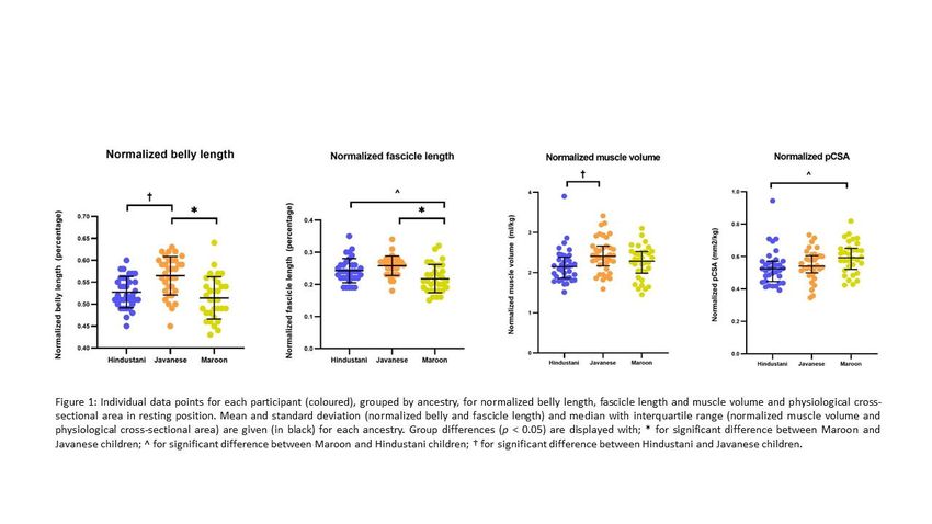
medRxiv preprint doi: https://doi.org/10.1101/2023.01.10.23284392; this version posted January 11, 2023. The copyright holder for this
preprint (which was not certified by peer review) is the author/funder, who has granted medRxiv a license to display the preprint in
                                                               perpetuity.
                                  It is made available under a CC-BY-NC-ND 4.0 International license .

   ABSTRACT

   Introduction: Muscle ultrasonography is frequently used to improve the understanding of
   musculoskeletal impairments in children with cerebral palsy. So far, most studies on muscle
   morphology and architecture have included typically developing children and children with cerebral
   palsy with similar ancestry, being mainly Caucasian. Less is known about differences in muscle
   morphology and architecture between children with different ancestral backgrounds. Therefore, the
   aim of this study was to compare muscle morphology and architecture of the medial gastrocnemius
   and Achilles tendon of Surinamese typically developing children from African, South Asian and
   Southeast Asian descent.

   Method: This explorative cohort study included 100 typically developing children identified as Maroon
   (Ghana, African descent), Hindustani (India, South Asian) or Javanese (Indonesia, Southeast Asian),
   aged 5 to 10 years. A conventional B-mode 2D ultrasound was used to define anatomical cross-
   sectional area (aCSA), fascicle length and pennation angle (architectural parameters). The muscle belly
   length, volume and physiological cross-sectional area (pCSA), as well as the tendon length
   (morphological parameters) were defined using 3D freehand ultrasound, which combines B-mode 2D
   ultrasound with 3D motion tracking. Muscle and tendon lengths were normalized to the total muscle
   tendon unit (MTU) lengths and fascicle lengths to muscle belly lengths, while volume, aCSA and PCSA
   were normalized to body mass. One-way Anova with post hoc t-tests were used to investigate
   differences between the ancestral groups. A two-way repeated measures Anova was used to define
   whether the extensibility of the muscle tendon, belly and fascicle differed between ancestral groups
   for the three conditions, i.e. when applying 0Nm, 1Nm and 4Nm ankle dorsiflexion torque.

   Results: The ancestral subgroups included 34 Hindustani, 34 Javanese and 32 Maroon children.
   Normalized belly length was 11% shorter in Maroon and 7% shorter in Hindustani children compared
   to Javanese children (p =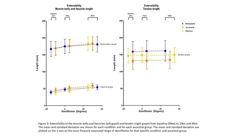
medRxiv preprint doi: https://doi.org/10.1101/2023.01.10.23284392; this version posted January 11, 2023. The copyright holder for this
preprint (which was not certified by peer review) is the author/funder, who has granted medRxiv a license to display the preprint in
                                                               perpetuity.
                                  It is made available under a CC-BY-NC-ND 4.0 International license .

   Discussion: Ancestry-specific reference data of the morphology of the medial gastrocnemius and
   Achilles tendon are needed when investigating altered muscle morphology in children with cerebral
   palsy. The current study showed differences in morphology (muscle belly-, tendon length and muscle
   volume, aCSA) and architecture (pCSA, fascicle length and deeper pennation angle) between children
   with different ancestry. These differences were most pronounced for Javanese compared to Maroon
   or Hindustani children. Future studies should report the ancestral background when describing muscle
   morphology and architecture of children and ancestral specifications in normative databases should
   be included.

   INTRODUCTION

   Muscle ultrasonography is increasingly used to improve the understanding of musculoskeletal
   impairments in children with spastic Cerebral Palsy (SCP) (Williams et al., 2021). During growth,
   adaptations in skeletal muscles, i.e., an increase in muscle length and anatomical cross-sectional area
   (aCSA), resulting in increasing muscle volume, occur in both typically developing (TD) children and
   children with SCP (Benard et al., 2011; Modlesky and Zhang, 2019; Bell et al., 2021; Handsfield et al.,
   2022). However, these increases are lower in children with SCP compared to TD children (Barber et
   al., 2016), while muscle tendons were found to be longer (Barrett and Lichtwark, 2010). For a good
   understanding of the musculoskeletal impairments in children with SCP, proper normative reference
   data of muscle morphology in TD children are important.

   Most of the current studies on muscle morphology and architecture in children with SCP compared to
   TD children have been published in North America, Europe, the UK, Australia, and Korea (Williams et
   al., 2021). While the ancestry of the enrolled children of these studies was not systematically
   mentioned, it may be assumed that data have been primarily reported for Caucasian children. Less is
   known about differences in muscle morphology between children with different ancestral
   backgrounds, such as African, East Asian and South Asian descent.

   To the best of our knowledge, only two previous studies compared muscle morphology and
   architecture between children of different ancestry (Kunimasa et al., 2022). Song et al. (2002) reported
   smaller total muscle volume of both the upper and lower limbs in East Asian and Caucasian children
   compared to children of African descent (Song et al., 2002). This finding was not confirmed by
   Kunimasa et al. (2022), who recently reported smaller muscle thickness for the medial gastrocnemius
   (MG) in Kenyan (African) children compared to Japanese (East-Asian) children (Kunimasa et al., 2022).
   The latter study also observed shorter fascicle and longer Achilles tendon lengths, with no differences
medRxiv preprint doi: https://doi.org/10.1101/2023.01.10.23284392; this version posted January 11, 2023. The copyright holder for this
preprint (which was not certified by peer review) is the author/funder, who has granted medRxiv a license to display the preprint in
perpetuity.
It is made available under a CC-BY-NC-ND 4.0 International license .
in pennation angle, in the Kenyan compared to Japanese children (Kunimasa et al., 2022). Most of
these morphological and architectural differences were already observed at an age of 4 years. Due to
the limited number of studies on ancestry-specific paediatric muscle morphology with contradicting
results, there is a need for more data on muscle morphology parameters of TD children with different
ancestral backgrounds.
Most previous studies that compared muscle morphology and architecture between ancestral groups,
as well as between TD with SCP children of one specific ancestral group, focused only on one or a
minimal selection of outcome parameters. However, an assessment of all relevant macroscopic
parameters is required for a good understanding of the differences between groups. For example,
morphological parameters, such as muscle volume or thickness, should be combined with
architectural parameter, such as fascicle length, to derive the physiological cross-sectional area
(pCSA). Furthermore, fascicle length should be accompanied by data on muscle belly and tendon
length to understand the potential contributions to altered joint range of motion. Moreover, the MG
lengths may be assessed at different ankle joint positions (i.e., when stretched by applying a specific
joint torques), to improve the understanding of the compliance of the muscle and tendon in TD
children. Integrated assessments that combine all morphological and architectural parameters may
help to understand differences between ancestral groups and altered muscle properties in children
with SCP.
The scarce results reported on ancestry-specific muscle morphology and architecture question the
validity of pooling muscle morphology and architecture data of children with different ancestral
backgrounds and suggest the need for an ancestral-specific reference database. This is especially
relevant for regions with a population including different ancestral backgrounds, such as Suriname,
where the research on children with SCP inherently includes children of mainly Indian (South Asian),
Ghana (African) and Indonesian (Southeast Asian) descent. It is crucial to know the differences
between children from different ancestral populations, before comparing muscle morphology and
architecture of SCP and TD children.
Therefore, the aim of this explorative cohort study was to compare the muscle morphology and
architecture of the MG of a large group of Surinamese TD children from African, South Asian and
Southeast Asian descent, from 5 to 10 years old. The ancestral-specific morphological and
architectural features for TD children were delineated and the comprehensive reference paediatric
database is put available online to be used for future studies (link to dataset will be added when the
study is published).medRxiv preprint doi: https://doi.org/10.1101/2023.01.10.23284392; this version posted January 11, 2023. The copyright holder for this
preprint (which was not certified by peer review) is the author/funder, who has granted medRxiv a license to display the preprint in
perpetuity.
It is made available under a CC-BY-NC-ND 4.0 International license .
METHOD
Study design and participants
This exploratory cohort study included TD children aged 5 to 10 years old. The ancestral background
of the child was identified as Maroon (Ghana, African descent), Hindustani (India, South Asian) or
Javanese (Indonesia, Southeast Asian) if at least three of the grandparents had the same ancestral
background, while all other cases were categorized as ‘mixed’ group (Krishnadath et al., 2015). For
homogeneous variation of age and sex in each age group, we aimed for an equal age and gender
distribution per ancestral group. Children were included if at least three of the grandparents were
categorized as Javanese, Hindustani or Maroon. Children were excluded in case of 1) mixed ancestral
background, 2) having a history of neurological problems or developmental disorders, 3) previous
lower leg musculoskeletal injuries, such as fractures or 4) doing organized sports three times or more
in a week.
Ethical approval was given by the Medical Ethical Committee of the Ministry of Health (reference VG
003-17). Approval to perform the measurements at schools was given by the Ministry of Education,
Science and Culture (NB/mz/Ag. 5653). Informed consent was obtained in writing from the
parent/guardian of all children as well as the children’s verbal consent before inclusion.
Recruitment and sample size determination
Recruitment and measurements of the children were performed from March 2021 to April 2022.
Almost all children were recruited via primary schools and seven children were recruited via family
and friends of co-workers of the Faculty of Medical Sciences of the Anton de Kom University of
Suriname. All children were living in the capital city of Suriname, Paramaribo.
For sample size calculation, the effect size was estimated using the available literature data on
differences in muscle mass, measured by dual energy X-ray absorptiometry, in children with different
ancestral backgrounds from United States of America (effect size: 0.94 for girls and 0.41 for boys (Silva
et al., 2010)) and the available literature data for the differences in adult Achilles tendon length among
adults (effect size: 0.80 (Kunimasa et al., 2014) and 1.3 (Refsdal, 2017)). These previous studies suggest
an averaged large effect size of 0.86. Based on a sample size calculation (independent t-test – power
0.80, alpha 0.0167 (corrected for 3 groups), effect size 0.86, G-power version 3.1.9.6) each ancestral
group should include 30 children. We finally aimed to recruit 30 to 35 children per ancestral
population, including 5 children for each of the 6 age-year categories, from 5 to 10 years old.medRxiv preprint doi: https://doi.org/10.1101/2023.01.10.23284392; this version posted January 11, 2023. The copyright holder for this
preprint (which was not certified by peer review) is the author/funder, who has granted medRxiv a license to display the preprint in
perpetuity.
It is made available under a CC-BY-NC-ND 4.0 International license .
Data collection and processing
In total, 5 schools gave permission for the measurements. At these schools, informed consent forms,
as well as questionnaires on ancestral background of the grandparents, history of neurological or
developmental disorders and frequency of weekly sport activities were handed out to the parents of
the children that could meet the inclusion criteria. If children met de inclusion criteria and the parents
and child agreed to participate, they were measured at school. For 7 children who were recruited via
family and friends of co-workers of the Faculty of Medical Sciences, the measurements were
performed at the Human Motion Laboratory at the Faculty of Medical Sciences of the Anton de Kom
University of Suriname. The left and right leg were randomly selected by use of a coin toss.
Anthropometric data were collected before the ultrasound assessment, while plantar flexor strength
was assessed afterwards. Children lay prone during the collection of muscle morphological and
architectural data and were instructed to relax while reading a book or watching a video. In case of
body segment movements or muscle contraction of the participant, the acquisition was repeated. The
duration of the measurement session lasted 45 minutes per child. All data were collected and
processed by one assessor (first author, FW), who was trained and experienced in conducting clinical
assessments, had 4 years of experience in performing ultrasound measurements, and was assisted by
at least one physiotherapy student or colleague. During the data processing, anonymously name files
for ancestry were used to ensure that FW was blinded for the ancestral background of the child.
Anthropometric measurements
Anthropometric measurements included height, body weight, maximal passive ankle dorsiflexion, calf
circumference and lower leg length (for more details, supplementary I). The position of the knee and
ankle joint during all ultrasound conditions was measured using a universal goniometer (7" Baseline
Plastic goniometer, Fabrication Enterprises). The ankle position was recorded with an interval of five
degrees for each different condition, representing a plantarflexion position with a negative angle and
a dorsiflexion with a positive angle.
Muscle morphology and architecture outcomes
The morphological and architectural parameters were assessed in resting position (RP) of the ankle
joint, i.e., with the foot hanging down freely at the end of a triangular pillow (supplementary II, Figure
1a). A conventional B-mode 2D ultrasound was used to define architectural parameters, namely the
aCSA, fascicle length and (deeper and superficial) pennation angle (for more details, supplementary
II). To define the morphological parameters, namely muscle volume, pCSA, belly length, tendon length
and MTU length, a previously described 3DfUS technique (Cenni et al., 2016) which combines B-modemedRxiv preprint doi: https://doi.org/10.1101/2023.01.10.23284392; this version posted January 11, 2023. The copyright holder for this
preprint (which was not certified by peer review) is the author/funder, who has granted medRxiv a license to display the preprint in
perpetuity.
It is made available under a CC-BY-NC-ND 4.0 International license .
2D ultrasound with 3D motion tracking, was used. The ultrasound collection, the 3D reconstructions
of the muscle and tendon, as well as all processing of the data, were performed with the open source
STRADWIN software (for more details, supplementary II).
Muscle volume and the aCSA and pCSA were normalized to body mass and muscle tendon and muscle
belly length were normalized to muscle-tendon unit (MTU) length. Fascicle length was normalized to
muscle belly length, since muscle belly length was different between ancestral groups.
Extensibility
To assess the extensibility of the muscle tendon, belly and fascicle, the muscles and tendons were
assessed in three other conditions. Therefore, the foot was placed in a custom-made set-up
(supplementary III, Figure 1) for applying a predefined torque of 0Nm, 1Nm and 4Nm on the ankle
joint (for more details, supplementary III). In the baseline condition (Baseline, 0Nm), the foot was
placed in the set-up and was hanging down freely at the end of a triangular pillow. This baseline
condition was always performed first, and was followed by a second and third condition, whereby
1Nm and 4 Nm ankle dorsiflexion torque was applied on the ankle, respectively (supplementary III,
Figure 2). In each condition, the muscle tendon, muscle belly and fascicle length was assessed. The
1Nm condition was chosen as a condition in between baseline and 4NM, in which the ankle joint was
often around neutral, i.e. 0 degrees of dorsiflexion in most children. The condition 4Nm was similar to
the ankle torque applied by Weide et al. (2015) and was in most cases the maximal tolerated range of
motion towards dorsiflexion.
Muscle strength
Isometric plantar flexion strength was measured with a MicroFed dynamometer (Hogan Health
Industries, Inc. 8020 South 1300 West, West Jordan, USA) held manually by the assessor at 75% of the
foot length. A mean of the maximal force (N) of three trials was used for further analysis and multiplied
by 75% of the foot length (expressed in meter, m) to calculate the ankle torque (expressed Nm). Body
weight was used for normalisation of ankle torque (for more details, supplementary IV)
Statistical analysis
Statistical analysis was performed in Jeffreys's Amazing Statistics Program (JASP) version 0.14.1. The
Pearson’s Chi-Squared test was performed to evaluate equal distribution of boys and girls between
ancestral groups. Normal distribution of data was assessed with the Shapiro-Wilk test and visual
inspection of the plots for each ancestral group and each individual parameter. The data were
described by means and standard deviation (SD) in case they were normally distributed, or by medians
and interquartile ranges (IQR) in case they were not normally distributed. When data was normallymedRxiv preprint doi: https://doi.org/10.1101/2023.01.10.23284392; this version posted January 11, 2023. The copyright holder for this
preprint (which was not certified by peer review) is the author/funder, who has granted medRxiv a license to display the preprint in
perpetuity.
It is made available under a CC-BY-NC-ND 4.0 International license .
distributed, the one-way Anova was used to investigate differences between ancestral groups, while
the Kruskall Wallis H test was used when the data of one of the ancestral groups was not normally
distributed. If the one-way ANOVA or Kruskall Wallis H test was significant, a post hoc t-test or Mann-
Whitney U test was used with a Bonferroni correction for multiple testing, to define differences
between the three ancestral groups. A two-way repeated measures Anova was used to define if there
were differences in the extensibility of the muscle tendon, belly and fascicle between ancestral
populations from baseline (0Nm) to 1NM and 4Nm. Level of significance, after correcting for multiple
testing was set to p < 0.05.
RESULTS
One hundred Hindustani (n=34), Javanese (n=34) and Maroon (n=32) children were included.
Participants were distributed over the age-range 5 to 10 and Pearson’s Chi-Square (Χ2 (2, N = 100) =
0.602, p = .740) showed equal distributions of boys and girls between ancestral groups
(supplementary table I).
There was a difference between ancestral groups for height (p = 0.023) and lower leg length (p = 0.003)
(table 1). The post hoc pairwise group comparisons showed that the Maroon children were taller (p =
0.011) compared to Hindustani children, with a longer lower leg length compared to Hindustani (p =
0.007) and Javanese children (p = 0.003). Lower leg circumference, absolute and normalized
plantarflexion strength were not different between groups (all p values ≥ 0.055).
Table 1: Group data for the anthropometric parameters of each ethnic group
Hindustani (n=34) Javanese (n=34) Maroon (n=32)
Median (IQR) Median (IQR) Median (IQR) p-value
Age (years) 7.58 (2.8) 7.38 (2.6) 8.64(2.8) 0.168
Height (cm) 126.75 (15.6) 121.00 (19.2) 133.50 (17.4) 0.023*
Body weight (kg) 21.75 (7.5) 22.25 (12.3) 26.75 (11.8) 0.224
Lower leg length (cm) 30.75 (5.3) 29.50 (4.8) 34.00 (5.1) 0.003*^
Lower leg circumference (cm) 23.50 (4.0) 24.25 (6.5) 25.00 (4.1) 0.292
Plantarflexion torque (Nm) 11.13 (5.2) 11.03 (5.6) 14.84 (6.8) 0.055
Normalised plantarflexion torque (Nm/kg) 0.44 (0.2) 0.41 (0.2) 0.51 (0.1) 0.122
Abbreviations: IQR, interquartile range. Group differences (p < 0.05) are displayed with; * for significant difference between
Maroon en Javanese children; ^ for significant difference between Maroon en Hindustani children; † for significant difference
between Hindustani en Javanese children.medRxiv preprint doi: https://doi.org/10.1101/2023.01.10.23284392; this version posted January 11, 2023. The copyright holder for this
preprint (which was not certified by peer review) is the author/funder, who has granted medRxiv a license to display the preprint in
perpetuity.
It is made available under a CC-BY-NC-ND 4.0 International license .
The MTU length was different between groups (p = 0.001), with significantly longer MTU length in
Maroon compared to Javanese children (p =medRxiv preprint doi: https://doi.org/10.1101/2023.01.10.23284392; this version posted January 11, 2023. The copyright holder for this
preprint (which was not certified by peer review) is the author/funder, who has granted medRxiv a license to display the preprint in
perpetuity.
It is made available under a CC-BY-NC-ND 4.0 International license .
Extensibility
The most frequently recorded ankle angle for the position at 0Nm was 15 degrees of plantarflexion in
Hindustani children and 10 degrees of plantarflexion in Maroon and Javanese children. In the position
of 1Nm, the most common measured ankle angle was 0 degrees in Hindustani children and 5 degrees
dorsiflexion in the Maroon and Javanese children. The increase of ankle dorsiflexion from 1Nm to
4Nm, based on the most common recorded ankle angle, was 35 degrees in Hindustani children, 25
degrees in Javanese children and 20 degrees in Maroon children. Muscle belly, tendon and fascicle
length significantly increased and pennation angle significantly decreased when applying increasing
torques from baseline (0Nm) to 1Nm and 4Nm (all p valuesmedRxiv preprint doi: https://doi.org/10.1101/2023.01.10.23284392; this version posted January 11, 2023. The copyright holder for this
preprint (which was not certified by peer review) is the author/funder, who has granted medRxiv a license to display the preprint in
perpetuity.
It is made available under a CC-BY-NC-ND 4.0 International license .
et al., 2022). Lee et al. (2014) reported that African and Caribbean children (living in London) aged 5-
10 years old tended to be taller, heavier, with larger leg circumferences and relatively longer legs
compared to other ancestral groups, including South Asian children (Lee et al., 2014). While Lee et al.
(2014) investigated similar age groups, current data showed no differences in body weight and calf
circumference between ancestral groups. Longer leg lengths were also found in a recent study of
Kunimasa et al. (2022) in children with African compared to East Asian ancestry. Therefore, it was
considered important to normalize muscle length parameters when comparing muscle morphology
and architecture between ancestral groups, especially when including children with African ancestry.
Normalized tendon length of the MG muscle was 7-11% longer in both Hindustani (South Asian) and
Marron (African) children, compared to Javanese Southeast Asian) children. This is in line with a recent
study, where differences in tendon length between Japanese (East Asian) and Kenyan (African)
children were found, already at the age of 4 years old (Kunimasa et al., 2022). Similar percentages of
difference in Achilles tendon length (i.e., ≈10% longer tendon lengths) between Kenyan (African)
compared to Japanese (East Asian) adult runners has been reported (Kunimasa et al., 2014; Sano et
al., 2015). The current study results revealed that the difference in tendon length between south Asian
and African descent compared to southeast Asian descent exceeds the lower limit of difference (i.e.,
≈4%) found in tendon length between TD and SCP (Barrett and Lichtwark, 2010; Kruse et al., 2021).
Therefore, when comparing the tendon length of a group of TD and SCP children including different
ancestral groups (especially including the southeast Asian ancestry), group-matching needs to take
ancestral-variance into account.
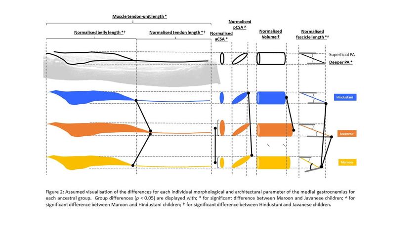
Normalized muscle volume and aCSA was significantly larger between Javanese children compared to
Hindustani children (for normalized muscle volume) and Maroon children (for normalized aCSA). A
greater muscle volume could have been expected in Javanese children, because of their longer muscle
belly length. On the other hand, no difference in normalized volume was found between Maroon and
Javanese children, while the muscle belly length was significantly shorter in Maroon compared to
Javanese children. Another contraction is the higher normalized pCSA in Maroon children compared
to the other ancestral groups. Yet, so far, no other studies have evaluated the MG volume between
children with different ancestry. While no differences in lower leg circumference between ancestries
were found in current study, Refsdal et al. (2017) described differences at different shank heights
between adults with African and Caucasian ancestry, which could suggest differences in the shape of
the shank. Also the contradictory combination of differences in normalized muscle volume and muscle
belly length found in the current study, suggest that children with different ancestry may have amedRxiv preprint doi: https://doi.org/10.1101/2023.01.10.23284392; this version posted January 11, 2023. The copyright holder for this
preprint (which was not certified by peer review) is the author/funder, who has granted medRxiv a license to display the preprint in
perpetuity.
It is made available under a CC-BY-NC-ND 4.0 International license .
different shape of the MG muscle belly. This could be further explored using statistical shape modeling
in future studies (Ghouth et al., 2022).
The muscle belly length of the MG is defined by the muscle architectural parameters fascicle length
and pennation angle. Normalized fascicle length was lower for Maroon compared to Hindustani and
Javanese children, while the deeper pennation angle was greater in Maroon children compared to
Javanese children. A deeper pennation angle was also found in a recent study of Kunimasa et al. (2022)
for children with African compared to East Asian ancestry. Despite the fact that the deeper pennation
angle is considered more sensible for errors than the superficial pennation angle due to the
unavoidable tilt of the probe during the acquisition (Bénard et al., 2009), the differences in the deeper
pennation angle were between ancestral groups. This may strengthen our earlier assumption of
muscle shape differences between children with different ancestral background. The results for
fascicle length were in line to the findings of Kunimasa et al. (2022), who also reported longer
normalized fascicle lengths in Kenyan children compared to Japanese children. In short, fascicle length
was the longest in Javanese children and smallest in Maroon children, which is in line with a longer
belly length in Javanese compared to Maroon children.
The significant increase in muscle belly, tendon and fascicle length and significant decrease in
pennation angle when a torque of 1 and 4Nm was applied compared to the condition of 0Nm torque,
are in line with the changes found during passive stretching of the MG (Bolsterlee et al., 2017; Kalkman
et al., 2018). According to models that present the stiffness caused by the arrangement of muscle
fascicles (and thereby the sarcomeres) (Kruse et al., 2021), the increase in fascicle length is assumed
to decrease the passive stiffness and thereby increase the extensibility. Although fascicle length was
significant different between ancestral groups, we found no differences in the extensibility of the
muscle belly, muscle tendon and fascicles between ancestral groups. This suggests that other
components, such as intramuscular connective tissue (Järvinen et al., 2002), may have contributed to
the observed extensibility.
We did not expect to find great differences in extensibility of the tendon between typically developing
children with different ancestry. Weide et al. (2020) found no differences in extensibility of the MTU
from baseline to 4Nm between TD and SCP children, which was in contrast to other studies that found
less muscle belly and more muscle tendon extensibility in SCP compared to TD children (Theis,
Mohagheghi and Korff, 2016; Kalkman et al., 2018; Weide et al., 2020). The contradicting results could
have been caused by difference in methods. The current study and Weide et al. (2020) measured the
extensibility statically and the other studies defined extensibility during dynamic stretching. However,
the current study data created a unique norm-database for the extensibility of the muscle belly,medRxiv preprint doi: https://doi.org/10.1101/2023.01.10.23284392; this version posted January 11, 2023. The copyright holder for this
preprint (which was not certified by peer review) is the author/funder, who has granted medRxiv a license to display the preprint in
perpetuity.
It is made available under a CC-BY-NC-ND 4.0 International license .
muscle tendon and fascicle length that can be used as a reference when investigating the extensibility
parameters of SCP children.
Studies in adults have indicated differences in the muscle morphology between Caucasian, African
and East Asian descendants. One study reported smaller muscle thickness, shorter fascicle length and
longer tendon length of the MG in African compared to Caucasian descendants (Refsdal, 2017). When
comparing African descendants to East Asian descendants, shorter fascicle lengths, greater pennation
angles and longer Achilles tendon lengths were found in African descendants compared to East Asian
descendants (Sano et al., 2015; Kunimasa et al., 2022). These results are in line with the current
findings. It remains unknown if the differences found between the ancestral groups in Suriname can
be generalized to ancestral groups in other countries. We advise that future studies report the
ancestral background when describing muscle morphology of children and adults and that ancestral
specifications are well-considered when establishing and using normative databases.
Limitations
Besides ancestral differences, also nutritional and other environmental factors can influence muscle
growth. Therefore, we included only children living in the capital city of Paramaribo to minimalize
cultural differences between ancestral groups (e.g. beliefs and traditional food) and living
circumstances (e.g. housing, possibility of schools, access to sports clubs). Although we do not expect
great differences by these factors from 5-11 years old, it would be interesting to take these factors in
account in further studies or studies with older children.
This study did not focus on the relation of muscle morphology to function and performance, while the
moments arm at the joints can differ between ancestral groups (Hanson et al., 1999; Kunimasa et al.,
2022). This is of interest for further research, since ancestry-specific features of the MG muscle
morphology may potentially be related to force production and performance in walking and running
(McCarthy et al., 2006; Sano et al., 2014; Butler and Dominy, 2016).
Conclusion
The current study showed differences in MG morphology (muscle belly-, tendon length and muscle
volume, aCSA) and architecture (pCSA, fascicle length and deeper pennation angle) between children
with different ancestry, the Maroon (Ghana, African), Hindustani (India, South Asian) and Javanese
(Indonesia, Southeast Asian), aged 5 to 10 years old. These differences were present between all
groups, but were most pronounced in Javanese children compared to Maroon or Hindustani children.medRxiv preprint doi: https://doi.org/10.1101/2023.01.10.23284392; this version posted January 11, 2023. The copyright holder for this
preprint (which was not certified by peer review) is the author/funder, who has granted medRxiv a license to display the preprint in
perpetuity.
It is made available under a CC-BY-NC-ND 4.0 International license .
The current results revealed that ancestry-specific reference data are needed when investigating
altered muscle morphology in neurological or neuromuscular pathologies, such as cerebral palsy. We
recommend that future studies report the ancestral background when describing muscle morphology
and architecture of children and that ancestral specifications in normative databases should be
included.
References
Barber, L. . et al. (2016) ‘Calf muscle growth in ambulant children with hemiplegia and diplegia
cerebral palsy age 2 to 9 years’, Developmental Medicine and Child Neurology. L.A. Barber,
University of Queensland, Brisbane, Australia, 58, p. 82. doi: 10.1111/dmcn.13069.
Barrett, R. S. and Lichtwark, G. A. (2010) ‘Gross muscle morphology and structure in spastic cerebral
palsy: a systematic review’, Developmental Medicine and Child Neurology. R.S. Barrett, School of
Physiotherapy and Exercise Science, Griffith University Gold Coast Campus, Queensland 4222,
Australia, School of Physiotherapy and Exercise Science, Griffith University Gold Coast Campus,
Queensland 4222, Australia, 52(9), pp. 794–804. doi: 10.1111/j.1469-8749.2010.03686.x.
Bell, M. et al. (2021) ‘Typical m. triceps surae morphology and architecture measurement from 0 to
18 years: A narrative review’, Journal of Anatomy, (October), pp. 1–15. doi: 10.1111/joa.13584.
Benard, M. R. et al. (2011) ‘Effects of growth on geometry of gastrocnemius muscle in children: A
three-dimensional ultrasound analysis’, Journal of Anatomy, 219(3), pp. 388–402. doi:
10.1111/j.1469-7580.2011.01402.x.
Bénard, M. R. et al. (2009) ‘Anatomical information is needed in ultrasound imaging of muscle to
avoid potentially substantial errors in measurement of muscle geometry.’, Muscle & nerve. United
States, 39(5), pp. 652–665. doi: 10.1002/mus.21287.
Bolsterlee, X. B. et al. (2017) ‘How does passive lengthening change the architecture of the human
medial gastrocnemius muscle ?’, pp. 727–738. doi: 10.1152/japplphysiol.00976.2016.
Butler, E. E. and Dominy, N. J. (2016) ‘Architecture and functional ecology of the human
gastrocnemius muscle-tendon unit’, Journal of Anatomy, 228(4), pp. 561–568. doi:
10.1111/joa.12432.
Cenni, F. et al. (2016) ‘The reliability and validity of a clinical 3D freehand ultrasound system’,
Computer methods and programs in biomedicine, 136, pp. 179–187.medRxiv preprint doi: https://doi.org/10.1101/2023.01.10.23284392; this version posted January 11, 2023. The copyright holder for this
preprint (which was not certified by peer review) is the author/funder, who has granted medRxiv a license to display the preprint in
perpetuity.
It is made available under a CC-BY-NC-ND 4.0 International license .
Ghouth, S. G. Bin et al. (2022) ‘OPEN A statistical shape model of soleus muscle morphology in
spastic cerebral palsy’, Scientific Reports. Nature Publishing Group UK, pp. 1–10. doi:
10.1038/s41598-022-11611-z.
Handsfield, G. G. et al. (2022) ‘Muscle architecture, growth, and biological Remodelling in cerebral
palsy: a narrative review’, BMC Musculoskeletal Disorders. BioMed Central, 23(1), pp. 1–17. doi:
10.1186/s12891-022-05110-5.
Hanson, P. et al. (1999) ‘Anatomical differences in the psoas muscles in young black and white men’,
J. Anat, 194, pp. 303–307. doi: 10.1046/j.1469-7580.1999.19420303.x. PMID: 10337963; PMCID:
PMC1467925.
Järvinen, T. A. H. et al. (2002) ‘Organization and distribution of intramuscular connective tissue in
normal and immobilized skeletal muscles’, Journal of Muscle Research & Cell Motility, 23(3), pp.
245–254. doi: 10.1023/A:1020904518336.
Kalkman, B. M. et al. (2018) ‘Muscle and tendon lengthening behaviour of the medial gastrocnemius
during ankle joint rotation in children with cerebral palsy’, (April), pp. 1367–1376. doi:
10.1113/EP087053.
Krishnadath, I. S. et al. (2015) ‘A National Surveillance Survey on Noncommunicable Disease Risk
Factors: Suriname Health Study Protocol’, JMIR Res Protoc, 4(2), p. e75. doi: 10.2196/resprot.4205.
Kruse, A. et al. (2021) ‘Stimuli for Adaptations in Muscle Length and the Length Range of Active
Force Exertion—A Narrative Review’, Frontiers in Physiology, 12(October). doi:
10.3389/fphys.2021.742034.
Kunimasa, Y. et al. (2014) ‘Specific muscle-tendon architecture in elite Kenyan distance runners’,
Scandinavian Journal of Medicine and Science in Sports, 24(4). doi: 10.1111/sms.12161.
Kunimasa, Y. et al. (2022) ‘tendon architecture in Kenyans and Japanese : Potential role of genetic
endowment in the success of elite Kenyan endurance runners’, (January), pp. 1–12. doi:
10.1111/apha.13821.
Lee, S. et al. (2014) ‘Ethnic Variability in Body Size , Proportions and Composition in Children Aged 5
to 11 Years : Is Ethnic-Specific Calibration of Bioelectrical Impedance Required ?’, pp. 1–17. doi:
10.1371/journal.pone.0113883.
McCarthy, J. et al. (2006) ‘Ethnic Differences in Triceps Surae Muscle Tendon Complex and Walking
Economy’, Journal of Strength and Conditioning Research, 20(3), pp. 511–518.medRxiv preprint doi: https://doi.org/10.1101/2023.01.10.23284392; this version posted January 11, 2023. The copyright holder for this
preprint (which was not certified by peer review) is the author/funder, who has granted medRxiv a license to display the preprint in
perpetuity.
It is made available under a CC-BY-NC-ND 4.0 International license .
Modlesky, C. M. and Zhang, C. (2019) ‘Muscle Size, Composition, and Architecture in Cerebral Palsy’,
in Miller, F. et al. (eds) Cerebral Palsy. Cham: Springer International Publishing, pp. 1–16. doi:
10.1007/978-3-319-50592-3_14-1.
Refsdal, A. S. (2017) Jump performance in Maasai jumpers and Caucasian controls. Available at:
https://nih.brage.unit.no/nih-xmlui/handle/11250/2436204.
Sano, K. et al. (2014) ‘Can measures of muscle–tendon interaction improve our understanding of the
superiority of Kenyan endurance runners?’, European Journal of Applied Physiology, 115(4), pp. 849–
859. doi: 10.1007/s00421-014-3067-7.
Sano, K. et al. (2015) ‘Can measures of muscle–tendon interaction improve our understanding of the
superiority of Kenyan endurance runners?’, European Journal of Applied Physiology, 115(4), pp. 849–
859. doi: 10.1007/s00421-014-3067-7.
Silva, A. M. et al. (2010) ‘Ethnicity-related skeletal muscle differences across the lifespan’, American
Journal of Human Biology, 22(1), pp. 76–82. doi: 10.1002/ajhb.20956.
Song, M. Y. et al. (2002) ‘Prepubertal Asians have less limb skeletal muscle’, Journal of Applied
Physiology, 92(6), pp. 2285–2291. doi: 10.1152/japplphysiol.01066.2001.
Theis, N., Mohagheghi, A. A. and Korff, T. (2016) ‘Mechanical and material properties of the
plantarflexor muscles and Achilles tendon in children with spastic cerebral palsy and typically
developing children’, Journal of Biomechanics. T. Korff, Centre for Sports Medicine and Human
Performance, Brunel University, Middlesex, United Kingdom, 49(13), pp. 3004–3008. doi:
10.1016/j.jbiomech.2016.07.020.
Weide, G. et al. (2020) ‘Gastrocnemius Medialis Muscle Geometry and Extensibility in Typically
Developing Children and Children With Spastic Paresis Aged 6-13 Years.’, Frontiers in physiology, 11,
p. 528522. doi: 10.3389/fphys.2020.528522.
Williams, S. A. et al. (2021) ‘Measuring skeletal muscle morphology and architecture with imaging
modalities in children with cerebral palsy: a scoping review’, Developmental Medicine and Child
Neurology, 63(3), pp. 263–273. doi: 10.1111/dmcn.14714.You can also read