Nasopharyngeal Pneumococcal Colonization Density Is Associated With Severe Pneumonia in Young Children in the Lao People's Democratic Republic
←
→
Page content transcription
If your browser does not render page correctly, please read the page content below
The Journal of Infectious Diseases
   MAJOR ARTICLE

Nasopharyngeal Pneumococcal Colonization Density Is
Associated With Severe Pneumonia in Young Children in
the Lao People’s Democratic Republic
Olivia J. J. Carr,1,2 Keoudomphone Vilivong,3 Laddaphone Bounvilay,3 Eileen M. Dunne,2 Jana Y. R. Lai,4 Jocelyn Chan,2,5 Malisa Vongsakid,3
Anisone Changthongthip,3 C. Siladeth,3 Belinda Ortika,2 Cattram Nguyen,2,5 Mayfong Mayxay,3,6,7 Paul N. Newton,3,7,8 Kim Mulholland,2,5,9 Lien A. H. Do,2,5
Audrey Dubot-Pérès,3,7,10 Catherine Satzke,2,5,11, David A. B. Dance,3,7,8 and Fiona M. Russell2,5
1
 University of Tasmania, Hobart, Tasmania, Australia, 2Murdoch Children’s Research Institute, Melbourne, Victoria, Australia, 3Lao-Oxford-Mahosot Hospital-Wellcome Trust Research Unit,
Microbiology Laboratory, Mahosot Hospital, Vientiane, Lao People’s Democratic Republic, 4World Health Organization, Lao People’s Democratic Republic, 5Department of Paediatrics, University of
Melbourne, Parkville, Victoria, Australia, 6Institute of Research and Education Development, University of Health Sciences, Ministry of Health, Vientiane, Lao People’s Democratic Republic, 7Centre

                                                                                                                                                                                                       Downloaded from https://academic.oup.com/jid/article/225/7/1266/6273782 by guest on 26 June 2022
for Tropical Medicine and Global Health, Nuffield Department of Clinical Medicine, University of Oxford, Oxford, United Kingdom, 8Faculty of Infectious and Tropical Diseases, London School of
Hygiene and Tropical Medicine, London, United Kingdom, 9Department of Infectious Disease Epidemiology, London School of Hygiene and Tropical Medicine, London, United Kingdom, 10Unité des
Virus Émergents (UVE: Aix-Marseille Univ-Institut de Recherche pour le Développement 190-Inserm 1207), Marseille, France, and 11Department of Microbiology and Immunology, Peter Doherty
Institute for Infection and Immunity, University of Melbourne, Parkville, Australia

   Background. No studies have explored the association between pneumococcal nasopharyngeal density and severe pneumonia
using the World Health Organization (WHO) 2013 definition. In Lao People’s Democratic Republic (Lao PDR), we determine the
association between nasopharyngeal pneumococcal density and severe pneumonia in children.
   Methods. A prospective observational study was undertaken at Mahosot Hospital, Vientiane, from 2014 to mid-2018. Children
pneumonia definition is important for these settings as, to our to the 2013 WHO definition: cough and/or difficulty breathing knowledge, no studies have explored this association [11]. and tachypnea (≥50 breaths per minute for children aged The Lao People’s Democratic Republic (Lao PDR) is an LMIC 2–11 months and ≥40 breaths per minute for children aged in Southeast Asia with a population of approximately 7 mil- 12–59 months) with any 1 of oxygen saturation
probe based real-time PCRs for the detection of the patho-              Unit, Oxford University); Ethics Research Committee (2013.30.
gens. Samples collected after December 2014 were tested                 LAO.2.EIP; Western Pacific Regional Office of WHO); National
by probe-based real-time RT-PCR using the iTaq Universal                Ethics Committee for Health Research (number 057/2013;
Probes One-Step reverse transcriptase kit (Bio-Rad) and pri-            Ministry of Health, Lao PDR); and University of Tasmania Human
mers/probe as previously described [19], from 10 µL of nu-              Research Ethics Committee (HREC number H0018186).
cleic acids, in a final reaction volume of 30 µL. A sample was
considered positive for RSV if the real-time PCR assay had a            RESULTS
cycle threshold value of 2 groups.                              mococcal carriers who had complete data for all variables in the
   To determine the association between pneumococcal density            multivariable model and were therefore included in the analysis.
and severe pneumonia, logistic regression was used to model se-         At the univariable level, there was some evidence of a positive
vere pneumonia as a function of pneumococcal carriage density           association between pneumococcal density and severe pneu-
and other covariates, reported as odds ratios (ORs) and 95% con-        monia in children who carried pneumococci (OR, 1.2 [95% CI,
fidence intervals (CIs). Only pneumococcal carriers and those           1.0–1.5]; P = .074). The youngest age group, 2–5 months, was
with complete data were included in this analysis. To assess for        4.2 times more likely to have severe pneumonia than the 24-
potential collinearity between covariates, correlation between          to 59-month age group (OR, 4.2 [95% CI, 2.9–6.1]; P < .001).
potential confounders was determined using Pearson r. The               Other variables strongly associated with severe pneumonia at
multivariable model was built using variables found to be signifi-      the univariable level included being from a minority ethnic
cant at the univariable level (P < .2) and those identified a priori.   group, living in Vientiane capital, having a cigarette smoker in
Variables identified a priori were visualized using a directed acy-     the house and being coinfected with RSV. When assessed for
clic graph (DAG) (http://www.dagitty.net/) and included all pos-        correlation, no variables were found to be strongly correlated.
sible variables that were related to severe pneumonia and were             Three hundred and seventy-two pneumococcal carriers were
potential confounders (Supplementary Figure 1). The DAG and             included in the analysis, of which 94 (25.3%) had severe pneu-
the univariable analyses identified the following variables for in-     monia. Of this subset, the median age was 15 months (IQR,
clusion in the multivariable model: age, ethnicity, residential lo-     9–25 months) and 217 (58.3%) were considered vaccinated using
cation, other children
Table 1. Characteristics of Study Participants in Children Hospitalized With Acute Respiratory Infections in Lao People’s Democratic Republic, by
Pneumonia Severity (N = 1268)

                                                             All Participants                       Severe Pneumoniaa                                Other ARI
Characteristics                                                (N = 1268)                                (n = 410)                                   (n = 858)                P Valueb

Demographics
     Age, mo, median (IQR)                                   15 (8–25)                                  11 (6–20)                                  17 (10–28)
Table 2. Associations Between Potential Confounders and Severe Pneumoniaa in 2- to 59-Month-Old Children Admitted to Hospital With Acute Respiratory
Infections in Lao People’s Democratic Republic (n = 372)b
Unadjusted OR Adjusted ORc
Variable (95% CI) P Value (95% CI) P Value
Main variable
Pneumococcal density (carriers only)d 1.2 (1.0–1.5) .074 1.4 (1.1–1.8) .020
Demographics
Age, mo
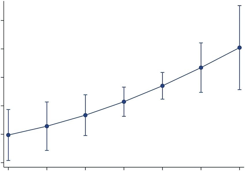
2–5 4.2 (2.9––6.1)Invasive lung aspirate studies are required to prove a causal
0.5 link between pneumococcal colonization density and pneumo-
Probability of severe pneumonia
coccal pneumonia, but these studies are invasive and difficult
0.4
P = .020 to do. A study from The Gambia found that lung and pleural
aspirates were able to identify the pathogens responsible for ra-
0.3
diologically confirmed pneumonia and found S. pneumoniae
0.2 responsible for 25% of cases [25]. The PERCH study found
that in lung and pleural aspirates, bacterial pathogens (most
0.1 often S. pneumoniae [lung aspirate] and Staphylococcus au-
reus [pleural fluid]) were more commonly detected than viral
0 pathogens [7]. Hence, the value of using pneumococcal density
2 3 4 5 6 7 8
as a diagnostic assay would be if it were able to identify addi-
Pneumococcal density (log10 ge/mL)
tional pneumococcal cases that were not radiologically or lab-
oratory confirmed, which represents the vast majority of cases
Downloaded from https://academic.oup.com/jid/article/225/7/1266/6273782 by guest on 26 June 2022
Figure 1. Probability of severe pneumonia [11] in children aged 2–59 months of pneumococcal pneumonia [26]. As such, PERCH found that
admitted to hospital with an acute respiratory infection and pneumococcal car-
riage, Lao People’s Democratic Republic, by pneumococcal density in log10 ge- a pneumococcal nasopharyngeal density >6.9 log10 copies/mL
nome equivalents/mL. Severe pneumonia was defined as cough and/or difficulty was associated with radiologically confirmed pneumonia, very
breathing and tachypnea (≥50 breaths per minute for children aged 2–11 months severe pneumonia, and hypoxic pneumonia, but performance
and ≥40 breaths per minute for children aged 12–59 months) with any 1 of oxygen
saturationonly 1 nasopharyngeal swab on admission was collected per directors of the Microbiology Laboratory, the staff of the lab-
child. Recent research has found that pneumococcal density oratory and the Lao-Oxford-Mahosot Hospital–Wellcome
may vary throughout a colonization episode [27]. Therefore, Trust Research Unit (LOMWRU), particularly the ARIVI
taking only a single nasopharyngeal swab may not capture peak team, for their technical help and support. We thank Associate
pneumococcal density. However, in our study, duration of ill- Professor Chanphomma Vongsamphan, the Former Director of
ness was not associated with severity: median was 3 days (IQR, Department of Health Care, Ministry of Health, and Associate
2–5 days) among severe, and 4 days (IQR, 2–5 days) in other ARI Professor Bounkong Syhavong, Minister of Health, Lao PDR,
(P = .922), so duration of illness is unlikely to be a confounder. for their support for the work of LOMWRU. We also thank
We found that there was no difference between the duration of all the study, laboratory, and administrative staff at the Centre
illness between severe pneumonia and other ARI cases prior to for International Child Health, Department of Paediatrics,
admission; therefore, it is likely that the swab was taken at a sim- University of Melbourne, Australia; Murdoch Children’s
ilar time from the onset of symptoms for the severe pneumonia Research Institute, Australia; and the Expanded Programme
and other ARI groups. Third, children with pneumococci in the on Immunization, Ministry of Health Lao PDR, including the
nasopharynx who are coinfected with respiratory viruses tend to late Dr Anonh Xeuatvongsa at the national immunization pro-
Downloaded from https://academic.oup.com/jid/article/225/7/1266/6273782 by guest on 26 June 2022
have high nasopharyngeal pneumococcal densities [8, 10, 28]. In gram. RSV testing was funded by the Institute of Research for
our study, we used the RSV data in the regression model and we Development, Aix-Marseille University, Wellcome Trust of
adjusted for RSV as it is a potential confounder. During this ob- Great Britain.
servation period we also tested for influenza but only detected 33 Financial support. This analysis was part of a study funded
cases. Influenza may affect density but as there were so few cases, by the Bill & Melinda Gates Foundation (grant number
we did not adjust for influenza in the analysis as it is very unlikely OPP1115490). F. M. R. is supported by National Health and
to have affected our results as influenza was rare. Finally, as there Medical Research Council (NHMRC) fellowships. P. N. N. and
were missing data, we could not run the regression model on the D. A. B. D. are supported by the Wellcome Trust, which also
whole data set. However, we found no major differences between provides core funds to LOMWRU. C. S. was supported by an
those with complete and missing data, suggesting no systematic Australian NHMRC Career Development Fellowship (award
differences between the 2 groups. Our study is strengthened by number 1087957) and a Veski Inspiring Women Fellowship.
using highly sensitive and specific laboratory techniques, and the Potential conflicts of interest. C. N., C. S., E. D. M., and L. A.
fact that our findings are consistent with studies that have used H. D. are investigators on a grant funded by Pfizer, outside the
more specific pneumococcal pneumonia definitions. submitted work. All other authors report no potential conflicts
Determining the etiology of pneumonia and effect of PCV on of interest.
pneumonia is very challenging as there is no case definition that All authors have submitted the ICMJE Form for Disclosure
is sensitive and specific enough to capture most cases. We have of Potential Conflicts of Interest. Conflicts that the editors
found pneumococcal colonization density to be a predictor of consider relevant to the content of the manuscript have been
severe pneumonia in pneumococcal carriers in our study set- disclosed.
ting. Although causality cannot be ascribed due to the study’s
observational design, further studies are warranted to deter- REFERENCES
mine whether this association exists in other settings, as this 1. O’Brien KL, Wolfson LJ, Watt JP, et al; Hib and Pneumococcal
could be a potential outcome to measure the impact of PCV Global Burden of Disease Study Team. Burden of disease
implementation for epidemiological purposes. Our findings caused by Streptococcus pneumoniae in children younger
contribute to a growing body of evidence demonstrating the than 5 years: global estimates. Lancet 2009; 374:893–902.
positive association between pneumococcal density and severe 2. Bryce J, Boschi-Pinto C, Shibuya K, Black RE; WHO Child
pneumonia, and provide insight in the understanding of pneu- Health Epidemiology Reference Group. WHO estimates of
mococcal colonization density and the role it plays in pneumo- the causes of death in children. Lancet 2005; 365:1147–52.
coccal disease. 3. Bogaert D, De Groot R, Hermans PW. Streptococcus
pneumoniae colonisation: the key to pneumococcal disease.
Notes Lancet Infect Dis 2004; 4:144–54.
Author contributions. O. J. J. C. and F. M. R. had full access to 4. Simell B, Auranen K, Käyhty H, Goldblatt D, Dagan R,
all the data in the study and take responsibility for the integrity O’Brien KL; Pneumococcal Carriage Group. The funda-
of the data and the accuracy of the data analysis. mental link between pneumococcal carriage and disease.
Acknowledgments. We would like to thank the patients and Expert Rev Vaccines 2012; 11:841–55.
their families for participation in this study. We are very grateful 5. van der Poll T, Opal SM. Pathogenesis, treatment, and
to the directors and staff of Mahosot Hospital; the late Dr prevention of pneumococcal pneumonia. Lancet 2009;
Rattanaphone Phetsouvanh; and Dr Manivanh Vongsouvath, 374:1543–56.
1272 • jid 2022:225 (1 April) • Carr et al6. Chan J, Nguyen CD, Dunne EM, et al. Using pneumococcal 17. Dunne EM, Manning J, Russell FM, Robins-Browne RM,
carriage studies to monitor vaccine impact in low- and Mulholland EK, Satzke C. Effect of pneumococcal vac-
middle-income countries. Vaccine 2019; 37:6299–309. cination on nasopharyngeal carriage of Streptococcus
7. Baggett HC, Watson NL, Deloria Knoll M, et al. Density pneumoniae, Haemophilus influenzae, Moraxella
of upper respiratory colonization with Streptococcus catarrhalis, and Staphylococcus aureus in Fijian children. J
pneumoniae and its role in the diagnosis of pneumococcal Clin Microbiol 2012; 50:1034–8.
pneumonia among children agedYou can also read