Navigation-assisted fluoroscopy in minimally invasive direct lateral interbody fusion: a cadaveric study
←
→
Page content transcription
If your browser does not render page correctly, please read the page content below
Navigation-assisted fluoroscopy in minimally invasive
direct lateral interbody fusion: a cadaveric study
Jonathan E. Webb, Gilad J. Regev, Steven R. Garfin and Choll W. Kim

Int J Spine Surg 2010, 4 (4) 115-121
doi: https://doi.org/10.1016/j.esas.2010.09.002
http://ijssurgery.com/content/4/4/115
This information is current as of June 29, 2022.

   Email Alerts     Receive free email-alerts when new articles cite this article. Sign up at:
                    http://ijssurgery.com/alerts

The International Journal of Spine Surgery
2397 Waterbury Circle, Suite 1,
Aurora, IL 60504, Phone: +1-630-375-1432
© 2010 ISASS. All Rights Reserved.
                     Downloaded from http://ijssurgery.com/ by guest on June 29, 2022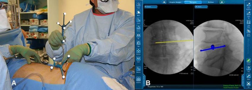
Available online at www.sciencedirect.com

                                                         SAS Journal 4 (2010) 115–121
                                                                                                                                www.sasjournal.com
                                                                           MIS
     Navigation-assisted fluoroscopy in minimally invasive direct lateral
                     interbody fusion: a cadaveric study
                Jonathan E. Webb, MD a, Gilad J. Regev, MD a, Steven R. Garfin, MD a,
                                   Choll W. Kim, MD, PhD a,b,*
                                                   a
                                                     University of California, San Diego, San Diego, CA
                                          b
                                              Spine Institute of San Diego at Alvarado Hospital, San Diego, CA

Abstract

Background: Minimally invasive surgery (MIS) is dependent on intraoperative fluoroscopic imaging for visualization, which significantly
increases exposure to radiation. Navigation-assisted fluoroscopy (NAV) can potentially decrease radiation exposure and improve the
operating room environment by reducing the need for real-time fluoroscopy. The direct lateral interbody fusion (DLIF) procedure is a
technique for MIS intervertebral lumbar and thoracic interbody fusions. This study assesses the use of navigation for the DLIF procedure
in comparison to standard fluoroscopy (FLUORO), as well as the accuracy of the NAV MIS DLIF procedure.
Methods: Three fresh whole-body cadavers underwent multiple DLIF procedures at the T10-L5 levels via either NAV or FLUORO.
Radiation exposure and surgical times were recorded and compared between groups. An additional cadaver was used to evaluate the
accuracy of the NAV system for the DLIF procedure by measuring the deviation error as the surgeon worked further from the anterior
superior iliac spine tracker.
Results: Approach, discectomy, and total fluoroscopy times for FLUORO were longer than NAV (P ⬍ .05). In contrast, the setup time was
longer in NAV (P ⫽ .005). Cage insertion and total operating times were similar for both. Radiation exposure to the surgeon for NAV was
significantly less than FLUORO (P ⬍ .05). Accuracy of the NAV system was within 1 mm for L2-5.
Conclusion: Navigation for the DLIF procedure is feasible. Accuracy for this procedure over the most common levels (L2-5) is likely
sufficient for safe clinical application. Although initial setup times were longer with NAV, simultaneous anteroposterior and lateral imaging
with the NAV system resulted in overall surgery times similar to FLUORO. Navigation minimizes fluoroscopic radiation exposure.
Clinical significance: Navigation for the DLIF procedure is accurate and decreases radiation exposure without increasing the overall
surgical time.
© 2010 SAS - The International Society for the Advancement of Spine Surgery. Published by Elsevier Inc. All rights reserved.

Keywords: Navigation; Direct lateral interbody fusion; Accuracy; Minimally invasive spine surgery

   Minimally invasive surgery (MIS) techniques for the                           ization of the surgical field, thereby requiring increased
spine have been developed with the goal of minimizing                            dependence on intraoperative fluoroscopic guidance during
soft-tissue trauma associated with spinal surgery.1 The di-                      the procedure.3 Frequent use of fluoroscopy during the pro-
rect lateral interbody fusion (DLIF) technique is a relatively                   cedure results in increased radiation exposure to the patient
new MIS technique that uses a lateral, retroperitoneal ap-                       and surgical team. Reliance on cumbersome protective gear
proach for anterior interbody fusion and deformity correc-                       and frequent repositioning of the C-arm to obtain multipla-
tion.2 However, as with many MIS techniques, the smaller                         nar images further interferes with and obstructs the sur-
incisions used for the DLIF procedure limit direct visual-                       gical field.3,4 Because of these issues with the C-arm and
                                                                                 radiation exposure, there is renewed interest in the use of
   Partially funded by the National Institutes of Health Summer Research         navigation-assisted fluoroscopy (NAV) to decrease the
Training Grant (T35 HL07491).                                                    use of intraoperative fluoroscopy.5– 8 NAV systems use
 * Corresponding author: Choll W. Kim, MD, PhD, Spine Institute of San           fixed-reference frame markers on the patient and special-
Diego, Center for Minimally Invasive Spine Surgery at Alvarado Hospital,
6719 Alvarado Rd, Ste 308, San Diego, CA 92120; Phone: 619-265-7912;
                                                                                 ized surgical instruments that are simultaneously cap-
Fax: 619-265-7922.                                                               tured and tracked by an intraoperative camera attached to
   E-mail address: chollkim@smiss.org                                            a navigation computer. The computer overlays the posi-
1935-9810 © 2010 SAS - The International Society for the Advancement of Spine Surgery. Published by Elsevier Inc. All rights reserved.
doi:10.1016/j.esas.2010.09.002

                                       Downloaded from http://ijssurgery.com/ by guest on June 29, 2022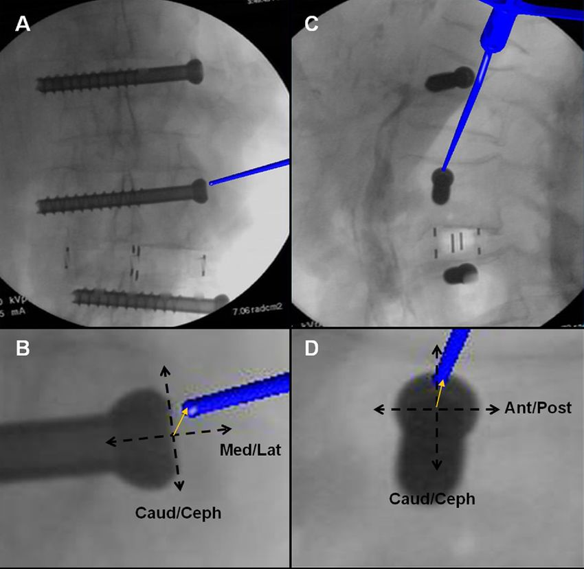
116                                         J.E. Webb et al. / SAS Journal 4 (2010) 115–121

tion of the instruments onto a set of fluoroscopic images             endplate dilator bone screws. Discectomy time began with
obtained at the beginning of the procedure. This enables              initial annulus incision and ended at initiation of cage in-
the surgeon to observe real-time instrument location dur-             sertion. Cage insertion time was recorded from beginning of
ing the procedure.                                                    cage insertion to verification of alignment by fluoroscopy.
   The use of NAV technology can minimize the use of                  Total operating time was defined as initial image library
real-time intraoperative fluoroscopy, thereby decreasing ra-          obtainment to removal of DLIF retractors. The total fluo-
diation exposure to the surgical team.7 By use of fluoro-             roscopy time was determined automatically by the internal
scopic images that are taken at the beginning of the surgery,         timer of the C-arm. Radiation exposure was evaluated by
navigation also minimizes the encroachment of the C-arm               use of radiation-detection badges worn by the surgeon an-
into the surgical field.                                              teriorly and exterior to the lead protective thyroid shield.
   The introduction of any new surgical technique is often            Separate radiation badges were worn for each single-level
accompanied by concerns from surgeons that may limit                  DLIF procedure. Control radiation badges were placed both
wide use. A recent survey study conducted by our group                inside and outside of the operating room.
suggests that concerns from surgeons about NAV include
the increased operative time, cost, lack of necessity, unre-          NAV MIS DLIF technique (NAV)
liable accuracy, and intraoperative problems.9 The purpose
                                                                          All specimens were thawed and positioned on a radiolu-
of this study is to assess the effectiveness and accuracy of
                                                                      cent table in the left lateral decubitus position, with the
NAV when used during the DLIF procedure.
                                                                      greater trochanter located at the table’s bend. Specimens
                                                                      were fixed to the table with tape, and the table was mechan-
Materials and methods                                                 ically bent at the hip to improve access to the lumbar spine
Study design                                                          by moving the superior iliac spine (Fig. 1). The ASIS was
                                                                      palpated and a 0.5-cm incision made for insertion of the
   Unilateral DLIF procedures (Medtronic Spine, Memphis,              navigation (NAV) tracker pin. A standard OEC 9800 C-arm
Tennessee) were performed on 3 cadaveric specimens. Pro-              (GE Healthcare Technologies, Waukesha, Wisconsin) was
cedures were done via either intraoperative NAV or stan-              fitted with a navigation tracker to allow image capture by
dard fluoroscopy (FLUORO) techniques. Just prior to each              the StealthStation Treon navigation computer (Medtronic
level of each cadaver, the type of intraoperative guidance to         Navigation). The C-arm was used to obtain true anteropos-
be used was randomly selected and divided into either the             terior (AP) and lateral images of a specific level while the
NAV group or FLUORO group. The surgeon was notified at                surgeon was distant from the machine. The images were
the time of randomization whether navigation or fluoros-              uploaded to the NAV system, and all surgical instruments
copy was to be used by a research assistant. After each               were registered. Image library acquisition was accom-
procedure, final C-arm images were obtained to confirm                plished for each level in which navigation was used. The
satisfactory implant position and spinal alignment. An NAV            C-arm was then removed from the surgical field for the
system (StealthStation Treon; Medtronic Navigation, Lou-              remainder of the procedure. The disk center was used as the
isville, Colorado) was used for all NAV procedures (as                surgical target to plan the skin incision (Fig. 2). A 2.5-cm
described later). The StealthStation equipment was set up             longitudinal incision was made through the skin at each
and run by trained research personnel and Medtronic rep-              level in which the DLIF procedure was accomplished, with
resentatives, exactly as it would be during an actual patient         subsequent blunt dissection by use of Mayo scissors through
surgery. All DLIF procedures were performed by a single               the lateral abdominal wall. The retroperitoneal space was
surgeon who had approximately 1 year of experience with               entered by finger dissection. The lateral aspect of the trans-
NAV with the DLIF technique. Specific operative steps                 verse process was palpated and the psoas muscle gently
were timed by a separate observer. Each specimen was                  cleared of intervening tissue. A navigated initial dilator was
positioned and draped only once, so recorded setup times do           then inserted through the psoas muscle to the disk center. A
not include these steps. Setup time for both groups started           long guidewire was passed through the cannulated portion
with image library acquisition, including locating the disk           of the navigated dilator and was inserted approximately 2
center, and ended at the time of the lateral skin incision. For       cm into the disk.
NAV, setup time also included insertion of a patient refer-               The approach to the T10-L1 levels was intrathoracic.
ence tracker in the anterior superior iliac spine (ASIS),             Imaging was used in an analogous fashion to identify the
registration of NAV instruments, and manipulation of the              optimum skin incision site. Dissection was carried down
C-arm. The NAV patient reference tracker was inserted                 through the intercostal space and the lung parenchyma
once into each specimen. Three values were obtained for               displaced by finger dissection followed by insertion of
NAV tracker pin insertion, and the mean time was used to              the dilators. Similar to the technique at the caudal levels, the
determine the length of all NAV group setup times.                    dilators were passed along the posterior chest wall (in the
   Exposure and approach times were recorded and were                 lumbar spine, the posterior abdominal wall) and thereafter
defined as beginning with the lateral skin incision and end-          centered on the target disk space. This allowed the dilator to
ing with the insertion of the last of the cephalad and caudal         pass along the diaphragmatic sulcus and displace the dia-

                                  Downloaded from http://ijssurgery.com/ by guest on June 29, 2022
J.E. Webb et al. / SAS Journal 4 (2010) 115–121 117
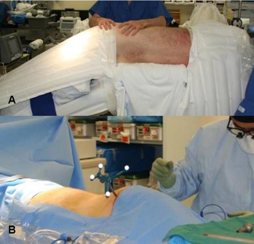
Fig. 1. NAV DLIF setup. Specimens were oriented in the left lateral decubitus position with the surgical table bent to provide great access to the lumbar spine
(A). A navigation tracker pin is placed in the anterior superior iliac spine for optical navigation by the StealthStation Treon (B).
phragm anteriorly. At T12-L1, the crus of the diaphragm tached and tightened to hold the retractor in the desired posi-
covers the disk space. Once the retractor was positioned, the tion. The dilators were removed, and 1 retractor bone screw
retractor blade bone pin was inserted into T12. Electrocau- was inserted into the superior endplate of the inferior vertebra.
tery was used to incise the crus, which was then swept under The retractor was opened by direct vision until sufficient disk
the caudal retractor blade by use of endoscopic peanut visualization was obtained, with the surgeon being alert to the
elevators. The L1 retractor bone pin was then inserted. presence of any nerves in the field. A second retractor bone
The navigated dilator was removed, and a series of tubular screw was inserted into the inferior endplate of the superior
dilators were placed over the guidewire to dilate through the vertebra. Finally, any remaining psoas muscle or tissue was
psoas muscle before introduction of the tubular retractor. The cleared by suction in a gentle sweeping motion.
tubular retractor was inserted over the largest tubular dilator After the placement of the tubular retractor, the lateral
via a twisting motion. A table-mounted holding arm was at- disk annulus was exposed and incised with an annulotomy
Fig. 2. Surgeon’s intraoperative use and perspective of navigation system. The surgeon plans the skin incision with the NAV Pointer (A) by simultaneously
visualizing the disk center in real time with the navigation computer’s virtual instrument overlay over the previously obtained anterior/posterior and lateral
images (B).
Downloaded from http://ijssurgery.com/ by guest on June 29, 2022118 J.E. Webb et al. / SAS Journal 4 (2010) 115–121
knife. A subtotal discectomy was performed. Disk debride- posterior images), and cephalad-caudad (via lateral fluoros-
ment was accomplished by use of a rotating shaver and disk copy images) deviation of the NAV Pointer tip from the
removed with pituitary rongeurs and curettes. Once ade- center of the screw head was measured by Photoshop soft-
quate discectomy had been achieved, the contralateral an- ware (Adobe, Inc., San Jose, California). Deviation distance
nulus was released with a long Cobb elevator. After con- was determined by use of an image standard ratio of the
tralateral annulus release, endplates were decorticated, and known screw shaft width in millimeters to the width of
the disk space was dilated with smooth metal sizers to the screw in the image in pixels. The image deviation
determine the necessary cage height for adequate correction. distance in pixels was then multiplied by this standard ratio
Cage length selection was made ensuring adequate span of to determine the deviation distance in millimeters. All de-
the apophyseal ring by use of the NAV Pointer by adding viation distances at each level, at each stage in the proce-
length to the pointer tip with the software. The surgeon then dure, and in all 3-dimensional planes were then averaged
inserted the intervertebral cage, keeping the inserter instru- and reported as a single average deviation value for a
ment perfectly upright, ensuring proper alignment and ori- specific procedure level.
entation of the cage. Both the discectomy and placement of
the graft were done solely via navigation guidance. At the
Statistical methods
end of the procedure, the C-arm was reintroduced and flu-
oroscopic images were taken to verify graft position. The All statistical comparisons were made by use of the
procedure ended with slow removal of the tubular retractor. Student t test. Significance was set at P ⬍ .05.
A final set of fluoroscopic images was then obtained.
MIS DLIF technique (FLUORO)
Results
For the FLUORO group, setup was similar to the NAV
group with a few exceptions. Setup for this group did not There were 21 unilateral DLIF procedures performed
include placement of a reference tracker. A set of new on 3 cadaveric specimens from T10-11 to L4-5: 11 in the
fluoroscopic images were taken to locate the disk by use of NAV group and 10 in the FLUORO group (Table 1).
the C-arm at the beginning of each level. Surgical approach, Comparisons between NAV and FLUORO for the DLIF
positioning of the retractors, discectomy, and insertion of procedure show statistically significant differences for
the intervertebral cage were analogous to NAV by use of setup, approach, discectomy, and total fluoroscopy times
multiple lateral and under-the-table AP fluoroscopic images (Fig. 4).
for instrument guidance. Therefore the C-arm remained in Our results show that mean approach times for the
the surgical field throughout the procedure.
FLUORO group were significantly longer than approach
NAV accuracy times for NAV (19.61 ⫾ 2.52 minutes vs 15.91 ⫾ 4.08
minutes, P ⫽ .024). Likewise, when comparisons were
A fourth specimen was used to evaluate the accuracy of made between discectomy times for the 2 groups, signifi-
the NAV system. All procedures were conducted by the cantly more time was required for discectomy with
same technique as previously described for NAV DLIF. FLUORO than for NAV (8.43 ⫾ 1.99 minutes vs 5.98 ⫾
Image library acquisition was done for each level, and
1.88 minutes, P ⫽ .009). In contrast, the setup time required
instruments were reregistered at each level as well. Four
for NAV was significantly longer than the setup time for
4.77-mm-diameter cannulated screws were placed in the
FLUORO (5.81 ⫾ 2.65 minutes vs 3.01 ⫾ 0.84 minutes,
L2-5 vertebral bodies. Accuracy data were collected at 4
P ⫽ .005). When mean cage insertion and total operating
stages of the NAV MIS DLIF procedure: after retractor
times were compared between groups, no statistically sig-
deployment, after Cobb release of the contralateral annulus,
after cage sizing, and after cage insertion. The surgeon nificant differences were obtained.
exposed the screw head, and the tip of the NAV Pointer was Fluoroscopy time used for FLUORO, as determined by
placed in the center of the screw head, at which time a the internal timer on the C-arm, was nearly twice as long as
virtual image was taken of the NAV Pointer tip location by with NAV (43.7 ⫾ 16.6 seconds vs 24.0 ⫾ 10.8 seconds per
the navigation computer. This protocol was repeated 3 times level, P ⫽ .004). Radiation exposure for NAV was nearly
at each designated stage of the procedure, with the surgeon undetectable (0.82 ⫾ 0.6 mrem per level), unlike exposures
removing the pointer from the field and repositioning the seen for FLUORO (4.80 ⫾ 3.08 mrem per level) (P ⫽
probe in the same known position. StealthStation images .0005).
were saved each time the pointer was returned to the field Accuracy data were recorded from a separate specimen
for each level, and these images were then used for devia- at 4 stages of each NAV DLIF procedure, and overall mean
tion measurements. values were determined per level from L4-5 to L1-2 (Fig.
The deviation distance of each navigation computer im- 5). Accuracy results were as follows: L4-5, 0.78 ⫾ 0.33
age was then measured (Fig. 3). Anterior-posterior (via mm; L3-4, 0.97 ⫾ 0.12 mm; L2-3, 0.86 ⫾ 0.08 mm; and
lateral fluoroscopy images), medial-lateral (via anterior- L1-2, 1.38 ⫾ 0.44 mm.
Downloaded from http://ijssurgery.com/ by guest on June 29, 2022J.E. Webb et al. / SAS Journal 4 (2010) 115–121 119
Fig. 3. Accuracy determination by use of navigation images. Accuracy measurements were made from intraoperative navigation images (A, C). Deviation
distances in 3 dimensions were determined by measuring the NAV Pointer tip deviation from the center of a screw head previously inserted into each vertebral
body (B, D).
Discussion provided by NAV is the likely factor that makes the ap-
proach, guidewire insertion, and discectomy times shorter
The DLIF technique is a recently developed spinal fusion than FLUORO. In addition, with the C-arm removed from
procedure using MIS techniques. The purpose of this study the surgical field for most of a navigation-assisted proce-
was to evaluate the effectiveness and accuracy of NAV for dure, the surgeon can more easily maneuver about the sur-
the DLIF procedure.
Our results show that use of NAV for the MIS DLIF
procedure is feasible. The data indicate that although initial
setup time is longer for NAV MIS DLIF, the total operating
times were not significantly different between the groups
because of the shorter approach and discectomy times for
the NAV group. The simultaneous AP and lateral imaging
Table 1
Spinal level in NAV and FLUORO groups
Spinal level NAV group FLUORO group
T10-11 3 0
T11-12 1 2
T12-L1 2 1
L1-2 1 2 Fig. 4. Comparison of surgical times. Various surgical step times for both
L2-3 3 0 the FLUORO group and the NAV group were compared. The FLUORO
L3-4 1 2 group had significantly longer approach, discectomy, and C-arm usage
L4-5 0 3 times, whereas the NAV group had a significantly longer setup time (P ⬍
Total 11 10 .05). Cage insertion times and total surgical times were not significantly
different between the 2 groups.
Downloaded from http://ijssurgery.com/ by guest on June 29, 2022120 J.E. Webb et al. / SAS Journal 4 (2010) 115–121
team to ionizing radiation. Likewise, previous studies have
shown that radiation exposure from spinal procedures is
much greater than exposure for surgery of the extremities
because of differences in tissue mass. Spinal images require
significantly more energy to penetrate tissues and thus lead
to greater generation of ionizing radiation.4,10 –12 Thus ef-
forts to limit radiation exposure during spinal procedures
remain paramount. According to the Nuclear Regulatory
Commission, the yearly exposure limits to the whole body,
eyes, and hand are 5,000, 15,000, and 50,000 mrem, respec-
tively. On the basis of our cadaveric data, each level leads
to 4.8 mrem of exposure. This coincides with 1,042 levels
Fig. 5. Accuracy of navigation-assisted DLIF procedure, showing mean per year for maximum whole-body exposure. Taking into
3-dimensional intraoperative NAV Pointer deviation distances for NAV
DLIF procedure. Navigation is accurate within 1 mm for the common
account that a significant portion of the surgeries would
DLIF levels of L2-5. A slight decrease in accuracy is seen as the NAV involve more than a single level, as well as the likelihood
Pointer moves further from the NAV tracker pin in the ASIS. that in the actual clinical setting, more imaging would be
used, the maximum exposure limit could be reached by an
gical field. Navigation-assisted procedures do, however, re- active spine surgeon. With navigation-assisted MIS tech-
quire proper setup of the navigation system, which includes niques, this radiation exposure can be dramatically de-
insertion of the navigation tracker pin and registration of the creased. During NAV DLIF, the C-arm is used at the be-
surgical instruments. These extra steps lead to longer setup ginning of the procedure to obtain an image library and is
times for NAV. Overall, however, this does not significantly not used again until the procedure is complete. During this
change the total operating times compared with FLUORO. time, the surgical team steps away from the C-arm. By
Accuracy data show that NAV MIS DLIF is accurate to eliminating radiation exposure, the surgeon and surgical
less than 1 mm over the most commonly indicated levels for team can avoid the use of cumbersome protective lead gear.
the DLIF procedure (L2-5). Although the error at L1-2 is This study shows that navigation technology is a useful
significant (1.38 ⫾ 0.44 mm), it was relatively similar to the tool for use with the MIS DLIF technique. However, it is
internal system error reported by the StealthStation Treon; important to note that this study only used a single surgeon
that level was 1.3 mm. The accuracy data show that as the who was experienced in MIS procedures and the use of
NAV Pointer progresses away from the ASIS NAV tracker navigation technologies. Therefore time data from this
pin, there is a slight increase in the positional error between study may not represent surgical times for all surgeons with
the navigation virtual image and the radiographic image. a variety of experience with these techniques. Likewise, the
This minor error increase is expected as the navigation surgeon in this study was familiar with interpreting real-
instruments move further from the fixed tracker pin. How- time simultaneous AP and lateral images produced by the
ever, our study shows that the error seen for the most navigation system. Surgeons not familiar with mentally
common DLIF levels of L2-5 is acceptable, at less than 1 interpreting navigation images into the reality of the surgi-
mm. Another interesting issue that arises when measuring cal field may require more time to complete each MIS DLIF
distances on radiographic films is the concept of parallax. In procedure.
our study, to minimize parallax, the surgical target site was Another key limitation of this study is that neurophysi-
centered for each level and a new image library was ob- ologic monitoring cannot be used in the cadaveric speci-
tained at the beginning of each level. Along these lines, the men. In the actual clinical setting, readjustment of the initial
NAV Pointer tip, from which 3-dimensional distances were dilator must be performed, especially at L4-5, despite an
measured, was an electronic overlay made by the Stealth- optimal position on fluoroscopy. The need to make changes
Station computer, thus minimizing image perspective is- to the position of the initial dilator solely because of trig-
sues. gering of the neuromonitoring system would add more time
It is important to note that this study analyzed the accu- to the approach. Finally, our statistical analyses used the
racy of the NAV MIS DLIF technique in a single specimen Student t test to compare 2 groups. Given the number of
at only 4 lumbar levels. New images were obtained after parameters and their interdependence, other statistical meth-
cage insertion at each level to account for changes in disk ods may be used that may alter the statistical significance of
space height due to cage insertion. In addition, as a surgeon the study.
corrects deformities from caudal to cephalad, movement of Although the use of navigation technology for MIS of
the spine due to cage insertion may, theoretically, alter the the spine is promising, surgeon perceptions about the use of
accuracy of the navigation system. spinal navigation have recently been studied and interest-
Minimally invasive spine surgery relies heavily on flu- ingly show that the majority of those queried are concerned
oroscopy for guidance and instrument placement for each about the increased operative time and cost, lack of neces-
procedure. This reliance exposes the surgeon and surgical sity, unreliable accuracy, and intraoperative problems with
Downloaded from http://ijssurgery.com/ by guest on June 29, 2022J.E. Webb et al. / SAS Journal 4 (2010) 115–121 121
navigation technology.9 However, navigation technology 2. Ozgur BM, Aryan HE, Pimenta L, Taylor WR. Extreme lateral inter-
has been widely used in the fields of neurosurgery, ortho- body fusion (XLIF): a novel surgical technique for anterior lumbar
interbody fusion. Spine J 2006;6:435– 43.
paedic surgery, and otolaryngology dating back to the
3. Holly LT, Foley KT. Intraoperative spinal navigation. Spine 2003;
1970s, with a large growth in technology development in 28(Suppl):S54 – 61.
the 1990s.13 Numerous studies have noted that although 4. Rampersaud YR, Foley KT, Shen AC, Williams S, Solomito M.
navigation-assisted technologies require longer setup and Radiation exposure to the spine surgeon during fluoroscopically as-
learning curves, the reproducibility and accuracy of the sisted pedicle screw insertion. Spine 2000;25:2637– 45.
surgical procedure improve with the use of naviga- 5. Assaker R, Reyns N, Vinchon M, Demondion X, Louis E. Transpe-
tion.3,5,14 –16 The intraoperative accuracy of image-guided dicular screw placement: image-guided versus lateral-view fluoros-
copy: in vitro simulation. Spine 2001;26:2160 – 4.
fluoroscopy for pedicle screw placement has been de-
6. Kim CW, Lee YP, Taylor W, Oygar A, Kim WK. Use of navigation-
scribed.17–19 Likewise, a previous study of navigation-as- assisted fluoroscopy to decrease radiation exposure during minimally
sisted MIS transforaminal lumbar interbody fusion (TLIF) invasive spine surgery. Spine J 2008;8:584 –90.
conducted by our group6 and a study conducted by Sasso 7. Mirza SK, Wiggins GC, Kuntz C, et al. Accuracy of thoracic vertebral
and Garrido20 note that navigation-assisted MIS techniques body screw placement using standard fluoroscopy, fluoroscopic image
for spinal fusion do not increase total surgical time. guidance, and computed tomographic image guidance: a cadaver
However, widespread adoption of spinal navigation tech- study. Spine 2003;28:402–13.
8. Vaccaro AR, Yuan PS, Smith HE, Hott J, Sasso R, Papadopoulos S.
nologies will likely require that the technology become
An evaluation of image-guided technologies in the placement of an-
more user-friendly, more reliable, more cost-effective, and terior thoracic vertebral body screws in spinal trauma: a cadaver study.
less prone to intraoperative malfunction. The added costs of J Spinal Cord Med 2005;28:308 –13.
new navigation technology in overall health care delivery 9. Choo AD, Regev G, Garfin SR, Kim CW. Surgeons’ perceptions of
must be further analyzed. Such analyses will likely have a spinal navigation: analysis of key factors affecting the lack of adoption
variable impact based on differences specific to each prac- of spinal navigation technology. SAS J 2008;4:189 –94.
tice setting. DeLucia et al21 have previously described the 10. Theocharopoulos N, Damilakis J, Perisinakis K, Papadokostakis G,
Hadjipavlou A, Gourtsoyiannis N. Occupational gonadal and embryo/
fundamental difficulties with image-guided MIS proce-
fetal doses from fluoroscopically assisted surgical treatments of spinal
dures. Their conclusions emphasized the need for surgeon disorders. Spine 2004;29:2573– 80.
practice and familiarity with navigation systems to improve 11. Theocharopoulos N, Perisinakis K, Damilakis J, Papadokostakis G,
their “mental model of the surgical environment.” To pro- Hadjipavlou A, Gourtsoyiannis N. Occupational exposure from com-
vide more practice to surgeons, navigation-assisted MIS mon fluoroscopic projections used in orthopaedic surgery. J Bone Joint
training must be available to those who are interested. Fur- Surg Am 2003;85:1698 –703.
12. Theocharopoulos N, Damilakis J, Perisinakis K, Papadokostakis G,
ther studies that evaluate the clinical efficacy, success, and
Hadjipavlou A, Gourtsoyiannis N. Fluoroscopically assisted surgical
limitations of NAV for the DLIF technique are currently treatments of spinal disorders: conceptus radiation doses and risks.
being conducted by our group and will further illuminate its Spine 2006;31:239 – 44.
importance as a new, effective minimally invasive tech- 13. Amiot LP, Poulin F. Computed tomography-based navigation for hip,
nique for spinal fusion. knee, and spine surgery. Clin Orthop Relat Res 2004:77– 86.
14. Amiot LP, Lang K, Putzier M, Zippel H, Labelle H. Comparative
results between conventional and computer-assisted pedicle screw
Conclusion installation in the thoracic, lumbar, and sacral spine. Spine 2000;25:
606 –14.
MIS of the spine is a promising area of clinical practice
15. Fu TS, Chen LH, Wong CB, et al. Computer-assisted fluoroscopic
that has the potential to provide improved outcomes for navigation of pedicle screw insertion: an in vitro feasibility study. Acta
patients. However, these techniques pose new challenges. Orthop Scand 2004;75:730 –5.
With decreased visualization of the surgical field, the sur- 16. Smith HE, Vaccaro AR, Yuan PS, Papadopoulos S, Sasso R. The use
geon must rely more on an intraoperative C-arm to visualize of computerized image guidance in lumbar disk arthroplasty. J Spinal
key anatomic landmarks. With increased C-arm use, there is Disord Tech 2006;19:22–7.
increased exposure to ionizing radiation. This study has 17. Foley KT, Smith MM. Image-guided spine surgery. Neurosurg Clin
North Am 1996;7:171– 86.
shown that spinal navigation provides an alternative means
18. Merloz P, Tonetti J, Pillet L. Pedicle screw placement using image-
of visualizing the surgical anatomy while offering the sur- guided techniques. Clin Orthop 1998;354:39 – 48.
geon a more safe and ergonomically friendly environment 19. Quiñones-Hinojosa A, Kolen R, Jun P, Rosenberg WS, Weinstein PR.
in which to operate. The NAV MIS DLIF technique is a Accuracy over space and time of computer-assisted fluoroscopic nav-
promising method of performing minimally invasive lumbar igation in the lumbar spine in vivo. J Spinal Disord Tech 2006;19:
interbody fusions using spinal navigation. 109 –13.
20. Sasso RC, Garrido BJ. Computer-assisted spinal navigation versus
serial radiography and operative time for posterior spinal fusion at
References L5-S1. J Spinal Disord Tech 2007;20:118 –22.
21. DeLucia PR, Mather RD, Griswold JA, Mitra S. Toward the improve-
1. Fessler RG. Minimally invasive spine surgery. Neurosurgery 2002; ment of image-guided interventions for minimally invasive surgery:
51(Suppl):Siii–iv. three factors that affect performance. Hum Factors 2006;48:23–38.
Downloaded from http://ijssurgery.com/ by guest on June 29, 2022You can also read