ON THE ROLE OF PARVALBUMIN INTERNEURONS IN NEURONAL NETWORK ACTIVITY IN THE PREFRONTAL CORTEX - From the Department of Neuroscience Karolinska ...
←
→
Page content transcription
If your browser does not render page correctly, please read the page content below
From the Department of Neuroscience
   Karolinska Institutet, Stockholm, Sweden

ON THE ROLE OF PARVALBUMIN
 INTERNEURONS IN NEURONAL
   NETWORK ACTIVITY IN THE
     PREFRONTAL CORTEX

            Nicolas Gustavo Guyon

                Stockholm 2021
All previously published papers were reproduced with permission from the publisher.
Published by Karolinska Institutet.
Printed by Universitetsservice US-AB, 2021
© Nicolas Gustavo Guyon, 2021
ISBN 978-91-8016-156-5
Cover illustration: “Secret Garden” by Nirupa Rao, 2021
On the role of parvalbumin interneurons in neuronal
network activity in the prefrontal cortex

THESIS FOR DOCTORAL DEGREE (Ph.D.)

By

Nicolas Gustavo Guyon

The thesis will be defended in public at Eva & Georg Klein, Biomedicum, Solnavägen 9, Solna,
Friday the 21st of May 2021 at 14h00

Principal Supervisor:                         Opponent:
Associate Professor Marie Carlén              Assistant Researcher Kathleen K.A. Cho
Karolinska Institutet                         University of California, San Francisco
Department of Neuroscience                    Department of Psychiatry and Behavioral Sciences
Department of Biosciences and Nutrition
                                              Examination Board:
Co-supervisors:                               Associate Professor Paolo Medini
Professor Konstantinos Meletis                Umeå Universitet
Karolinska Institutet                         Department of Integrative Medical Biology
Department of Neuroscience
                                              Principal Investigator Mª Victoria Puig
Professor Karl Deisseroth                     Institut Hospital del Mar d'Investigacions
Stanford University                           Mèdiques
Dept. of Psychiatry and Behavioral Sciences   Dept. of Integrative Pharmacology and Systems
Department of Bioengineering                  Neuroscience
Howard Hughes Medical Institute
                                              Associate Professor Rochellys Diaz Heijtz
                                              Karolinska Institutet
                                              Department of Neuroscience
                                              INSERM – Université de Rouen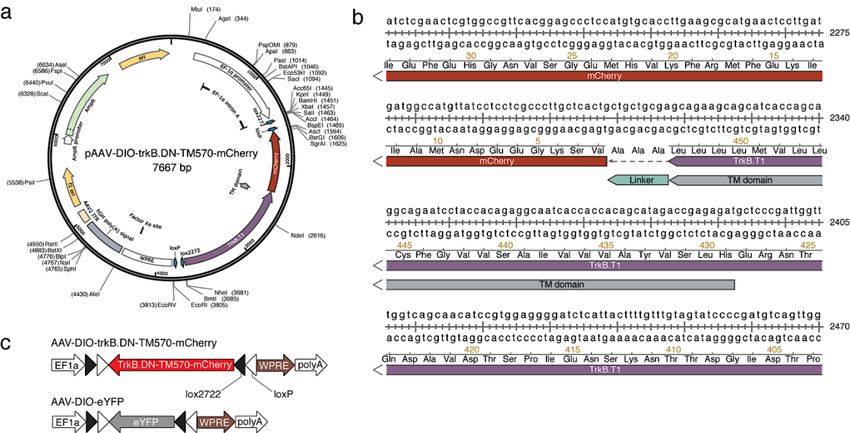
Minha alma é uma orquestra oculta; não sei que instrumentos tange e range,
cordas e harpas, tímbales e tambores, dentro de mim. Só me conheço como sinfonia.
                       – “Livro do Desassossego, por Bernardo Soares,” Fernando Pessoa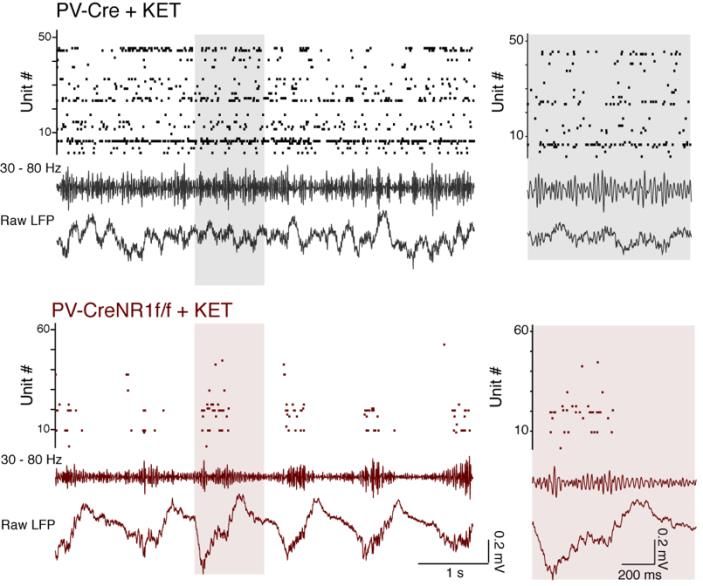
POPULAR SCIENCE SUMMARY OF THE THESIS
The brain is a complex, enigmatic organ composed of a multitude of neurons. These neurons
are interconnected, composing a networked web of connections that in the human brain goes
up to the trillions. The overall activity of these intermingled networks of neurons leads to the
body being able to react to its environment. One of the ways that neurons use their network of
connections to communicate with each other is by releasing chemicals called neurotransmitters.
For example, when a recipient neuron receives the excitatory neurotransmitter glutamate via
receptors placed on its surface, the neuron responds by eliciting an electrical signal called action
potential. We say that the neuron “fired”. If, on the other hand, the recipient neuron receives
the inhibitory neurotransmitter GABA, it will be less likely to fire. These cycles in excitation
and inhibition create voltage fluctuations outside the neurons that we call “brain oscillations”.

These oscillations are heavily studied because they tell us information about what the brain is
doing. For instance, they can help us infer how brain activity is organized temporally, how the
brain responds to sensory input, or how it elicits a movement. Brain oscillations do not look to
be very informative at first sight. However, after being decomposed into smaller components,
based on the frequency of their fluctuation, one can find that different frequencies will have a
bigger or smaller amplitude depending on the behavior of the individual being recorded.
Moreover, these various classes of oscillations look different if a person is awake or asleep,
giving us an insight into how the brain is operating during these specific states. The difference
in amplitude for specific oscillations usually depends on how the neurons are activated or
inhibited and the timing of that cycle — the faster they are activated/inhibited, the higher the
frequency — but also whether they fire in synchrony or more randomly.

Oscillations are rhythmic and reflect the synchronization of the neurons’ activity. This could
be compared to the noise made by people clapping at a concert at the end of a song. It starts by
being uncoordinated, but when the clapping becomes synchronized — because people are
clapping at the same time/frequency — the sound gets louder at that specific beat or frequency.
When recording the brain oscillations with electrodes, we can therefore infer the activity of
neurons by the impact of specific frequencies. For example, some sensory stimuli are known
to specifically increase signals that oscillate at 30-80 cycles per second (30-80 hertz, or Hz).

These oscillations are called gamma oscillations and are strongly correlated to cognitive
processes like attention, working memory and visual processing. Indeed, their amplitude
specifically and narrowly increases when a human or an animal performs a cognitive task.
Significantly, these evoked gamma oscillations are weaker during the performance of similar
tasks in patients with schizophrenia. However, when we observe the brain activity of these
same patients between two tasks or when at rest, a constant, wide-ranging and increased noise
in the gamma range is detected. These aberrant gamma oscillations have been replicated in
animal models of schizophrenia, in which neurons that release the inhibitory neurotransmitter
GABA are dysfunctional. But very little is currently known about how the inhibitory neurons
generate proper narrow gamma oscillation during a task and, at the same time, are paradoxically
associated, when dysfunctional, with increased broad noise in the gamma range during rest.

In Paper I, we tried to solve this issue by testing the suggestion that the broadband increase in
amplitude spanning the entire gamma-band might not always be a rhythm but could result from
asynchronous and noisy communication between neurons. For this, we recorded the brain
activity of transgenic mice that lacked a receptor essential for the proper activity of inhibitory
neurons. Since this receptor had been removed only in the inhibitory neurons, it allowed us to
precisely observe the effect of impairing the activity of these neurons. We found that the
activity recorded during rest was increased and associated with the neurons' asynchronous
activity.

It is as if these specific neurons were not able anymore to hear that other cells around them
were clapping on cue after the song was finished, making it difficult for them to follow the
rhythm of collective synchronization. This led to the inhibitory neurons in our concert room
clapping at random and making noise during the song as well. Importantly, we replicated this
noisy activity by applying ketamine locally to the brain of normal mice, which might explain
how ketamine mimics the symptoms of schizophrenia in humans. Surprisingly, similar
ketamine application in transgenic mice did not cause any such changes. We explain this by
suggesting that ketamine needs the inhibitory neuron receptor removed in the transgenic mice
to have an effect on the brain.

In Paper II, we expressed a modified receptor in the same inhibitory neurons as in Paper I,
this time with the help of a virus injected in a specific area of the brain called the prefrontal
cortex. The virus we generated induces the expression of a modified receptor that is usually
critical for how brain connections are maintained. The modified receptor was injected in adult
transgenic mice and competed with the normal receptor, making it less effective. Thus, this
approach allowed us to target the neurons we wanted to study in the specific brain area
important for social behavior, and avoid interference with other brain areas as well as with
developmental processes. This precision was necessary, as alterations specific to this receptor
in inhibitory neurons of the prefrontal cortex have been found in postmortem examination of
patients with neuropsychiatric disorders such as schizophrenia.

We found that the modified receptor altered the inhibitory neurons in which it was expressed
both morphologically and functionally. When recording the brain activity of transgenic mice
while socially interacting with other mice, we found an increased number of excitatory
neurons and abnormal gamma oscillations within the prefrontal cortex, which was correlated
with unusually aggressive behavior. These results suggest that the modified receptor in
inhibitory neurons reduced their inhibitory connection with excitatory neurons, allowing
them to be activated imprecisely.

In conclusion, abnormal gamma oscillations were observed in both studies. Changes in gamma
are widely reported in both animal models and human studies, but at the same time, the
heterogeneity of gamma-band abnormalities so far recorded has limited the translation of these
findings into clinical settings. A better understanding of how to interpret gamma oscillation
results may thus be a helpful guide in developing approaches where we can use gamma
oscillations to track changes due to disorders but also changes elicited by drugs. Moreover, the
results in this thesis contribute to our understanding of the biological mechanisms behind
neuronal and circuit modifications due to dysfunctional receptors implicated in
neuropsychiatry disorders, especially schizophrenia. This information can be used to develop
targeted diagnoses, as well as interventions aimed at more specifically treating cognitive
impairments seen in neuropsychiatry disorders.
Résumé grand public de la thèse
Le cerveau est un organe complexe composé d'une multitude de neurones. Ces neurones sont
amplement interconnectés, constituant un réseau de connexions qui, dans le cerveau humain,
peut atteindre les billions. L'activité de ces réseaux entremêlés de neurones guide le corps dans
son interaction avec son milieu. L'un des procédés que les neurones utilisent pour communiquer
entre eux consiste à libérer des composés chimiques appelés neurotransmetteurs. Par exemple,
lorsqu'un neurone reçoit du glutamate, un neurotransmetteur excitateur, via des récepteurs
placés à sa surface, cela provoque un signal électrique appelé potentiel d'action. Nous disons
que le neurone a « déchargé ». Si, en revanche, le neurone reçoit le neurotransmetteur inhibiteur
GABA, il sera moins susceptible de produire un potentiel d’action. Ces cycles d'excitation et
d'inhibition génèrent des fluctuations du signal électrique en dehors des neurones appelées «
oscillations cérébrales ».

Ces oscillations sont très étudiées car elles fournissent des informations sur ce que fait le
cerveau. Par exemple, elles peuvent nous aider à déduire comment l'activité cérébrale est
organisée temporellement, mais aussi comment le cerveau répond à des influx sensoriels ou
déclenche un mouvement. Les oscillations cérébrales ne paraissent pas très informatives à
première vue. Cependant, après avoir été découpées en composantes plus petites, en fonction
de la fréquence de leur fluctuation, on peut constater que différentes fréquences auront une
amplitude plus ou moins grande en fonction du comportement du sujet. De plus, ces distinctes
classes d'oscillations semblent différentes si un individu est éveillé ou endormi, ce qui nous
donne un aperçu du fonctionnement du cerveau pendant ces états spécifiques. La différence
d'amplitude pour des oscillations spécifiques dépend généralement de la façon dont les
neurones sont activés ou inhibés et de la durée de ce cycle - plus le cycle activation / inhibition
est rapide, plus la fréquence est élevée - mais aussi s'ils se déchargent de manière synchronisée
ou de manière plus aléatoire.

Les oscillations sont généralement rythmiques et reflètent la synchronisation de l’activité des
neurones. Cela pourrait être comparé au bruit fait par des gens applaudissant lors d'un concert
à la fin d’un morceau de musique. Cela commence par être désorganisé, mais lorsque les
applaudissements s’harmonisent - parce que les gens applaudissent en même temps / à la même
fréquence - le son devient plus intense à cette fréquence spécifique. Lors de l'enregistrement
des oscillations avec des électrodes, on peut donc déduire l'activité des neurones par l'intensité
des différentes fréquences. Par exemple, certains stimuli sensoriels sont connus pour augmenter
spécifiquement les signaux qui oscillent à 30-80 cycles par seconde (30-80 hertz ou Hz).

Ces oscillations sont appelées oscillations gamma et sont fortement corrélées à des processus
cognitifs tels que l'attention, la mémoire de travail et le traitement visuel. En effet, leur
amplitude augmente spécifiquement et étroitement lorsqu'un humain ou un animal effectue une
tâche cognitive. De manière significative, ces oscillations gamma évoquées sont plus faibles
lors de l'exécution de tâches similaires chez les patients atteints de schizophrénie. Cependant,
lorsque nous observons l'activité cérébrale de ces mêmes patients entre deux tâches ou au repos,
on peut détecter une activité bruyante constante et élevée correspondant plus ou moins aux
ondes gamma. Ces oscillations gamma aberrantes ont été répliquées dans des modèles animaux
de schizophrénie, dans lesquels les neurones qui libèrent le neurotransmetteur inhibiteur
GABA sont dysfonctionnels. Malgré cela on sait actuellement très peu de choses sur la façon
dont les neurones inhibiteurs génèrent les ondes cérébrales gamma pendant une tâche et en
même temps sont paradoxalement associés, lorsqu'ils sont dysfonctionnels, à une augmentation
d’un rythme gamma associé à du bruit de fond au repos.

Dans l'étude I, nous avons essayé de résoudre ce problème en testant l’hypothèse selon laquelle
l'augmentation de la fréquence élevée couvrant toute la bande gamma pourrait ne pas toujours
être un rythme mais pourrait être le résultat d'une communication asynchrone et bruyante entre
les neurones. Pour cela, nous avons enregistré l'activité cérébrale de souris transgéniques
dépourvues d'un récepteur important pour l’activité des neurones inhibiteurs. Ce récepteur,
n'ayant été éliminé que dans les neurones inhibiteurs, nous a permis d'observer spécifiquement
l'effet de modifier l'activité de ces neurones. Nous avons constaté que l'activité enregistrée au
repos était augmentée et associée à une activité asynchrone des neurones.

En somme, ce serait comme si ces neurones spécifiques n’étaient plus capables d’entendre que
d’autres cellules autour d’eux applaudissaient après la fin de la musique, ce qui les empêche de
suivre la cadence collective. Cela conduit les neurones inhibiteurs de notre salle de concert à
applaudir au hasard et à faire du bruit pendant que les musiciens jouent. Nous avons notamment
reproduit cette activité bruyante en appliquant de la kétamine localement sur le cerveau de
souris normales, ce qui pourrait expliquer comment la kétamine imite les symptômes de la
schizophrénie chez l'homme. De manière surprenante, une application similaire de kétamine
chez des souris transgéniques n'a pas provoqué de tels changements. Nous expliquons cela en
suggérant que la kétamine a besoin du récepteur neuronal inhibiteur éliminé chez les souris
transgéniques pour avoir un effet sur le cerveau.

Dans l'étude II, nous avons exprimé un récepteur modifié dans les mêmes types de neurones
inhibiteurs que dans l'article I, mais cette fois à l'aide d'un virus qui a été injecté dans une zone
spécifique du cerveau appelée cortex préfrontal. Le virus que nous avons généré induit
l'expression d'un récepteur modifié qui est généralement crucial pour la façon dont les
connexions cérébrales sont maintenues. Le récepteur modifié a été injecté à des souris
transgéniques adultes et est entré en compétition avec le récepteur normal, le rendant moins
efficace. Ainsi, cette approche nous a permis de cibler les neurones que nous voulions étudier
dans une zone spécifique du cerveau connue pour être importante pour le comportement social,
et d'éviter les interférences avec d'autres zones cérébrales ainsi qu'avec les processus de
développement. Il était important d’être précis, car des altérations spécifiques de ce récepteur
dans les neurones inhibiteurs du cortex préfrontal ont été retrouvées lors de l'examen post-
mortem de patients souffrant de troubles neuropsychiatriques tels que la schizophrénie.

Nous avons donc constaté que le récepteur modifié changeait les neurones inhibiteurs dans
lesquels il était exprimé à la fois morphologiquement et fonctionnellement. Lors de
l'enregistrement de l'activité cérébrale des souris transgéniques alors qu'elles interagissent
socialement avec d'autres souris, nous avons trouvé un nombre accru de neurones excitateurs
et des oscillations gamma anormales dans le cortex préfrontal, le tout corrélé à un
comportement inhabituellement agressif. Ces résultats suggèrent que le récepteur modifié dans
les neurones inhibiteurs a réduit leur connexion inhibitrice avec les neurones excitateurs, leur
permettant d'être activés de manière imprécise.
En conclusion, des oscillations gamma anormales ont été observées dans les deux études. Les
altérations des ondes cérébrales gamma sont largement rapportées dans les modèles animaux
et dans les études humaines, mais cependant l'hétérogénéité des anomalies correspondant aux
oscillations gamma observées jusqu'à présent a limité le potentiel translationnel de ces résultats
dans des contextes cliniques. Une meilleure conception des différentes manières d'interpréter
les résultats des oscillations gamma peut donc être utile dans le développement d'approches où
nous pouvons utiliser ces oscillations afin de suivre les changements dus aux troubles mais
aussi les changements induits par la médication. De plus, les résultats de cette thèse ont pour
but de contribuer à une plus grande compréhension des mécanismes biologiques à l'origine des
modifications neuronales et des circuits, dues à des récepteurs dysfonctionnels impliqués dans
les troubles neuropsychiatriques, dont la schizophrénie. Nous espérons que ces résultats
pourront être utilisés dans le développement de diagnostics plus ciblés, ainsi que
d’interventions visant à traiter plus spécifiquement les déficiences cognitives observées dans
les troubles neuropsychiatriques.
ABSTRACT
The prefrontal cortex (PFC) is an area important for executive functions, the initiation and
temporal organization of goal-directed behavior, as well as social behaviors. Inhibitory
interneurons expressing parvalbumin (PV) have a vital role in modulating PFC circuit plasticity
and output, as inhibition by PV interneurons on excitatory pyramidal neurons regulates the
excitability of the network. Thus, dysfunctions of prefrontal PV interneurons are implicated in
the pathophysiology of a range of PFC-dependent neuropsychiatric disorders characterized by
excitation and inhibition (E/I) imbalance and impaired gamma oscillations.

In particular, the hypofunction of receptors important for neurotransmission and regulating
cellular functions, such as the N-methyl-D-aspartate receptors (NMDARs) and the tyrosine
receptor kinase B (trkB), has been implicated in PV dysfunction. Notably, this hypofunction is
known to impair the normal development of PV interneurons. However, it can also affect adult
brain activity. The effects of altered receptors on PV interneurons are multiple, from impaired
morphological connectivity to disruption of intrinsic activity, but have not yet been fully
characterized. Moreover, the effects of deficits of PV neuron-mediated inhibition on neuronal
network activity are complex, involved with compensatory mechanisms, and not fully
understood either. For instance, the E/I imbalance due to PV inhibition has been suggested to
functionally disrupt the cortex, which can be observed through an abnormal increase in
broadband gamma activity. But as the synchronous activity of cortical PV interneurons is
necessary for the generation of cortical gamma oscillations, it is paradoxical that deficient PV
inhibition is associated with increased broadband gamma power.

This thesis aims to examine the role of PV interneurons in shaping neuronal network activity
in the mouse PFC by investigating the microscopic to macroscopic functional effects of
disrupting receptors necessary for the proper activity of PV interneurons.

In paper I, we observed that the increase of broadband gamma power due to NMDAR
hypofunction in PV neurons is associated with asynchronies of network activity, confirming
that dysfunction of neuronal inhibition can cause desynchronization at multiple time scales
(affecting entrainment of spikes by the LFP, as well as cross-frequency coupling and brain
states fragmentation). In Paper II, we prompted and analyzed the rippling effect of PV
dysfunction in the adult PFC by expressing a dominant-negative trkB receptor specifically in
PV interneurons. Despite avoiding interfering with the development of the brain, we found
pronounced morphological and functional alterations in the targeted PV interneurons. These
changes were associated with unusual aggressive behavior coupled with gamma-band
alterations and a decreased modulation of prefrontal excitatory neuronal populations by PV
interneurons.

Thus, the work presented in this thesis furthers our understanding of the role of PV function in
PFC circuitry, particularly of two receptors that are central to the role of PV interneurons in
coordinating local circuit activity. A better understanding of the potential mechanisms that
could explain the neuronal changes seen in individuals with neuropsychiatric dysfunctions
could lead to using gamma oscillations or BDNF-trkB levels as biomarkers in psychiatric
disorders. It also presents possibilities for potential treatments designed around reestablishing
E/I balance by modifying receptor levels in particular cell types.
LIST OF SCIENTIFIC PAPERS

 I. Network asynchrony underlying increased broadband gamma power.
    The Journal of Neuroscience. 2021 Mar 31;41(13):2944-2963.
    Nicolas Guyon, Leonardo Rakauskas Zacharias, Eliezyer Fermino de Oliveira,
    Hoseok Kim, João Pereira Leite, Cleiton Lopes-Aguiar, Marie Carlén

 II. Adult trkB signaling in parvalbumin interneurons is essential to prefrontal
     network dynamics.
     The Journal of Neuroscience. 2021 Apr 7;41(14):3120-3141.
     Nicolas Guyon, Leonardo Rakauskas Zacharias, Josina Anna van Lunteren,
     Jana Immenschuh, Janos Fuzik, Antje Märtin, Yang Xuan, Misha Zilberter,
     Hoseok Kim, Konstantinos Meletis, Cleiton Lopes-Aguiar, Marie Carlén,
CONTENTS
1 INTRODUCTION ......................................................................................................1
1.1 THE PREFRONTAL CORTEX ........................................................................1
1.1.1 General organization of the mPFC .........................................................2
1.1.2 Prefrontal cell-types ...............................................................................3
1.2 PARVALBUMIN INTERNEURONS ..............................................................4
1.3 PREFRONTAL CIRCUIT ACTIVITY .............................................................6
1.3.1 Excitatory/inhibitory balance .................................................................6
1.3.2 Neuronal ensembles ...............................................................................7
1.4 SYNCHRONY IN THE BRAIN .......................................................................8
1.4.1 Oscillatory activity .................................................................................8
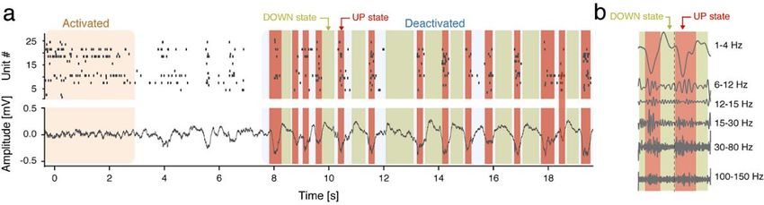
1.4.2 Cortical deactivated and activated states .............................................. 10
1.4.3 UP and DOWN states .......................................................................... 12
1.4.4 Cross frequency coupling..................................................................... 13
1.5 N-METHYL-D-ASPARTATE RECEPTOR................................................... 14
1.6 BDNF-TRKB SIGNALING ............................................................................ 17
1.6.1 trkB receptors....................................................................................... 18
1.6.2 BDNF-trkB signaling in prefrontal PV interneuron activity................. 19
1.7 REGULATION OF SOCIAL PROCESSING BY THE PREFRONTAL
PV INTERNEURONS .................................................................................... 21
2 RESEARCH AIMS................................................................................................... 23
3 MATERIALS AND METHODS .............................................................................. 25
3.1 SPECIFIC TARGETING AND MANIPULATION OF PV NEURONS ........ 25
3.1.1 Cre-lox system and transgenic animals ................................................ 25
3.1.2 NR1 floxed transgenic line................................................................... 25
3.1.3 Viral delivery ....................................................................................... 27
3.1.4 Viral expression of trkB.DN-mCherry ................................................. 27
3.2 MOLECULAR AND CELLULAR READOUTS ........................................... 28
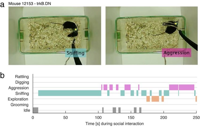
3.3 SOCIAL INTERACTION ............................................................................... 30
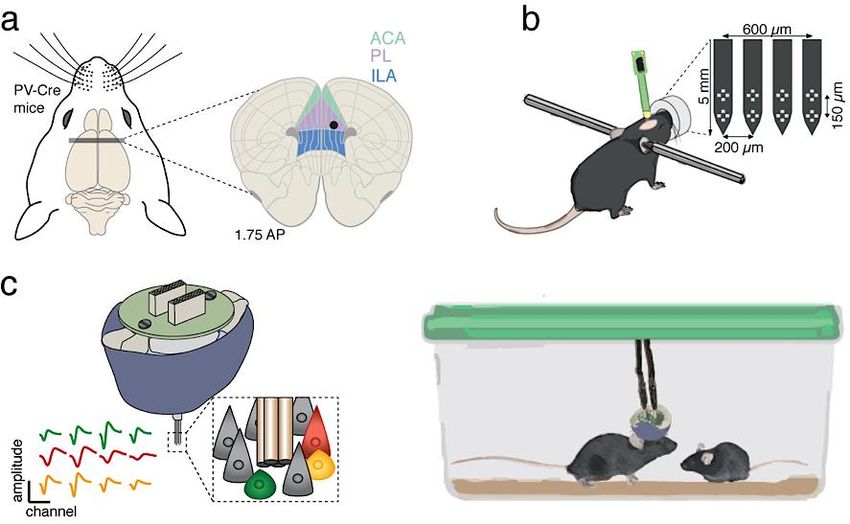
3.4 RECORDING THE ACTIVITY OF THE PFC ............................................... 31
3.4.1 Ex vivo electrophysiology .................................................................... 31
3.4.2 In vivo electrophysiology ..................................................................... 32
3.4.3 Electrophysiology data analysis ........................................................... 33
3.5 ETHICAL CONSIDERATIONS..................................................................... 36
3.5.1 On the need to open sources................................................................. 37
3.5.2 On the need to open access .................................................................. 37
4 RESULTS AND DISCUSSION ............................................................................... 39
4.1 NMDAR ACTIVITY IN PV NEURONS AND ASYNCHRONOUS
mPFC NEURONAL ACTIVITY .................................................................... 39
4.1.1 Altered cortical states ........................................................................... 39
4.1.2 Asynchronous neuronal activity ........................................................... 40
4.1.3 Disorganization of single-unit activity ................................................. 404.1.4 Diverse asynchronies caused by ketamine application or by the
removal of NMDAR from PV neurons ................................................ 41
4.2 BDNF-TRKB SIGNALING IN PV INTERNEURONS IN THE ADULT
mPFC .............................................................................................................. 43
4.2.1 Molecular and morphological alterations............................................. 43
4.2.2 Reduced sensitivity and firing activity of PV interneurons .................. 46
4.2.3 Social behavior dysfunctions ............................................................... 46
4.2.4 Altered prefrontal excitatory dynamics................................................ 47
4.2.5 On potential sexual dimorphic differences........................................... 50
5 CONCLUSION AND PERSPECTIVES .................................................................. 51
5.1 PV DYSFUNCTION AND ASYNCHRONOUS ACTIVITY ........................ 51
5.2 PV DYSFUNCTION AND CORTICAL STATES IMPAIRMENTS............. 53
5.3 PV DYSFUNCTION AND CIRCUIT ALTERATIONS ................................ 54
5.4 MANIPULATION OF RECEPTORS TO STUDY PV FUNCTION ............. 56
5.5 POTENCIAL CLINICAL RELEVANCE....................................................... 56
6 ACKNOWLEDGEMENTS...................................................................................... 59
7 REFERENCES ......................................................................................................... 61LIST OF ABBREVIATIONS
AAV Adeno-associated virus p75NTR p75 neurotrophin receptor
ACA Anterior cingulate area PCA Principal component analysis
AMPA α-amino-3-hydroxy-5-methyl-4- PCP Phenylcyclohexyl piperidine or
isoxazolepropionic acid Phencyclidine
BDNF Brain-derived neurotrophic factor PFC Prefrontal cortex
CFC Cross-frequency coupling PL Prelimbic cortex
DIO Double-floxed inverted open- PSD Power spectral density
reading-frame PV Parvalbumin
EEG Electroencephalograms
REM Rapid eye movement
E/I Excitatory/inhibitory snRNAseq Single nuclei RNA sequencing
EPSC Excitatory postsynaptic current trkB Tyrosine receptor kinase B
eYFP Enhanced yellow fluorescent trkB.DN Dominant-negative trkB
protein
trkB.FL Full-length trkB
FACS Fluorescence-activated cell
trkB.T Truncated trkB
sorting
VIP Vaso-intestinal peptide
GABA Gamma-Aminobutyric acid
VTA Ventral tegmental area
GAD Glutamic acid decarboxylase
GAT-1 GABA transporter-1
HFB High-frequency band
HFOs High-frequency oscillations
ILA Infralimbic area
IPSC Inhibitory postsynaptic current
LFP Local field potential
MK-801 Dizocilpine
mPFC Medial prefrontal cortex
NMDAR N-methyl-D-aspartate receptor
NR1 NMDA receptor subunit 1
NREM Non-rapid eye movement
ORB Orbital area1 INTRODUCTION
1.1 THE PREFRONTAL CORTEX
Located in the forefront of the brain, the prefrontal cortex (PFC) is a distinctive region involved
in various brain functions and processes linked to cognition and goal-oriented actions. The PFC
has been labeled a major evolutionary specialization, as its relative size peaks in primates – up
to 30% of the cortical domain is occupied by the PFC in humans (Carlén, 2017; Smaers et al.,
2017).
The work in this thesis has been performed in mice (Mus musculus) to, among other things,
make use of techniques allowing the spatially and timely restricted manipulation and recording
of cell-type-specific activity. I will therefore focus on the role of the PFC in this model
organism. However, a debate continues about the use of mice for studying the PFC, notably
whether one can translate the concept of prefrontal cortex between species, even though a
growing body of research has revealed functional homologies in rodents and primates (Carlén,
2017; Laubach et al., 2018). For example, hallmark functions of the PFC, like working
memory, attention, and behavioral flexibility, have been conceived based on findings in
primates and successfully replicated in rodents (Kamigaki and Dan, 2017; Kim et al., 2016b;
Liu et al., 2014). It is thus conceptually feasible to use the rodent PFC to shed light on the
functional properties of the primate brain, including the human brain (Carlén, 2017; Le Merre
et al., 2021). Besides, the mouse PFC is involved in sensory processing, the preparation of
motor functions, attention (Kim et al., 2016b), working memory (Kim et al., 2016a), and social
behavior (Felix-Ortiz et al., 2016; Levy et al., 2019; Yizhar and Levy, 2021; Yizhar et al.,
2011), among other cognitive behaviors (Le Merre et al., 2021).
Functionally, the PFC integrates internal and external information regarding the present state
in order to represent future goals and predict future actions. This capacity allows the temporal
organization of behavior in mammals as well as the initiation of goal-directed behaviors
(Fuster, 2015). Specifically, it is thought that sensory information flows from the periphery via
the thalamus and sensory cortical regions right up to the PFC, in a “bottom-up” fashion. In the
PFC, sensory information is then assimilated with information about the state and the goal, as
well as previous experience (Fuster, 2015). From the PFC, the information is then sent back to
other cortical regions, like the motor cortex, and to subcortical regions that are implicated in
the selection and execution of movement. This “top-down” or “executive” signal is thus
thought to be essential for guiding, biasing and modulating activity in downstream regions for
the appropriate action in response to a situation. For instance, pharmacological perturbation of
PFC activity causes disruptions of cortex-wide activity necessary for correctly performing a
task (Allen et al., 2017; Makino et al., 2017).
The pattern of connections to and from the medial prefrontal cortex reflects this functional
capacity to work as a highly integrative network. The primary inputs to the mouse PFC are
originated locally. However, the PFC is also densely interconnected with the rest of the cortex
and with numerous subcortical brain regions, receiving and projecting to a vast number of
regions in a reciprocal manner, making it the area with the highest proportion of feedback
projections (Ährlund-Richter et al., 2019; Harris et al., 2019; Le Merre et al., 2021). Common
connections to and from the PFC arise from the motor and sensory cortical regions but also
1regions involved in arousal, memory, emotional and social responses, like the basal forebrain, thalamus, amygdala, hippocampus dorsal raphe nucleus and locus coeruleus (Ährlund-Richter et al., 2019; Collins et al., 2018; Hoover and Vertes, 2007). Being an essential part of the integrative network underlying cognition, dysfunctions of the prefrontal cortex have been causally implicated in a multitude of neuropsychiatric disorders. Patients with prefrontal damage usually show signs of deficits in decision making, disrupted selective attention for relevant inputs, and increased distractibility by irrelevant stimuli, as well as impaired working memory (Lewis et al., 2005). For instance, epilepsy, autism spectrum disorder, and schizophrenia have been related to malfunctions in the PFC neuronal circuitry, particularly involving the disorganized firing of subsets of neurons, affecting its local and long- range connectivity (Cho et al., 2015; Homayoun and Moghaddam, 2007; Lewis et al., 2005; Schmitt et al., 2017; Yizhar et al., 2011). Research on the several mechanisms that could produce pathological changes in the PFC circuitry is essential to link these PFC dysfunctions to cognitive impairments in neuropsychiatric disorders (Gordon, 2016; Marín, 2012; Tang et al., 2021). 1.1.1 General organization of the mPFC The PFC can be said to be an “umbrella term” for cortical regions located in the forefront of the brain (Le Merre et al., 2021). The PFC has thus been historically divided into several sub- regions - divisions based mainly on anatomical and histological examinations of the brain. Mice possess fewer prefrontal regions than primates, and all regions in the prefrontal cortex of mice lack the layer IV (e.g. the regions are agranular). In rodents, the cortical regions thus identified as shaping the prefrontal cortex are the prelimbic area (PL), the infralimbic area (ILA), the anterior cingulate areas (ACA) and the orbital areas (ORB). Both the papers presented in this thesis use the term medial prefrontal cortex (mPFC) to depict the more medial regions of the mice PFC (ventral ACA, PL, ILA and medial ORB) (Figs. 1a, b). However, several ways of classifying the PFC still prevail today, primarily based on cytoarchitecture or connectivity (Ährlund-Richter et al., 2019), but no clear function has yet been given to each specific area – in humans, as in their homologous regions in rodents (Euston et al., 2012). More research is thus needed in order to define the functions of each sub-region of the PFC (Carlén, 2017). The cellular organization of the PFC is considered to be canonically organized by layers and by columns. Organization conserved not only between species, but also similar to other cortical areas (except for the lack of layer 4). However, although a general organization pattern is observed (Figs. 1b, c), a definite circuit has not yet been defined for the PFC (Douglas and Martin, 2007; Harris and Shepherd, 2015). Importantly, the syntax allowing the translation of this structural organization into function is still not entirely known. It has nevertheless been shown that cortical neurons within prefrontal columns are inter-connected, and receive thalamic inputs, across all layers (Constantinople and Bruno, 2013), while larger excitatory pyramidal neurons of the lower layers generate most of the output from the PFC to the thalamus and other subcortical parts of the brain. Placed among the pyramidal neurons, gamma- Aminobutyric acid (GABA)-ergic inhibitory interneurons are mostly found in layers 2 to 6, locally restricting where they spread both their axonal and dendritic arbors (Tremblay et al., 2016). Although highly interconnected locally, they still receive inputs from other cortical and subcortical regions (Ährlund-Richter et al., 2019). Furthermore, there are distinct recruitment 2
patterns of GABAergic interneurons by local excitatory networks, forming feedforward and
feedback inhibitory loops, as well as disinhibitory paths due to the significant interconnections
between inhibitory interneurons. The mechanistic underpinnings of how these various
inhibitory circuits are formed and how the neuronal circuits process different inputs and shape
reliable output patterns are not fully understood. Studies considering the morphology and
connection patterns of specific cell-types while investigating their function are necessary to
pinpoint their possible implications for proper mPFC function.

Figure 1 - Organization of the mPFC. (a) 3D representation of the organization of the different sub-regions
composing the mouse PFC. (b) Coronal section depicting the mPFC sub-regions from +1.70 to +2.00 antero-
posterior relative to Bregma. Colors represent the same regions as in (a). (c) Schematic illustration representing
the cellular organization and distinct layer profile of the mPFC of the mouse. PN: pyramidal neurons. 3D mouse
brain made with SBA Composer. Pyramidal neuron (doi.org/10.5281/zenodo.3925905) by Federico Claudi, as
well as the interneuron (doi.org/10.5281/zenodo.3925929) were adapted from scidraw.io.

1.1.2 Prefrontal cell-types
At the cellular level, the mouse prefrontal local circuitry consists of 44% of glia cells and
55% of neurons (Erö et al., 2018). The neurons can furthermore be separated into two main
populations depending on if they release either the excitatory (glutamate) or inhibitory
(GABA) neurotransmitters. The presence of a small percentage (around 1%) of
dopaminergic, serotonergic, or cholinergic neuromodulatory neurons is also suggested to be
present in the PFC, but is out of the scope of this thesis (Erö et al., 2018).

The glutamatergic principal excitatory neurons referred throughout this thesis as pyramidal
neurons constitute around 82% of the neurons in the PFC. They are thus the primary
component of the PFC, performing local computation and being the primary communicators
between different cortical areas, as well as with other regions of the brain.

Intertwined among the pyramidal neurons, inhibitory GABAergic interneurons are smaller
in number, representing 14% of the neurons in the PFC, but more diverse regarding
morphology, connectivity and physiology, as well as molecularly (Marín, 2012).
Interneurons contribute mostly to the local network, but some are known to send projections
to subcortical regions (Lee et al., 2014). The use of molecular markers, morphology, intrinsic
firing patterns, but also of the localization of synaptic targeting on pyramidal neurons
compartments, allows the further classification of interneurons into more precise cell-types,
sharing common molecular, morphological and circuitry traits (Fishell and Kepecs, 2020).

                                                                                                                3
As such, recent single-cell transcriptomics data showed that GABAergic interneurons could be divided into six main sub-classes, further separated into 61 types (Tasic et al., 2018). The most common class of GABAergic interneurons in the PFC are the parvalbumin (PV), somatostatin, and vaso-intestinal peptide (VIP) neurons (Fig. 1c) (Tremblay et al., 2016). PV interneurons make synaptic contact onto the soma or the initial segment of the axon of the pyramidal neurons, while somatostatin neurons target the dendrites of pyramidal neurons (Fishell and Kepecs, 2020). VIP expressing neurons send their inhibitory synapses onto other GABAergic interneurons, having a disinhibitory effect on the circuit. Of note, the PFC has a higher density of somatostatin neurons and a lower density of PV interneurons, unlike other cortical regions (Kim et al., 2017). However, the functional ramifications of such a difference are not known. All in all, interneurons provide inhibitory input important for feedback inhibition, information gating and other regulatory aspects of the microcircuit, making them vital to the control of excitability and oscillatory rhythms in the PFC. It is, therefore, necessary to characterize in vivo the activity of these different elementary neuronal components to improve our understanding of the local computations performed by cortical circuits. 1.2 PARVALBUMIN INTERNEURONS Among the several cell-types pertaining to the group of GABAergic inhibitory interneurons, the ones expressing the calcium-binding protein parvalbumin have been considerably studied due to their central role in several sets of PFC-dependent behaviors (Hu et al., 2014; Tremblay et al., 2016). This magnified interest was made possible by their relatively easy identification via their fast-spiking phenotype or the labeling with antibodies of the specific PV marker. Furthermore, the specific targeting of the promoter for the PV gene via the use of genetic and viral methods allows them to be labeled with fluorescent proteins or manipulated with optogenetics methods (e.g. light-manipulation of neurons that have been genetically modified to express light-sensitive receptors or channels) (Hu et al., 2014). The term “PV neuron” is used here to refer to all PV-positive neurons found in the brain, including subcortical areas, as these are not defined as interneurons. Whereas the term “PV interneurons” is used to refer specifically to PV-positive neurons found in the cortex, including the PFC. Therefore, quite a lot has been learned about the function of PV interneurons, notably in the cortex (Bartos et al., 2007; Cardin et al., 2009; Hu et al., 2014; Lewis et al., 2005). PV activity is crucial during development but also during the maintenance of cortical activity in the adult brain. Some attention has been focused on the proper function of PV interneurons in the PFC circuitry, as this has extensive implication in understanding normal cortical computation but also in understanding impaired circuit dynamics underlying neuropsychiatric disorders (Kim et al., 2016b; Lewis et al., 2005; Pafundo et al., 2018; Sohal et al., 2009). PV interneurons are categorized by their fast firing rates and their narrow-spiking shape. Their complex dendritic and axonal arborization allows them to integrate multiple layers inputs and, at the same time, modulate the activity of several pyramidal neurons. PV interneurons are densely interconnected through gap junctions and exert potent inhibition onto pyramidal neurons. These two features are assumed to help the PV interneurons to generate an innate firing range in the gamma range (30–80 Hz; Buzsáki and Draguhn 2004). They are therefore 4
known as potent regulators of local network activities (Hu et al., 2014), and synchronous
activation of PV interneurons is sufficient for the generation of gamma oscillations (Cardin et
al., 2009; Sohal et al., 2009). Furthermore, PV interneurons have been shown to mediate the
excitation–inhibition balance and regulate the timing of pyramidal neurons (Ferguson and Gao,
2018a; Hu et al., 2014; Moore et al., 2010; Yizhar et al., 2011).
More specifically, PV interneurons are characterized at the morphological level by their notable
axonal targeting near the soma of adjacent pyramidal neurons. This particularity allows them
to tightly control the output of pyramidal neurons, as they innervate them at the location where
action potentials are initiated (Hu et al., 2014). Furthermore, two sub-classes of PV
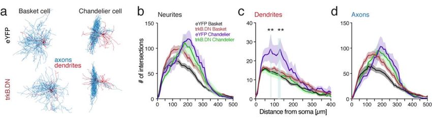
interneurons can be separated based on the axonal innervation area. The basket neurons have
their main direct inhibitory output onto the cell-body of the pyramidal neurons, their axons
forming a basket-like structure around the soma and proximal dendrites. In comparison,
chandelier neurons target the initial segment of the pyramidal neuron’s axon (Karube et al.,
2004).
Furthermore, PV interneuron axonal connections have been shown to be dense and nonspecific
(Karube et al., 2004; Packer and Yuste, 2011), allowing the PV interneuron to control local
circuits activity. For instance, PV’s axons show widespread arborization and have a high
number of boutons placed all along their extension, allowing them to make connections with a
large number of neurons. Thus, by balancing excitation within the area covered by their axons,
they form what is called a “blanket of inhibition” stretched over local pyramidal neurons
(Karnani et al., 2014). However, it has been demonstrated that PV interneurons adapt their
morphology and their synapses, depending on the local circuit activity (Dehorter et al., 2015;
Ferguson and Gao, 2018a), suggesting that despite making broad connections with a multitude
of neurons, they might be more specifically controlling the function of certain neuron types or
ensembles (Agetsuma et al., 2018; Fishell and Kepecs, 2020; Kim et al., 2016b; Kvitsiani et
al., 2013).
On the other end, cortical PV interneurons complex and long dendritic arborization allow them
to sample their inputs from local neurons. They receive numerous excitatory inputs from a large
population of local pyramidal neurons, as well as inhibitory inputs from other interneurons.
Moreover, helped by the fact that their dendritic arbor span across several cortical layers, PV
interneurons also receive inputs from diverse feedback and feedforward pathways originating
from numerous cortical and subcortical regions (Ährlund-Richter et al., 2019; Tremblay et al.,
2016).
Prefrontal PV activity is correlated with several cognitive behaviors, as PV firing activity can
be positively or negatively modulated during specific phases of a behavioral task (Lagler et al.,
2016), including during attention (Kim et al., 2016b), foraging (Kvitsiani et al., 2013) or social
behavior (Selimbeyoglu et al., 2017; Yizhar et al., 2011). The modulation of the activity of PV
interneurons is associated with the inhibition of certain pyramidal neurons and the increased
activity of other local pyramidal neurons (Kim et al., 2016b), suggesting that this specific
modulation of groups of neurons is essential for the proper delineation of neuronal
ensembles relevant for optimal performance during behavior. PV interneurons are thus
proposed to participate in neuronal ensembles formation by controlling the ensemble size
through inhibition of less efficiently recruited neurons (Holtmaat and Caroni, 2016).
51.3 PREFRONTAL CIRCUIT ACTIVITY 1.3.1 Excitatory/inhibitory balance Homeostasis is a process that allows a system or living organism to adjust its internal environment to resist and adapt to external forces of change through feedback control, acting comparably in the same way as thermostats or autopilots. There is a growing body of literature that recognizes the importance of homeostasis in local circuits of the brain via the balanced interaction between excitatory and inhibitory neurons to generate proper local operations and long-range neuronal communication (Hoftman et al., 2017; Pozo and Goda, 2010; Rich and Wenner, 2007; Turrigiano, 2011). The role of this close pairing between excitation and inhibition is not entirely clear, but it is thought to be necessary for how fast and accurate neurons can respond, as it could work as a fine-tuning mechanism at the network level (Okun and Lampl, 2008). Specifically, the excitatory output of neurons can be modulated by excitatory and inhibitory feedback from adjacent neurons, offering a more controlled local network response (Hennequin et al., 2017; Turrigiano and Nelson, 2004). Therefore, the E/I balance seems essential for how brain networks respond to stimuli by regulating the excitatory inputs, and how it communicates by controlling the neuronal output (Froemke, 2015; Turrigiano, 2011). Indeed, balanced inhibition is known to be important in shaping the tuning of neurons to specific sensory cues (Atallah et al., 2012; Tao et al., 2014). It is also central to information transmission by allowing activity to propagate through the network without losing or enhancing too much of the activity in the system. In the same fashion, E/I balance is known to be essential for brain plasticity and the capacity of the brain to change at the cellular and network level. For example, there is an over-excitation of the brain during its early development. But subsequently, the maturation of neurons that release inhibitory neurotransmitters leads to an increased inhibition that balances the E/I level. This stabilization of the neuronal networks continues and is shaped by environmental experiences during the critical period (Reh et al., 2020; Takesian and Hensch, 2013). Concomitantly, brain states, like sleep or wake, are known to gate the homeostatic processes that help in stabilizing neuronal circuits. This stability is maintained by controlling firing rates within a normal set-point range, but only during some brain states, for example, during sleep (Hengen et al., 2016; Tononi and Cirelli, 2014). The mechanisms underlying the maintenance of an accurate E/I balance are diverse and intricate. Previous research suggests that homeostatic regulation of neuronal firing could be achieved by two different mechanisms – either by synaptic changes that adjust the balance between excitatory and inhibitory inputs, or by intrinsic modification of the balance of inward and outward voltage-dependent currents (Turrigiano, 2011). The change in synaptic strength, also called synaptic scaling, is thus believed to be accompanied by changes in the accumulation of receptors like NMDA, AMPA, or trkB, at synaptic sites (Rich and Wenner, 2007; Turrigiano and Nelson, 2004), or by adjustment in neurotransmitters or neurotrophins content of synaptic vesicles (Pozo and Goda, 2010). Consequently, abnormalities in the neuron structure, dendritic arborization, deficits at the synapses including changes in the formation and placement of receptors, altered neurotransmitters synthesis and transport, or alterations in long-range communication between 6
brain structures, are known to be involved in the E/I imbalances that can result in various
malfunctions at the cellular and network level. This imbalance has been observed in multiple
psychiatric and neurological conditions such as autism, schizophrenia, and epilepsy, among
others (Hoftman et al., 2017; Lee et al., 2017a). For example, when inhibition is blocked
pharmacologically, improper inhibition leads to generalized neuronal firing, aberrant neuronal
oscillatory activity, cognitive deficits and several psychiatric comorbidities found in epilepsy
(Marín, 2012; Valero et al., 2017).

1.3.2 Neuronal ensembles
While there is no commonly accepted proper definition of neuronal ensemble, it can be
defined as a stable group of co-active neurons dynamically involved in particular neuronal
computations (Carrillo-Reid and Yuste, 2020a). Neurons belonging to a neuronal ensemble
fire together in a time window that allows the consolidation of the connections between them.
A sensory stimulus would be therefore denoted by the overall change of activity of a
population of neurons, instead of individual neurons, and different sensory stimuli could be
represented in the activation of different neuronal ensembles. Manipulation of neuronal
ensembles with stimulation of selected patterns of neurons with holographic optogenetics has
been shown to be sufficient to control behavior in a Go/No-Go task (Carrillo-Reid and Yuste,
2020b).
As neuronal ensembles are constituted of recurrent connections between excitatory neurons
and inhibitory interneurons, the activity of inhibitory interneurons can strengthen the
connectivity of most engaged neurons, while weakening less engaged neurons, leading to a
spatial definition of the ensemble. They can also help define the temporal aspect of the
activation of neuronal ensemble, or orchestrate the transitions between neuronal ensembles,
by firing at a different phase of oscillations or brain states (Buzsáki, 2010). In other words,
interneurons could orchestrate which and when neuronal ensembles play (Agetsuma et al.,
2018), and thus modifying interneurons function can alter neuronal ensembles (Agetsuma et
al., 2018; Hamm et al., 2017). Therefore, proper functioning of interneurons and balanced
excitatory/inhibitory activity might be critical for the temporal and spatial manutention of
neuronal ensemble dynamics (Agetsuma et al., 2018).
As referred previously, prefrontal cortical activity seems to follow the cellular organization of
the mPFC, going through layers and within columns. However, how prefrontal neurons are
recruited and maintained among different ensembles during behavior remains unclear. Several
lines of evidence suggest that active maintenance of a specific neuronal representation, or
ensemble, in the mPFC is necessary for the performance of behaviors. Previous works have
thus recorded sustained increase or decrease of the firing rate of a population of neurons during
a variety of behavioral tasks, notably during attentional processing (Fujisawa et al., 2008; Kim
et al., 2016b) or working memory (Kamigaki and Dan, 2017; Liu et al., 2014). Furthermore,
manipulating specifically the behaviorally responsive neuronal population, notably by
stimulating neighboring PV interneurons, is enough to disrupt the behavioral outcome (Kim et
al., 2016b). Of note, although the activity of distinct neuronal ensembles can be collectively
reflected in neuronal oscillations, these are temporally defined and lack spatial resolution, thus
representing a powerful but indirect way to measure neuronal ensemble activity (Buzsáki,
2010).

                                                                                                7
You can also read