Onchocerca lupi in imported dogs in the UK: implications for animal and public health - Research Square
←
→
Page content transcription
If your browser does not render page correctly, please read the page content below
Onchocerca lupi in imported dogs in the UK:
implications for animal and public health
John McGarry
University of Liverpool
Rossella Carrozza
Eye Veterinary Clinic Ltd
Claire Bradley
South Devon Referrals
Maria Latrofa
University of Bari
Benjamin Makepeace ( blm1@liverpool.ac.uk )
University of Liverpool
Domenico Otranto
University of Bari
Research Article
Keywords: Onchocerca lupi, zoonosis, risk from importation, public health, humans, dogs, One Health
Posted Date: May 4th, 2021
DOI: https://doi.org/10.21203/rs.3.rs-493136/v1
License: This work is licensed under a Creative Commons Attribution 4.0 International License.
Read Full License
Page 1/7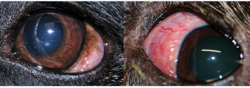
Abstract
The zoonotic nematode Onchocerca lupi is reported in two dogs in the UK, re-homed from Europe. One
dog developed an ocular nodule seven years after arrival from Spain, whereas the other dog, originally
from Romania, developed recurring nodular lesions in both eyes. In this dog, parasitism was particularly
invasive and resulted in unilateral enucleation. Increasingly, former stray dogs of unknown infection
status are entering the UK, raising veterinary and medical concerns.

Introduction
Environmental changes, anthropic behaviour, and animal movements in Europe over the past 20 years
have led to increased threats from a range of zoonotic viral, bacterial and protozoal vector-borne diseases
[1]. During the same timeframe, there has been an emergence of conditions caused by various vector-
transmitted nematodes, for which dogs and other carnivores act as reservoirs of zoonotic infection [2–3].
The movement of domestic dogs plays an important role in the epidemiology of these vector-borne
diseases; the latter including, for example, mosquito-transmitted Dirofilaria spp. [3]. The UK Animal and
Plant Health Agency recorded approximately 45,000 imported dogs in 2019, and this figure includes an
unknown number of former stray animals from European countries, whose numbers have increased year-
on-year for the past seven years. With a history of scavenging and exposure to biting disease vectors,
these so-called ‘Trojan dogs’ can be harbingers of unfamiliar, pathogenic parasites including several
types of vector-borne nematodes [3], for which treatment is not legally required before crossing borders.

An example of one such vector-borne nematode is the emerging canine eye worm Thelazia callipaeda,
which is now endemic throughout much of Europe and associated with corresponding cases in humans
[4]. The first reports of canine thelaziosis in the UK were recently diagnosed in dogs re-homed from Italy,
France and Romania [5], and coincidentally, that same year a case of human disease in an international
traveller was registered [6].

Another, more pathogenic canine eye worm has spread rapidly in several European countries - the filaria
Onchocerca lupi [7]. This is also zoonotic, with the first human case described only 10 years ago [8].
Subsequently, up to 18 patients have now been diagnosed [7] in countries where canine onchocerciasis
has become endemic, including the Southwest USA, where it is thought to have established following
transportation of an infected dog from Europe [9]. Adult O. lupi usually localize in the episcleral tissues of
infected dogs, and occasionally cats, whilst their microfilariae are located in the skin, particularly of the
head. However, animals with patent infections do not always display overt clinical signs. Indeed, in
endemic regions of Portugal, ocular nodules rarely form in dogs with mature, microfilariae-positive
infections [10]. Here, we describe the first two cases of imported canine onchocerciasis in the UK, which
presented with unusual pathogeneses. These raise concerns for animal and public health given the
potential for this parasite to establish in the UK through the increasingly popular practice of re-homing
dogs from other European countries.

                                                   Page 2/7
Case Reports
An eight-year-old male crossbreed was investigated in January 2021 for a conjunctival perilimbal mass in
the left eye, of two months’ duration (Fig. 1). The dog came from the Algarve region of Portugal aged one
year and has remained in the UK ever since. Closely associated with the ventral oblique muscle, the mass
was excised in its entirety and revealed multiple small white cysts containing parasite fragments (Fig. 2).
Histopathology of the cysts demonstrated parasitic granulomas associated with degenerate and partially
mineralised intralesional worms. Anthelmintic treatment was oral doxycycline for three months (targeting
the Wolbachia symbionts in adult worms) and monthly topical imidacloprid and moxidectin (Advocate®)
alongside monthly ivermectin. There were no further complications in this case.
The other dog was a seven-year-old female small crossbreed having entered in the UK as a former stray
from Romania in January 2020. Seven months after arrival, and with no history of other travel, a raised
tan, perilimbal mass (Fig. 1) was noted in the left eye. Due to progressive exophthalmos, the mass was
surgically explored, revealing long, thin nematode fragments, and as in the case above, histology also
revealed parasitic granulomas. This dog was already receiving monthly topical imadacloprid and
moxidectin (Advocate®) since arriving in the UK, and oral doxycycline and prednisolone therapy was
initiated. Three weeks following surgery, additional tan lesions appeared in the same eye and further
exploration revealed that the parasites had reached as far as the optic nerve and were strongly
associated with the extraocular muscles; given the extent of disease, the globe was enucleated. One
month later, the right eye developed conjunctival hyperaemia. Two injections of melarsomine
dihydrochloride were administered 24 hrs apart, alongside continued oral and topical prednisolone. Over
the following three weeks, however, tan nodules appeared which on surgical removal revealed numerous
live worms. As of April 2021, further lesions still remain.
Nematodes from both cases were morphologically identified as Onchocerca lupi based on the typical
appearance of their cuticle (Fig. 2), which has an arrangement of prominent ridges and internal striae
[11]. Identification was also confirmed by sequence analysis of cytochrome c oxidase subunit 1 (coxI)
and 12S rRNA gene fragments (10). BLAST analysis (http://blast.ncbi.nlm.nih.gov/ Blast.cgi) of
sequences showed a high nucleotide identity with those of O. lupi available in GenBank (99.85–100%,
coxI; 100%, 12S rRNA). Accession numbers deposited in GenBank are MW835250, MW835251,
MW829782 and MW829783.
Discussion
Onchocerca spp. have unusually long pre-patent periods of up to 18 months [12]. Although
autochthonous transmission of canine onchocerciasis in the UK cannot be excluded, it is highly likely that
the first dog was infected as a one-year-old in the Algarve region of Portugal, a known focus of
transmission [10]. The pre-patent period noted here of seven years is extremely long but is comparable to
a recent observation in Germany in a dog introduced from Greece, which developed eye problems six
years following rescue [13]. Such cases of prolonged parasite and nodule development highlight
Page 3/7challenges in case diagnosis and management of O. lupi, as well as in monitoring zoonotic disease
incursion.
The second case presents an unusually severe pathogenicity, with exophthalmos in one eye and disease
progression despite appropriate therapy. To the best of our knowledge, the appearance of new worms
following surgical removal of an existing nodule is a novel finding, and the invasive nature of nodule
growth necessitating globe removal is an exceptional outcome. However, subsequent nodule
development in the previously healthy contralateral eye, a matter of weeks after enucleation of the
infected eye, has been described previously [9] and suggests that nematodes may survive undetected
until they form overt nodular lesions.
In both cases, lesions became prominent, with timely veterinary interventions. However, as already
mentioned, not all dogs display overt clinical signs, especially when worms do not develop in the external
parts of the ocular apparatus [7]. In undetected covert infections of mature worms, microfilariae will
accumulate in the skin for a long time, allowing for potential parasite transmission.
The identity of vectors of this parasite remains unclear, but as for most Onchocerca spp., one or more
species of Simulium (blackflies) may have a role. In the UK, there are least six species of blackflies
recorded as biting both humans and dogs [14], of which S. reptans (west of England and Wales) and S.
tuberosum/S. variegatum (north of England and Scotland) are abundant [15]. Considering that the black
fly species composition is similar to that of countries in Europe where O. lupi is endemic and assuming
they could act as vectors, we hypothesise that local transmission in the UK could occur. An assessment
of the suitability of conditions in the UK for parasite circulation by modelling of climate, ecological and
other factors is therefore required.
In conclusion, it is apparent that Onchocerca lupi infection may only become evident in dogs many years
following importation. Nodules may be invasive and appear unpredictably with asynchronous
development. The popular trend to re-home dogs from O. lupi-endemic regions of Europe will increase the
risk of transmission of this parasite in the UK and presents a growing problem of One Health concern.
References
1. Medlock JM, Leach SA (2015) Effect of climate change on vector-borne disease risk in the UK.
Lancet Infect Dis 15: 721–30
2. Otranto D, Dantas-Torres F, Mihalca AD, Traub RJ, Lappin M, Baneth G (2017) Zoonotic Parasites of
sheltered and stray dogs in the era of the global economic and political crisis. Trends Parasitol
33(10):813–825. doi: 10.1016/j.pt.2017.05.013.
3. Wright I, Collins M, McGarry J, Teodoru S, Constantin SA, Corfield EL. et al (2020) Threat of exotic
worms in dogs imported from Romania. Vet Rec 187(9):348–349. doi: 10.1136/vr.m4207
4. Otranto D, Mendoza-Roldan JA, Dantas-Torres F (2021) Thelazia callipaeda. Trends Parasitol
37(3):263–264. doi: 10.1016/j.pt.2020.04.013
Page 4/75. Graham-Brown J, Gilmore P, Colella V, Moss L, Dixon C, Andrews M, et al (2017) Three cases of
imported eye worm infection in dogs: a new threat for the United Kingdom. The Vet Rec 181(13):
346–35. doi:10.1136/vr.104378
6. McGarry JW, Graham-Brown J, Pasztor M (2017) Threats of vector-borne zoonotic disease in Europe:
dogs, drosophilids, and oriental eye worm. Lancet Infect Dis 17(11): 1115–1117. doi:10.1016/S1473-
3099(17)30573-X
7. Rojas A, Morales-Calvo F, Salant H, Otranto D, Baneth G (2021) Zoonotic ocular onchocercosis by
Onchocerca lupi. Yale J Biol Med in press.
8. Otranto D, Sakru N, Testini G, Gürlü VP, Yakar K, Lia RP, Dantas-Torres F, Bain O (2011) Case report:
First evidence of human zoonotic infection by Onchocerca lupi (Spirurida, Onchocercidae). Am J
Trop Med Hyg 84(1):55–8. doi: 10.4269/ajtmh.2011.10-0465
9. Verocai GG, Conboy G, Lejeune M, Marron, F, Hanna P, MacDonald E, et al (2016) Onchocerca lupi
Nematodes in dogs exported from the United States into Canada. Emerg Infect. Dis 22 (8): 1477–
1479
10. Otranto, D, Dantas-Torres, F, Giannelli, A, Latrofa, MS, Papadopoulos, E, Cardoso, L. et al (2013)
Zoonotic Onchocerca lupi Infection in Dogs, Greece and Portugal, 2011–2012. Emerg Infect
Dis.19(12): 2000–2003.doi: 10.3201/eid1912.130264
11. Otranto D, Dantas-Torres, F, Cebeci, Z, Yeniad, B, Buyukbabani, N, Buyukbaba Bora O (2012) Human
ocular filariasis: further evidence on the zoonotic role of Onchocerca lupi. Parasites Vector 5:84
http://www.parasitesandvectors.com/5/1/84
12. Colella V, Lia RP, Di Paola G, Cortes H, Cardoso L, Otranto D. International dog travelling and risk for
zoonotic Onchocerca lupi (2018) Transbound Emerg Dis 65(4):1107–1109. doi: 10.1111/tbed.12842
13. Hodžić A, Hinney B, König S, Naucke TJ, Duscher G, Joachim A (2018) A case of ocular infection with
Onchocerca lupi in a dog from Germany. Transbound Emerg Dis. 65(1): 214–216. doi:
10.1111/tbed.12715
14. Crosskey RW (2005) A perspective on anthropophily in British blackflies (Diptera, Simuliidae) with
keys to the identification of the culprit species. Dipterist’s Digest 12 (1): 29–58
15. Edwards FW, Oldroyd H, Smart J (1939) British Blood-Sucking Flies. British Museum (Natural
History), London.
Declaration
Ethics approval and consent to participate
Owners of the dogs in this study have provided permissions for details of their pets to be published
through their veterinarians who are co-authors of this article.
Consent for publication. N/A. No human subjects.
Availability of data and material
Page 5/7Sequence data: Accession numbers deposited in GenBank are MW835250, MW835251, MW829782 and
MW829783.
Competing interests - None
Funding - None
Authors' contributions: John W. McGarry (identified the parasites, drafted and coordinated publication);
Rossella Carrozza (veterinarian investigating Case 1, clinical work); Claire Bradley (veterinarian
investigating case 2, clinical work); Maria Stefania, Latrofa (molecular identification of worms); Benjamin
L. Makepeace (worm morphology, specialist advice); Domenico Otranto (molecular identification of
worms, specialist advice). All authors contributed to the text and critical review of the MS.
Acknowledgements – None.
Figures
Figure 1
Lesions at presentation. Left, dog originally from Portugal and right, dog imported from Romania
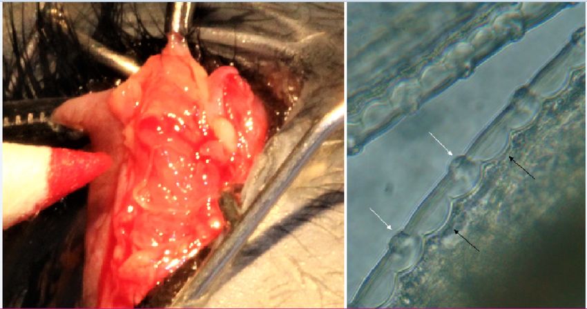
Page 6/7Figure 2
Nodule removed from dog originally from Roamania showing cysts and worms; right, characteristic
appearance of the cuticle of Onchocerca lupi, with arrows showing the typical arrangement of ridges and
internal striae
Page 7/7You can also read