OncoREXTM p53 Protein Array Standard Autoantibody Assay - Sengenics
←
→
Page content transcription
If your browser does not render page correctly, please read the page content below
OncoREX TM p53 Protein Array
Standard Autoantibody Assay
Wet Lab Protocol
February 2021
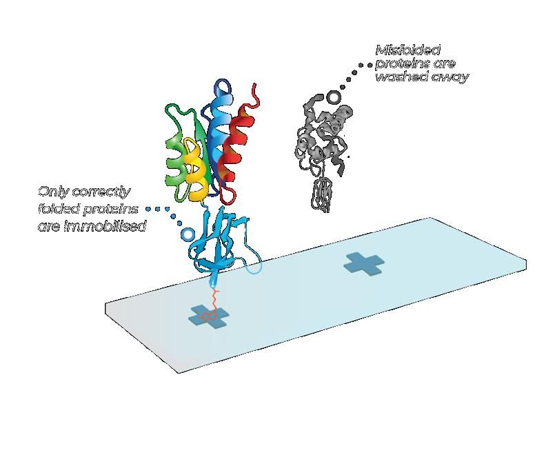
Misfolded
proteins are
washed away
Only correctly
folded proteins
are immobilised
World’s only technology
that consistently produces full-length, correctly folded, functional proteins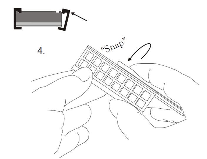
Document       SGN-OP-
                                       Standard Autoantibody Assay on                      No.            MSL-014
                                           p53 Protein Microarray                          Revision No.   1
                                                                                           Effective
                                              Wet Lab Protocol                             Date

 Contents

1.0 Purpose                                                                                               1

2.0 Scope                                                                                                 1

3.0 Reference Documents                                                                                   1

4.0 Relevant Personnel                                                                                    1

5.0 Definition                                                                                            1

6.0 Procedure                                                                                             2
  6.1 Labelling of Anti-Human IgG using PD-10 Columns                                                     2
  6.2 Preparation of Working Buffers                                                                      7
  6.3 Autoantibody Assay                                                                                  9

                                             © Sengenics 2008-2021
                 This is a controlled document. Printed copies are not valid without authorization.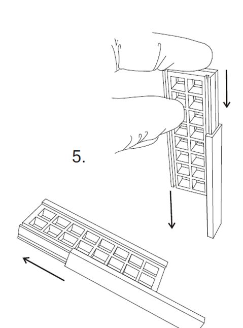
Document       SGN-OP-
                                      Standard Autoantibody Assay on                 No.            MSL-014
                                     OncoREXTM p53 Protein Microarray                Revision No.   1
                                                                                     Effective
                                             Wet Lab Protocol                        Date

1.0     Purpose
        This procedure defines the process to perform antibody assay on the OncoREX p53 Protein
        Array.

2.0     Scope
         This procedure applies to all inactivated or pre-pandemic serum and plasma samples that
         are to be assayed on the OncoREX p53 Protein Array.

3.0     Reference Documents (Other SOPs)
         3.1      Risk Assessments
                  a. SGN-OP-04_RA4_Autoantibody/Antibody Assay
                  b. SGN-OP-05_RA5_Decontamination

4.0     Relevant Personnel
          Microarray Service Lab personnel carrying out Autoantibody Assay on the OncoREX p53
          Protein Array.

5.0     Definition
         5.1    Inactivated samples refer to serum and plasma samples that are pre-treated with 1 %
                Triton X-100 for 2 hours at room temperature.
        5.2     RT: Room temperature

                                            © Sengenics 2008-2021
This is a controlled document. Printed copies are not valid without authorization.                      Page 1
Document       SGN-OP-
                                      Standard Autoantibody Assay on                      No.            MSL-014
                                     OncoREXTM p53 Protein Microarray                     Revision No.   1
                                                                                          Effective
                                             Wet Lab Protocol                             Date

6.0     Procedure
6.1       Labelling of Human IgG using PD-10 columns.
6.1.1      Required reagents, consumables and equipment

Table 1: List of reagents, consumables and equipment required for antibody labelling
                                                    REAGENTS
 Materials                                           Manufacturer          Catalogue number              Storage

 Polyclonal Rabbit Anti-Human IgG                Dako                           A042301                      4 °C

 Monoreactive Cy3 dye                            GE Healthcare               GEH-PA23001                     4 °C

 10x Phosphate Buffer Saline (PBS)               BioSynTech                    PB0344-1L                     RT

 Glycine                                         Sigma Aldrich                   G8898                       RT

 18.2MΩcm Mili-Q Water                           MiliPore                            -                       RT

                                                CONSUMABLES
 PD-10 columns                                   GE Healthcare                 17-0851-01                    RT

 0.5 mL non-translucent vials                    Fisher Scientific                   NA                      RT

 0.5 mL non-translucent screw cap                Fisher Scientific                   NA                      RT

 1.5 mL microcentrifuge tube                     General                             NA                      RT

 15 mL centrifuge tubes                          General                             NA                      RT

 Gloves                                          General                             NA                      RT

 10, 100 and 1000 µL tips                        General                             NA                      RT

 5 mL tips                                       Eppendorf                    0030000978                     RT

 Aluminium foil                                  Various                             NA                      RT

                                                   EQUIPMENT
 NanoDrop 8000 Spectrophotometer                 Thermo Scientific              ND-8000                      RT

 Shaker                                          JeioTech                        SK-300                      RT

 5 mL pipette                                    Eppendorf                    3120000070                     RT

 P10, P100 and P1000 pipette                     General                             NA                      RT

                                            © Sengenics 2008-2021
This is a controlled document. Printed copies are not valid without authorization.                           Page 2
Document SGN-OP-
Standard Autoantibody Assay on No. MSL-014
OncoREXTM p53 Protein Microarray Revision No. 1
Effective
Wet Lab Protocol Date
6.1.2 PPE Required
a. Gloves
b. Lab gown
c. Mask
d. Lab shoes
6.1.3 Protocol for Labelling Human IgG using PD-10 columns
6.1.3.1 Preparation of working reagent for antibody labelling
a. Prepare 1 L of 1x PBS by adding 10 mL of 10x PBS to 900 mL of distilled water (Mili-Q
water (MQH2O)).
b. Prepare 100 mL of 1 M glycine by dissolving 7.51 g of glycine powder with MQH20.
6.1.3.2 Determination of Anti-Human IgG concentration
a. Prepare Anti-Human IgG to determine the actual protein concentration and vortex to mix.
b. Open the spreadsheet ‘TEMPLATE for Cy3-IgG concentration calculator using NanoDrop’.
c. Open the ‘Protein concentration’ program in the NanoDrop software. Load 1.5 µL Mili-Q
water on the pedestal to wash. Wipe it off with KimWipes.
d. ‘Blank’ the NanoDrop using 1X PBS. Then clean with KimWipes.
e. Load 1.5 µL of Anti-Human IgG onto pedestal A and click ‘Measure’. Measure the Anti-
Human IgG concentration in duplicate.
f. Save the data file in the designated file and make note of the protein concentration and
absorbance at 280 nm.
g. In the excel spreadsheet ‘Template for Cy3-IgG concentration calculator using Nanodrop’,
enter the batch numbers of Anti-Human IgG and Cy3 dye accordingly that will be used in
the labelling process.
h. Enter Anti-Human IgG stock concentration given on the vial in mg.ml -1 into the spreadsheet.
i. Enter the protein concentration and 280 nm absorbance (A280 10mm) reading into the
spreadsheet to calculate the Anti-Human IgG concentration giving 1 AU.
j. Save the spreadsheet in a folder with the name of the reagent batch.
k. Acceptance criteria: The relative standard deviation of the determination must beDocument SGN-OP-
Standard Autoantibody Assay on No. MSL-014
OncoREXTM p53 Protein Microarray Revision No. 1
Effective
Wet Lab Protocol Date
6.1.3.3 Antibody labelling
a. Open the required number of Cy3 vials and place in a tube rack.
b. Add 1 mL of the 2mg/mL Anti-Human IgG stock solution to each of the dye vials
accordingly.
c. Vortex thoroughly to dissolve the dye.
d. Incubate reaction vials at room temperature for 70 minutes under aluminium foil on the
orbital shaker at speed 150 rpm. Keep the reaction mixtures under foil to reduce photo
bleaching.
e. Quench the labelling reactions by adding 10 µL 1 M glycine to each vial. Vortex to mix and
incubate on ice for 10 minutes on the shaker.
Note: PD-10 column purification can be carried out subsequently for each labelled antibody
solution. Keep the remaining solutions on ice until processing.
f. Prepare five empty 1.5 mL Eppendorf tubes per PD-10 column to collect the fractions of
eluate.
g. Prepare one PD-10 column by equilibrating with 25 mL 1x PBS. Drain the liquid (25 mL of
1x PBS) to the surface of the column. Caution: Do not allow column to dry out.
h. Add 1 mL of each labelling reaction to a column and allow immersing. Discard the flow
through.
i. Add 1.5 mL of 1x PBS. Try to get a distinct clear band between the volumes.
j. Then further add 3.5 mL 1x PBS to flush out IgG-Cy3 from the PD-10 column. Get ready to
collect the eluates in an empty 1.5 mL Eppendorf tube.
k. Start collecting the liquid as soon as it starts to become light pink in colour (Figure 1).
Collect fractions of 0.5 mL volume in each 1.5 mL Eppendorf tube. Stop collecting fractions
once the pink colour gets darker. Keep the fractions on ice until all the PD-10 columns have
been eluted (if it involves more than one PD-10 column).
Figure 1 Distinct fraction between IgG-Cy3 and unbound Cy3
© Sengenics 2008-2021
This is a controlled document. Printed copies are not valid without authorization. Page 4Document SGN-OP-
Standard Autoantibody Assay on No. MSL-014
OncoREXTM p53 Protein Microarray Revision No. 1
Effective
Wet Lab Protocol Date
6.1.3.4 Measuring labelled protein concentration
a. Vortex the collected fractions to mix.
b. Open the ‘Protein – Labelled’ program in the NanoDrop software.
c. Use 1.5 µL of 1x PBS as a blank.
d. Then add 1.5 L from tube 1 and click ‘Measure’. Wipe-off sample with KimWipes and
repeat this process for all the fractions that have been collected.
e. Save the data file and make note of the absorbance at 280 nm and 550 nm.
f. Open the spreadsheet ‘TEMPLATE for Cy3-IgG concentration calculator using NanoDrop’.
g. Enter the protein concentration and absorbance at 280 nm and 550 nm into the
spreadsheet to calculate the IgG concentration and labelling ratio.
h. If the absorbance readings are outside the range of 0.2-1.2, adjust the dilutions so that the
readings fall within this range and repeat the measurements.
i. The spreadsheet calculates the concentrations and labelling ratio according to the
following equations:
i. Antibody concentration calculation:
Concentration Cy3-anti-IgG (mgmL-1) = (A280-(0.08 x A552)) x Dilution x E
Concentration giving 1 AU at 280 nm for anti-IgG = E
ii. Fluorophore concentration calculation:
Concentration Cy3 (M)= A552*Dilution/150000
Molar extinction coefficient at 552 nm for Cy3 dye = 150000 M-1cm-1.
iii. Recovery calculation:
Recovery (%) = [Volume of pooled IgG-Cy3 (µL) x average concentration (mgml-1)] /
[amount of antibody used in labelling reaction (µL) x unlabelled antibody concentration
(mgmL-1)]
iv. Labelling ratio calculation:
Labelling ratio (nmole fluorophore (mg antibody)-1) = Concentration Cy3 x 106 /
Concentration Cy3- IgG
j. The template will help determine which of the fractions from each column should be pooled
together into a 15 mL Falcon tube.
© Sengenics 2008-2021
This is a controlled document. Printed copies are not valid without authorization. Page 5Document SGN-OP-
Standard Autoantibody Assay on No. MSL-014
OncoREXTM p53 Protein Microarray Revision No. 1
Effective
Wet Lab Protocol Date
6.1.3.5 Combine fractions of Cy3-Anti Human IgG
a. Pool the desired fractions into a 15 mL Falcon tube.
b. Perform another protein concentration determination on the final pooled eluate sample
and enter the readings into the template.
c. Enter the total volume of the pooled eluate sample into the template.
d. The template will determine the volume of 1x PBS to add to the pooled IgG-Cy3 to achieve
the target protein concentration if needed.
e. Add the required volume of 1x PBS if necessary. Then perform a protein concentration
determination on this normalized solution.
f. Enter the values for the normalized solutions into the template.
Note:
The actual protein concentration should be higher than the target concentration at this point
and a series of dilutions will need to be performed to achieve the target concentration.
It is better to perform these dilutions in gradual steps to avoid overshooting the target and
ending up with a labelled protein at a lower target value. Approximately three dilution
steps will need to be performed.
g. Enter the absorbance readings for the last dilution into the final product section of the
template where the pass or fail criteria will be determined.
h. Acceptance criteria: The relative standard deviation of the determination must beDocument SGN-OP-
Standard Autoantibody Assay on No. MSL-014
OncoREXTM p53 Protein Microarray Revision No. 1
Effective
Wet Lab Protocol Date
6.2 Preparation of working buffers
6.2.1 Required reagents, consumables and equipment
Table 2 List of reagents, consumables and equipment required for antibody labelling
REAGENTS
Materials Manufacturer Catalogue number Storage
10x Phosphate Buffer Saline (PBS) BioSynTech PB0344-1L RT
Bovine Serum Albumin (BSA) Sigma Aldrich A3059-100G 4 °C
Triton X-100 Sigma Aldrich T9284-100ML RT
18.2 MΩ-cm Mili-Q Water MiliPore - RT
CONSUMABLES
Weighing boat Various NA RT
50 mL tube Various NA RT
5 mL tip Eppendorf 0030000978 RT
100 µL Various NA RT
EQUIPMENT
Laboratory balancer Mettler Toledo JP1203C RT
Magnetic stirrer Heidolph 505-30000-00 RT
Magnetic stirring bar Various NA RT
Spatula Various NA RT
Measuring jug, 5 L Various NA RT
5 mL pipette Eppendorf 3120000070 RT
100 µL pipette Various NA RT
100 mL beaker Various NA RT
© Sengenics 2008-2021
This is a controlled document. Printed copies are not valid without authorization. Page 7Document SGN-OP-
Standard Autoantibody Assay on No. MSL-014
OncoREXTM p53 Protein Microarray Revision No. 1
Effective
Wet Lab Protocol Date
6.2.2 Preparation of working buffers
6.2.2.1 Array wash (SAB Buffer)
Table 3: Reagent composition for SAB wash buffer
Serum Albumin Buffer (SAB) – 2 Litres
% Weight of component
Component Volume to add (mL)
(v/v; w/v) (g)
10x Phosphate Buffer Saline
10% 200 -
(PBS)
Triton X-100 0.1% 2 -
Bovine Serum Albumin (BSA) *0.1% - 2
Mili-Q Water (18.2MΩ) Make up to a final volume of 2 L
a. Collect 1.6 L of Mili-Q Water (18.2 MΩ-cm) in a clean 5 L measuring jug.
b. Add 200 mL of 10x PBS into the 5 L measuring jug.
c. Then, add 2 mL of Triton X-100.
d. Weigh out 2 g of BSA in a weighing boat using a smaller bench top balance.
e. Add the BSA to the PBS triton mixture (prepared from steps (a) to (c)).
f. Put the measuring jug on a magnetic stirrer plate and introduce a magnetic stirring bar to
the jug. Begin to stir the buffer. Continue stirring until the reagents are thoroughly mixed.
g. Add Mili-Q water to make up a final volume of 2 L.
h. Store the buffer at 4 °C.
© Sengenics 2008-2021
This is a controlled document. Printed copies are not valid without authorization. Page 8Document SGN-OP-
Standard Autoantibody Assay on No. MSL-014
OncoREXTM p53 Protein Microarray Revision No. 1
Effective
Wet Lab Protocol Date
6.3 Autoantibody Assay
6.3.1 Required reagents, consumables and equipment
Table 4 List of reagents, consumables and equipment required for antibody assay
REAGENTS
Catalogue
Materials Manufacturer Storage
number
Human serum test samples NA NA -20/-80 °C
Human serum control Sigma Aldrich H4522-20ML -20/-80 °C
In-house
Cy3- Anti- Human IgG NA -20 °C
production
18.2MΩcm water NA NA RT
In-house
Serum Assay Buffer (SAB) NA 4 °C
production
CONSUMABLES
Evergreen
30 mL Pap jars FIS#05-557-2 RT
Scientific
1.5 mL tube General NA RT
96 well plates (Canonical or U - bottom) General NA RT
10/200/1000 µL tip General NA RT
Solution basins/reservoir General NA RT
EQUIPMENT
Refrigerated incubator shaker JeioTech/Medline SI-600R RT
Shaker JeioTech/Medline SK-300 RT
20°C water bath General NA RT
Vortex General NA RT
Microcentrifuge 13,000 g General NA RT
Microcentrifuge with MTP adapter General NA RT
Multi-8 channel pipette, 200 µL General NA RT
10/200/1000 µL Pipette General NA RT
Pap Jar racks (24 places) General NA RT
Polyacetyl rack BRAND BR471400 RT
© Sengenics 2008-2021
This is a controlled document. Printed copies are not valid without authorization. Page 9Document SGN-OP-
Standard Autoantibody Assay on No. MSL-014
OncoREXTM p53 Protein Microarray Revision No. 1
Effective
Wet Lab Protocol Date
Polyacetyl trough BRAND BR471500 RT
50 mL laboratory dispenser Various NA RT
Blunt forceps/spatula General NA RT
Volumetric flask glass 200 mL General NA RT
2 L bottle General NA RT
Lab timer General NA RT
MiliQ Water Purification System General NA RT
Biological Safety Cabinet General NA RT
Agilent G4900DA/
Microarray scanner RT
Technologies G2505 C
ProPlate® 8 Well Multi-Array Slide
Grace Bio-Labs 246858 RT
System
6.3.2 Preparation of working reagent
a. Each assay can accommodate up to 24 slides (8 wells per slide) containing 191 test samples
and 1 pooled normal (human serum control). One technical laboratory personnel will
handle one assay at a time.
b. Pour approximately 200 mL cold Serum Albumin Buffer (SAB) into a slide trough/dish and
keep at 4 °C until required.
c. Pour 2 L of SAB into a 2 L Duran bottle and equilibrate to 20 °C for 30 min in a designated
circulating water bath. Fix a 50 mL laboratory dispenser to the bottle before use.
d. Pour 1 L of SAB into a 1 L Duran bottle and equilibrate to 20 °C for 30 min in a designated
circulating water bath. Fix a 5 mL laboratory dispenser to the bottle before use.
e. Label the 15 mL Falcon tubes from number 1 up to 24 and place in order in a polystyrene
tube rack.
f. Pipette 4.5 mL of SAB into each tube using the designated 5 mL Eppendorf laboratory
dispenser.
g. Label the Quadriperm plates with consecutive numbers corresponding to the number of
samples (up to 24) on the bottom of the plates.
h. Place 24 Pap jars into a suitable rack and label each tube with consecutive numbers
corresponding to the number of samples (up to 24).
© Sengenics 2008-2021
This is a controlled document. Printed copies are not valid without authorization. Page 10Document SGN-OP-
Standard Autoantibody Assay on No. MSL-014
OncoREXTM p53 Protein Microarray Revision No. 1
Effective
Wet Lab Protocol Date
6.3.3 Autoantibody Assay Protocol
6.3.3.1 Sample Dilution
a. All sample dilutions must be performed in a BSCL2 cabinet.
b. Ensure all sample addition procedures are accompanied by another operator to act as a
“buddy” system to ensure all samples are correctly added to the designated well as written
in sample manifest form. The second operator will observe and record all sample dilutions
performed by the first operator.
Note: You may increase the slide capacity if you feel comfortable doing so.
c. Check each sample visually to ensure that each of the tubes has sufficient serum (1.5 µL) for
assay. Place in the JeioTech shaking incubator set at 20 °C to thaw for 30 mins.
d. Then, vortex mix each sample for a count of three at full speed.
e. Centrifuge for 3 minutes at 13,000 g. Disinfect the centrifuge with 70 % ethanol if spillage
occurs.
f. Dilute the serum/plasma in SAB buffer to provide the assay solutions. Take the first sample,
call out the sample ID on the tube, and pipette 1.5 µL of the sample into 300 µL of SAB
Buffer in tube number 1.
g. Vortex to mix for a count of three at maximum speed. Place the vortexed tube in a different
tube rack to avoid confusion with unused buffer tubes.
Note: Teammate to check that the correct samples are added to the correct tubes and
mark the batch records accordingly.
© Sengenics 2008-2021
This is a controlled document. Printed copies are not valid without authorization. Page 11Document SGN-OP-
Standard Autoantibody Assay on No. MSL-014
OncoREXTM p53 Protein Microarray Revision No. 1
Effective
Wet Lab Protocol Date
6.3.3.2 Incubation with diluted samples
a. Take out the slide dish and rack containing 200 mL cold SAB.
b. According to the total slide number to be used, randomly pick Pap jars containing 2 protein
array slides.
c. Take each slide in turn from their storage buffer by gripping the array between thumb and
index finger at the labelled end of the slide.
d. Drain excess liquid from the slide by touching the edge of the array on the rim of the Pap
jar.
e. Lift the rack from the slide dish and place the slide in slot 2 with the barcoded side facing
towards slot 1. Then place the rack back in the slide dish.
f. Add each slide in turn to the rack from left to right, making sure the slides are all in the
same orientation.
g. When all the slides have been added, gently shake the rack up and down five times to aid
mixing of the slide buffer interface.
h. Put the lid on the slide dish and shake on an orbital shaker at 50 rpm for 5 minutes.
Note: Washing time that exceeds 5 minutes is not critical.
a. Place several layers of white laboratory tissue onto the bench surface and cover this with
three layers of KimWipes.
b. Assemble the ProPlate® 8-Well Gasket by following the illustrated instructions in Appendix
1.
Important Note: The assembly of the ProPlate® 8-Well Gasket with a glass slide needs to be
done cautiously. It needs a certain pressure to clip the gasket and the slide using the
stainless-steel clip. Practice the clipping of gasket with blank glass slides first to familiarize
with the pressure needed to clip the module and to avoid breakage of protein array slide.
SAB buffer residues on glass slides will help to slide the clip smoothly.
c. Pipette 250 µL of each diluted sample from the 1.5 mL tubes into their corresponding wells.
d. Place the slides immediately into an empty chamber or any container with flat surface.
e. Set a timer to countdown for 2 hours after addition of the first array. Gently swirl the plate
to cover the slide with incubation solution.
f. After addition of all slides, scan the barcode on each slide and it will automatically log into
the relevant batch record.
g. Place the cover and incubate in the shaking incubator at 50 rpm, 20 °C for 2 hours.
© Sengenics 2008-2021
This is a controlled document. Printed copies are not valid without authorization. Page 12Document SGN-OP-
Standard Autoantibody Assay on No. MSL-014
OncoREXTM p53 Protein Microarray Revision No. 1
Effective
Wet Lab Protocol Date
Note: It is advisable to assemble and process ONE slide at a time. Please ensure that sample
plate and slide is in correct position.
Note: Ensure that the arrays are kept horizontal at all times to prevent slopping of solutions
between wells. Handle the arrays very gently to prevent slopping or splashing of contents
between wells.
Figure 2 Sengenics OncoREX p53 Array 8-plex slide layout
© Sengenics 2008-2021
This is a controlled document. Printed copies are not valid without authorization. Page 13Document SGN-OP-
Standard Autoantibody Assay on No. MSL-014
OncoREXTM p53 Protein Microarray Revision No. 1
Effective
Wet Lab Protocol Date
6.3.3.3 Washing after Sample Incubation
a. Towards the end of the incubation period, fill Pap jars with 30 mL of SAB according to the
total number of arrays.
b. Wash 1: When the incubation time has finished, discard the diluted samples, remove each
clip from the gasket and wash each array individually in a Pap jar.
c. Cap the Pap jar and invert four times before placing in order in the Pap jar rack on the
shaker and shake at 50 rpm.
d. Start a timer to countdown 20 min after addition of the first array.
e. Process the remaining slides in order and place each in the Pap jar rack on the shaker whilst
shaking at 50 rpm as they are prepared.
f. Wash 2: After the 20-minute incubation has finished, take the first array and pour off the
wash solution into an empty beaker then dispense another 30 mL of SAB into the tube at
the back of the array. Invert the Pap jar four times and place in the Pap jar rack on the
shaker at 50 rpm. Start the timer to count down 20 min.
g. Wash 3: When the second wash step is nearly finished, prepare a slide staining box with a
rack and add 200 mL of SAB. When the second washing has finished, take the first Pap jar
and pour off the buffer. Take the array between index finger and thumb and place in slot
2 of the slide rack with the barcoded side facing towards slot 1. Place the rack back in the
SAB.
h. Start the timer to count down 20 minutes and add the remaining arrays sequentially until
all slides have been transferred. Ensure the slides are all in the same orientation and order.
Replace the slide rack in buffer between the additions of each array.
i. When all the arrays have been added, gently shake the slide rack up and down five times
to aid mixing. Place the lidded box on a shaker for the remainder of the incubation time
at 50 rpm.
© Sengenics 2008-2021
This is a controlled document. Printed copies are not valid without authorization. Page 14Document SGN-OP-
Standard Autoantibody Assay on No. MSL-014
OncoREXTM p53 Protein Microarray Revision No. 1
Effective
Wet Lab Protocol Date
6.3.3.4 Incubation with Cy3-Anti-Human IgG
a. When the third washing step is nearly done, measure 200 mL of SAB at 20 °C into a
volumetric flask, add in the Cy3-Anti-Human IgG (1:1000) solution. Mix well by repeated
inversions. Pour the solution into a fresh slide staining box (without a rack) and cover until
required.
b. Place a wad of KimWipes on top of the work bench. Ensure that the bench does not
become contaminated with the buffer.
c. After the third wash is finished, lift the rack of arrays from the wash solution and place them
on the wad of KimWipes.
d. Bang the slide rack gently on the wipes five times to remove excess wash buffer.
Immediately place the arrays in the mixture of Cy3-Anti-Human IgG solution.
e. Shake the rack up and down five times to help in the mixing of the probing solution at the
surface of the arrays. Be careful not to shake the arrays out of the racks. Set a timer to
count down for 2 hours.
f. Lid the box and shake in the shaking incubator at 50 rpm, 20 °C for the remainder of the 2-
hour incubation.
6.3.3.5 Washing after Cy3-Anti-Human IgG incubation
a. Towards the end of the incubation period, fill a slide staining box with 200 mL of fresh SAB
buffer (20 °C).
b. Wash 1: When the incubation has finished, lift the slide rack from its incubation solution and
place into the fresh SAB wash solution.
c. Shake the rack gently up and down five times. Replace the lid and shake for 5 min at 50
rpm at room temperature.
d. Wash 2: After wash 1 has finished, lift the slide rack out of the dish and pour off the buffer
into a beaker. Pour in 200 mL of fresh SAB buffer (20 °C).
e. Shake the rack gently up and down five times. Replace the lid and shake for 5 min at 50
rpm at room temperature.
f. Wash 3: After wash 2 has finished, lift the slide rack out of the dish and pour off the buffer
into a beaker. Pour in 200 mL of fresh SAB buffer (20 °C).
g. Shake the rack gently up and down five times then replace the lid and shake for 5 min at
50 rpm at room temperature.
h. When the third wash has finished, lift the slide rack out of the dish and pour off the SAB.
Fill the box with high purity water.
i. Place the slide rack in the water and shake gently up and down five times. Then pour off
the high purity water. Repeat this step three times to ensure the buffer components are
washed away from the slide rack and arrays.
j. Place a wad of KimWipes on a clean bench and also in a clean and dry staining box.
© Sengenics 2008-2021
This is a controlled document. Printed copies are not valid without authorization. Page 15Document SGN-OP-
Standard Autoantibody Assay on No. MSL-014
OncoREXTM p53 Protein Microarray Revision No. 1
Effective
Wet Lab Protocol Date
k. Remove the slide rack from the dish and bang gently five times on the wad of KimWipes to
remove excess water.
l. Place the slide rack in the fresh staining box on top of the KimWipes.
6.3.3.6 Drying down the slides
a. Lid the box and dry the arrays by centrifugation for 4 minutes at 400 g.
Note: Add a balancing box if necessary.
6.3.3.7 Scanning
a. Place the slides in the slide holder with the barcoded side facing upward. Close and lock
the cassette lid.
b. Place the slide holder into the Agilent slide carousel.
c. Scan the slides at 10 µm resolution, 16-bit.
© Sengenics 2008-2021
This is a controlled document. Printed copies are not valid without authorization. Page 16Document SGN-OP-
Standard Autoantibody Assay on No. MSL-014
OncoREXTM p53 Protein Microarray Revision No. 1
Effective
Wet Lab Protocol Date
Appendix 1
Illustrated instructions to assemble the ProPlate® Slide Module (e.g. showed in images below is
based on a 16-well gasket. However, the method is applicable to 2-, 4-, 8- and 24-well gaskets)
Remove release liner to
expose the silicone gasket.
Place the Sengenics
OncoREX p53 Protein Array
(barcoded-side facing
down) over the silicone
gasket, aligning edges of
the slide with the edges of
the upper structure.
Press gently on the back
side of the slide to adhere
slide to gasket.
© Sengenics 2008-2021
This is a controlled document. Printed copies are not valid without authorization. Page 17Document SGN-OP-
Standard Autoantibody Assay on No. MSL-014
OncoREXTM p53 Protein Microarray Revision No. 1
Effective
Wet Lab Protocol Date
Place the stainless steel clip
onto the slide assembly by
snapping onto the long
edge of the module.
Grasping the assembly,
place the flat inner edge of
the clip over the glass slide
and press the clip into the
groove in the upper
structure surface.
Slide the clip with the
assembly until the end.
Alternatively, clips may be
pressed against the bench
top to facilitate application.
Repeat assembly step for
each gasket.
© Sengenics 2008-2021
This is a controlled document. Printed copies are not valid without authorization. Page 18You can also read