Ovarian microcystic stromal tumor with omental metastasis: the first case report and literature review
←
→
Page content transcription
If your browser does not render page correctly, please read the page content below
Man et al. Journal of Ovarian Research    (2021) 14:73
https://doi.org/10.1186/s13048-021-00812-1

 CASE REPORT                                                                                                                                          Open Access

Ovarian microcystic stromal tumor with
omental metastasis: the first case report
and literature review
Xiaxia Man1†, Zhentong Wei1†, Baogang Wang2, Wanying Li1, Lingling Tong3, Liang Guo3* and Songling Zhang1*

  Abstract
  Background: Microcystic stromal tumor (MCST) of the ovary is an extremely rare subtype of sex cord-stromal
  neoplasm first described by Irving and Young in 2009. Tumors from all previously reported cases (fewer than 40
  total) were benign, but one was a case of ovarian MCST that reoccurred.
  Case presentation: Herein, we present a unique single case of ovarian MCST with omental metastasis in a
  47-year-old Chinese female along with its histologic and immunohistochemical profile and genetic
  alterations. The tumor exhibited the previously described classic microscopic features and immunoprofiles of
  MCST. The tumorlet in the omentum presented the same histological structures and characteristically
  expressed β-catenin protein (localized in the nucleus). Molecular analysis identified a point mutation
  (c.98C > G) in exon 3 of CTNNB1.
  Conclusions: To the best of our knowledge, no such report has been documented for ovarian MCST with
  omental metastasis. The study may provide new insights into the tumor biology of MCST and provide a
  better understanding of this rare entity.
  Keywords: β-Catenin, Microcystic stromal tumor, Ovary, Immunophenotype, Metastasis

* Correspondence: guo_liang@jlu.edu.cn; slzhang@jlu.edu.cn
†
 Xiaxia Man and Zhentong Wei contributed equally to this work.
3
 Department of Pathology, the First Hospital of Jilin University, Xinmin Street
1,Changchun, 130021 Jilin, People’s Republic of China
1
 Department of Oncologic Gynecology, The First Hospital of Jilin University,
Xinmin Street 1,Changchun, Jilin 130021, People’s Republic of China
Full list of author information is available at the end of the article

                                          © The Author(s). 2021 Open Access This article is licensed under a Creative Commons Attribution 4.0 International License,
                                          which permits use, sharing, adaptation, distribution and reproduction in any medium or format, as long as you give
                                          appropriate credit to the original author(s) and the source, provide a link to the Creative Commons licence, and indicate if
                                          changes were made. The images or other third party material in this article are included in the article's Creative Commons
                                          licence, unless indicated otherwise in a credit line to the material. If material is not included in the article's Creative Commons
                                          licence and your intended use is not permitted by statutory regulation or exceeds the permitted use, you will need to obtain
                                          permission directly from the copyright holder. To view a copy of this licence, visit http://creativecommons.org/licenses/by/4.0/.
                                          The Creative Commons Public Domain Dedication waiver (http://creativecommons.org/publicdomain/zero/1.0/) applies to the
                                          data made available in this article, unless otherwise stated in a credit line to the data.
Man et al. Journal of Ovarian Research     (2021) 14:73                                                                              Page 2 of 7

Introduction                                                              carcinoembryonic antigen levels were normal. Given this
In 2009, Irving and Young originally described a new                      mass, the patient underwent laparotomy and left salpingo-
distinct histopathologic subtype of neoplasm called                       oophorectomy. At operation, the mass had a bosselated,
microcystic stromal tumor (MCST) of the ovary [1]. The                    smooth surface without obvious evidence of peritoneal in-
distinctive histological characteristics include microcys-                volvement. The result of intraoperative frozen biopsy was a
tic, solid cellular regions and a hyalinized fibrous stroma,              sex cord stromal tumor, and a granulosa cell tumor could
with immunohistological features of diffuse and strong                    not be excluded. Accordingly, staging laparotomy, including
positive staining for CD10 and β-catenin (localized in                    hysterectomy, bilateral salpingo-oophorectomy, bilateral
the nucleus). Genetically, alterations in the CTNNB1                      pelvic lymphadenectomy, omentectomy and staging bi-
gene or in other genes involved in the Wnt/β-catenin                      opsies, was performed. After the immunohistochemical
pathway are involved in MCST tumorigenesis. Previously,                   examination, the patient was finally diagnosed with
all cases of MCST worldwide were described as having                      MCST, and the pelvic lymph nodes were free from tu-
benign biological behavior, but one case of ovarian                       mors. However, an unexpected typical omental metas-
MCST that presented with recurrence has been identi-                      tasis was confirmed according to randomized omentum
fied [2]. Recently, we encountered a case of ovarian                      pathological sections. Because of the possibility of fa-
MCST that exhibited features similar to those reported,                   milial adenomatous polyposis (FAP), the patient was re-
but omental metastasis was unexpectedly identified in                     ferred for combined upper (Fig. 1b) and lower (Fig. 1c)
the postoperative histopathological specimen. This was                    gastrointestinal endoscopy. Eventually, FAP was ruled
the first case of MCST with omental metastasis, which                     out. No further oncologic therapy was administered.
indicates undetermined potential or even malignant                        She is currently disease-free at 10 months postoperation
biological actions.                                                       and is scheduled for follow-up after 19 months.

Case presentation
The study was approved by the Institutional Review                        Pathologic findings
Board of The First Hospital, Jilin University (IRB No.                    A nodular mass measuring 95 × 65 × 58 mm with an in-
2019–302) and performed in accordance with the princi-                    tact capsule was sent to the pathology department at our
ples of the Declaration of Helsinki. Written informed                     hospital. The cut surface was solid, soft and tan to gray
consent was obtained from the patient for publication of                  in color. There were small multiple cystic changes in the
this case report and any accompanying images.                             focal areas of the tumor with gelatinous material. In the
   A 47-year-old Chinese female, gravida 1, para 1                        low power field view, the tumor was predominantly
(G1P1) with no pertinent past medical history, was ad-                    microcystic with a partial/focal solid region (Fig. 2a).
mitted to our hospital because of abdominal discomfort                    Under higher magnification, the solid area was com-
for 1 mo. Physical examination revealed a solid and cys-                  posed of medium-sized tumor cells with eosinophilic
tic mobile mass in the left adnexal regions. Abdominal                    cytoplasm. The tumor was separated by fibers with
CT imaging revealed a 89 × 68 mm sized left ovarian                       hyalinization. Mitosis was scarce. Small nucleoli and
mass with solid and cystic portions, which raised the                     intracytoplasmic vacuoles could be seen. There was a
possibility of a sex cord stromal tumor, and it was                       tumorlet (2 mm in diameter) in the omentum with the
suspected to be a malignant epithelial tumor (Fig. 1a).                   same microcystic structure and histologic features of
Preoperative      serum     CA-125,     CA-199      and                   tumor cells as the left ovary (Fig. 2b).

 Fig. 1 Clinical evaluation of the patient. a Image of the tumor. Computed tomography showed an 89 × 68 mm solid-cystic mass in the left ovary.
 b Image from the colonoscopy. The colonic mucosa was smooth, and no nodules or polyps were found. c Image from the gastroscopy. The
 gastric mucosa is smooth, and no nodules or polyps are found

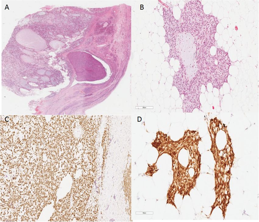
Man et al. Journal of Ovarian Research     (2021) 14:73                                                                               Page 3 of 7

 Fig. 2 Histology and immunohistochemistry of the tumor. a Whole slide scan showing a cystic growth pattern with a partial solid area. H&E, × 4
 magnification. b Tumorlet in the omentum. H&E, × 200 magnification. c Positive immunohistochemical staining for β-catenin (nuclear and
 cytoplasmic). The tumor cells are nuclear and cytoplasmic positive while the normal ovarian stromal cells on the right margin of the graph are
 positive on the membrane. Envision × 100 magnification. d The tumorlet in the omentum was positive for β-catenin (nuclear and cytoplasmic).
 Envision × 200 magnification

Immunohistochemical studies                                               Molecular studies
Surgical specimens were fixed in 10% neutral-buffered                     Genomic DNA was extracted from 5 mm-thick un-
formalin and routinely processed. Paraffin-embedded                       stained sections cut from formalin-fixed paraffin-
blocks were sectioned (3 mm-thick) and stained with                       embedded tumor blocks using an Ezup Column Animal
hematoxylin and eosin. Immunohistochemistry was per-                      Tissue Genomic DNA Extraction Kit (B518251, Sangon
formed using paraffin-embedded tissue samples using                       Biotech, Shanghai, China) according to the manufac-
the streptavidin-peroxidase method. Primary antibodies                    turer’s instructions. Exon 3 of CTNNB1 was amplified
were purchased from GSGB-BIO (Beijing, China) and                         by PCR using the following specific primer pairs: 5′-
Maxvision (Fuzhou, China) and used according to the                       GATTTGATGGAGTTGGACATGG-3′ (sense) and 5′-
manufacturer’s instructions.                                              GCTACTTGTTCTTGAGTGAAGG-3′ (antisense). The
  The sex cord tumor markers calretinin and inhibin-α                     PCR products were confirmed by agarose gel electro-
were both negative; however, CD10 and β-catenin                           phoresis, purified using the DNA Clean/Extraction Kit
(nuclear and cytoplasmic) (Fig. 2c) were both positive.                   (B518141, Sangon Biotech, Shanghai, China), and sub-
The other positive markers were WT-1, SF-1, cyclin D1,                    mitted for direct sequencing using BigDye Terminator
AR, ER, PR, vimentin, and CD99 (perinuclear dot-like).                    v1.1 (Applied Biosystems, Carlsbad, CA, USA) accord-
Focal areas of the tumor were positive for CK7, CK-pan,                   ing to the manufacturer’s protocol. The sequencing
CD56, Syn, and SMA. The tumor was negative for                            products were ethanol-precipitated before running on
SALL4, CD34, and E-cadherin. The Ki-67 index was                          a 3730XL Genetic Analyzer (Applied Biosystems, Fos-
very low, and MMR protein expression was intact                           ter City, CA, USA), and the resulting sequence data
(Supplementary Fig. 1). The reticular fibers around                       were analyzed using Chromas software. Each mutation
individual tumor cells were identified by reticulin                       was verified in both the sense and antisense direc-
staining. The tumorlet in the omentum was positive                        tions and was evaluated independently by two
for β-catenin (nuclear and cytoplasmic) (Fig. 2d).                        investigators.
Man et al. Journal of Ovarian Research    (2021) 14:73                                                              Page 4 of 7

  DNA sequencing analysis revealed a missense mutation,             index is low, indicating a low proliferation rate of the
c.98C > G, in exon 3 of CTNNB1 (Fig. 3), which caused               tumor [15]. However, tumorlet metastasis in the omen-
the replacement of serine with cysteine (UCU > UGU) at              tum suggests undetermined biological behavior.
codon 33 and led to the loss of the glycogen synthase                 Concerning the molecular mechanism of MCST,
kinase (GSK)-3β phosphorylation site in β-catenin.                  Maeda first reported a point mutation in exon 3 of
                                                                    CTNNB1 in two cases [5]. Given the rarity of this tumor
Discussion                                                          and the limited investigation of genomic and immuno-
Ovarian MCST is a rare variant of the pure stromal                  histochemical profile, we compared our results with
tumor that was first described by Irving and Young in               others reported and summarized their similarities. The
2009 [1]. Additional studies have been published since              Genetic characteristics of 38 cases of ovarian MCST
then, and demonstrated the unique histologic and                    have been summarized (Table 2). According to our
immunohistochemical features of MCST, including the                 retrospective study, in 26 of 38 cases in which CTNNB1
involvement of the Wnt/β-catenin pathway in the patho-              mutations were detected in the original study and all
genesis. Nevertheless, to date, less than 40 cases of               cases but one exhibited nuclear and cytoplasmic β-
MCST have been reported worldwide, all of which are                 catenin immunoreactivity [1–14],which indicates the im-
described as having benign biological behavior, but one             portant role of Wnt/β-catenin in the MCST. In addition,
patient experienced recurrence [1–14].                              APC mutations were identified in 5 women with MCST,
   The tumor of the present case includes all classical             4 of whom showed clinical features of FAP [2, 4, 13, 14],
histologic features of MCST, such as microcysts, solid              which explained the strong nuclear immunostaining for
cellular regions, and hyalinized fibrous stroma. The                β-catenin, although in the absence of β-catenin muta-
markers of sex cord tumors (calretinin and inhibin-α)               tions, further indicating that the Wnt/β-catenin/APC
and germ cell tumors (SALL4 and OCT3/4) are negative,               pathway mediated the occurrence and development of
while CD10 and β-catenin (nuclear and cytoplasmic) are              MCST. In the present study, however, an oncogenic mis-
characteristically positive. Other positive markers re-             sense mutation (c.98C > G) in CTNNB1 was detected.
ported in previous literature, such as WT-1, SF-1, cyclin           Unfortunately, APC mutational status was not checked.
D1, AR, ER, PR, vimentin, and CD99 (perinuclear dot-                The patient denied a family history of FAP, and no
like), are also expressed in our case. The expression pro-          polyps were found on gastrointestinal endoscopy. Hence,
files of relative markers from references listed in present         we speculate that APC gene mutations are unlikely to be
article are summarized in Table 1. Both morphology and              present. In 2018, McCluggage et al. clarified that ovarian
immunoprofile exclude other microcystic tumors of the               MCST may be an extracolonic manifestation of FAP and
ovary but MCST. Tumors in the omentum share the                     that APC mutations occur in a minority of MCSTs and
same histologic features and β-catenin immunoexpres-                are mutually exclusive to CTNNB1 mutations [4].
sion as those in the left ovary, which is highly indicative         Accordingly, it is also possible that alterations in the
of omentum metastasis. Mitosis is rare, and the Ki-67               other genes involved in the Wnt/β-catenin pathway are
                                                                    involved in its tumorigenesis. It is unclear whether there
                                                                    is a common morphology of MCST in different genetic
                                                                    backgrounds [16]. Therefore, it is beneficial for all
                                                                    females with MCST to be evaluated for FAP, including
                                                                    an assessment of different genes involved in the Wnt/β-
                                                                    catenin signaling pathway.
                                                                      The behavior of MCST is not well known because of
                                                                    limited case reports and follow-ups, but the available
                                                                    information suggests that MCST is likely benign. The
                                                                    most unique aspect of the present case revealed a 2-
                                                                    mm-diameter tumorlet in the omentum. Both morph-
                                                                    ology and immunophenotype are identical to the
                                                                    primary ovarian MCST, which indicated that MCST is
                                                                    not a purely benign ovarian tumor, as previously
                                                                    believed. Previous studies have elucidated the crucial
                                                                    role of Wnt/β-catenin in the MCST. β-Catenin-mediated
                                                                    migration and adhesion is linked on the one hand to
 Fig. 3 Molecular analysis of the tumor. Sequence chromatogram of   stimulating the expression of protooncogenes due to its
 the case harboring a point mutation in exon 3 of
                                                                    nuclear accumulation and on the other to E-cadherin
 CTNNB1 (c.98C > G)
                                                                    stabilization [17]. The E-cadherin/β-catenin complex
Man et al. Journal of Ovarian Research
Table 1 Immunohistochemical characteristics
Ref. β-catenin CD10 CyclinD1 WT-1 CD99 vimentin Calretinin inhibin-α AR ER PR AE1/AE3 CK7 Syn CD56 SALL4 E-cadherin SMA CD34
[1, 3] 15/15 16/16 15/15 15/15 2/15 16/16 1/16 W 1/16 W 0/0 1/15F 0/15 0/0 0/0 6/15 2/15 0/0 0/15 0/0 0/0
[2] 1/1 1/1 0/0 0/0 1/1 1/1 0/0 0/1 0/0 0/0 0/0 0/1 0/1 0/1 0/1 0/0 0/0 0/0 0/0
(2021) 14:73
[6] 6/6 6/6 0/0 0/6 3/5 6/6 0/6 0/6 0/0 0/0 2/5F 0/0 0/0 0/0 1/5F 0/0 0/5 0/0 0/0
[5] 2/2 2/2 0/0 2/2 0/2 2/2 0/2 0/2 0/0 0/2 0/2 1/2 0/2 0/2 0/2 0/2 0/0 0/2 0/2
[7] 1/1 1/1 0/0 1/1 0/0 1/1 0/1 0/1 0/0 0/0 0/0 0/0 0/0 0/0 0/0 0/0 0/1 0/0 0/0
[8] 2/2 2/2 2/2 2/2 0/2 2/2 0/2 0/2 0/2 0/2 0/2 0/0 0/0 0/2 0/2 0/2 0/2 0/2 0/2
[9] 2/2 2/2 0/0 0/0 2/2 2/2 0/0 0/0 0/0 0/0 0/0 0/0 0/0 0/0 0/0 0/0 0/0 0/0 0/0
[10] 1/1 1/1 0/0 1/1 0/0 1/1 1/1F 1/1F 0/0 0/0 0/0 0/1 0/0 0/0 0/0 0/0 0/0 0/0 0/0
[11] 1/1 1/1 0/0 0/0 0/0 1/1 1/1 + W 0/1 0/0 0/0 0/0 0/1 0/0 0/0 0/0 0/0 0/0 0/0 0/0
[12] 4/4 3/3 4/4 4/4 1/1 0/0 0/4 0/4 2/2 0/3 0/2 0/0 0/1 0/0 2/2F 0/0 0/0 0/0 0/1
[13] 1/1 1/1 0/0 0/0 0/0 1/1 0/1 0/1 0/0 0/1 0/1 0/1 0/0 0/0 0/0 0/0 0/0 0/0 0/0
[14] 1/1 1/1 1/1 1/1 0/0 1/1 0/1 0/1 0/0 0/0 0/0 0/1 0/0 0/1 0/1 0/0 0/0 0/0 0/0
Total 37/37 37/37 22/22 26/26 9/28 34/34 3/35 2/36 2/2 1/23 2/27 1/7 0/4 6/21 5/28 0/4 0/23 0/2 0/5
W Weak expression, F Focal expression
Page 5 of 7Man et al. Journal of Ovarian Research (2021) 14:73 Page 6 of 7
Table 2 Genetic characteristics
References Case CTNNB1 Mutation APC Mutation Comorbid FAP Follow up
time (m)
Maeda et al. [5] 1 c.98C > G p.S33C NK NK 14
2 c.98C > G p.S33C NK NK 4
Irving et al. [3] 3 c. 95A > T, p.D32V – NK NK
4 c. 104 T > G, p.I35S – NK NK
5 c. 95A > T, p.D32V – NK NK
6 c.110C > G, p.S37C – NK NK
7 c. 109 T > C, p.S37P – NK NK
8 c. 110C > G, p.S37C – NK NK
c. 95A > T, p.D32V
9 c. 101G > A, p.G34E – NK NK
10 c. 95A > T,p.D32V NK
11 – c. 1620_1621insA, p.Q541Tfs*19 NK NK
12 c.98C > G, p.S33C – NK
13 – – NK
14 c. 101G > A, p.G34E – NK
15 – c.1257delC, p.T419fs NK
c.1449 T > A, p.C483*
16 NK NK NK
17 – – NK
M Yang et al. [11] 18 NK NK NK NK
Y N et al. [7] 19 c.97 T > C p.S33P NK NK NK
S H Lee et al. [13] 20 – c.2376_2378delGCAinsCC, Y NK
(p.lys792Asnfs*28).
c.3796G > A, p.D1266N
c.1540delG, (p.Ala514 Profs*9).
Bi et al. [6] 21 c.122C > T p.T41I NK NK 60
22 Wild-type NK NK 18
23 c.110C > G p.S37C NK NK 7
24 c.101G > A p.G34E NK NK NK
25 c.97 T > C P.S33P, NK NK 59
26 Wild-type NK NK 2
Podduturi et al. [10] 27 c.101 G > A, p.G34E NK NK NK
J H Lee et al. [9] 28 c.98C > G; p.S33C NK NK NK
29 c.98C > G; p.S33C NK NK NK
K Na et al. [8] 30 c.122C > T p.T41I NK NK 57
31 c.88_99delTACCTGGACTCT NK NK 20
p.Y30_S33del
W G McCluggage 32 c.100G > A,p.G34R NK NK NK
et al. [12]
33 c.98C > G,p.S33C NK NK NK
34 wid-type NK NK NK
35 c.97 T > G,pS33A NK NK NK
C Liu et al. [14] 36 – Intron 6,c.730-1G > T Y NK
Y. Zhang et al. [2] 37 – c.1590C > T, p.G530E Y 108
Man et al. (this case) 38 c.98C > G,p.S33C – N 19
A/Ala Alanine, S Serine, C Cysteine, D Aspartic acid, V Valine, I Isoleucine, P/Pro Proline, G Glycine, H Histamine, lys Lysine, L Leucine, Y Tyrosine, E Glutamic acid, T
Thorenine, R Arginine, Q Glutamine, Asn Asparagines, del Deletion, ins Insertion, fs Frame shift, NK Not known, N No, Y Yes, FAP Familial adenomatous polyposisMan et al. Journal of Ovarian Research (2021) 14:73 Page 7 of 7
affects cell adhesion and may regulate cancer invasion and Received: 12 January 2021 Accepted: 14 April 2021
seeding metastasis. In the current case, mutation of β-
catenin may affect cell adhesion, which results in the References
detachment of tumor cells and causes omental deposits. 1. Irving JA, Young RH. Microcystic stromal tumor of the ovary: report of
On the basis of these pathological findings and molecular 16 cases of a hitherto uncharacterized distinctive ovarian neoplasm.
Am J Surg Pathol. 2009;33(3):367–75. https://doi.org/10.1097/PAS.
alterations, we assume that MCST more likely belongs to 0b013e31818479c3.
an underrecognized tumor of undetermined potential. 2. Zhang Y, Tao L, Yin C, Wang W, Zou H, Ren Y, et al. Ovarian microcystic
Not much is known regarding the biophysical behavior stromal tumor with undetermined potential: case study with molecular
analysis and literature review. Hum Pathol. 2018;78:171–6. https://doi.org/1
of MCST because of its rarity. Here, we present a rare 0.1016/j.humpath.2018.02.012.
case of ovarian MCST with omental metastasis, which 3. Irving JA, Lee CH, Yip S, Oliva E, McCluggage WG, Young RH. Microcystic
alerts us to the undetermined potential, and even the stromal tumor: a distinctive ovarian sex cord-stromal neoplasm
characterized by FOXL2, SF-1, WT-1, Cyclin D1, and β-catenin nuclear
malignant biological behavior, of MCST. More cases and expression and CTNNB1 mutations. Am J Surg Pathol. 2015;39(10):1420–6.
molecular studies will be necessary to further warrant https://doi.org/10.1097/PAS.0000000000000482.
this speculation. 4. McCluggage WG, Irving JA, Chong AS, Clarke BA, Young RH, Foulkes WD,
et al. Ovarian microcystic stromal tumors are characterized by alterations in
the Beta-catenin-APC pathway and may be an Extracolonic manifestation of
Abbreviation familial adenomatous polyposis. Am J Surg Pathol. 2018;42(1):137–9. https://
MCST: Microcystic stromal tumor doi.org/10.1097/PAS.0000000000000981.
5. Maeda D, Shibahara J, Sakuma T, Isobe M, Teshima S, Mori M, et al. β-
Catenin (CTNNB1) S33C mutation in ovarian microcystic stromal tumors. Am
J Surg Pathol. 2011;35(10):1429–40. https://doi.org/10.1097/PAS.0b013e31
Supplementary Information 822d6c71.
The online version contains supplementary material available at https://doi.
6. Bi R, Bai QM, Yang F, Wu LJ, Cheng YF, Shen XX, et al. Microcystic
org/10.1186/s13048-021-00812-1.
stromal tumour of the ovary: frequent mutations of β-catenin
(CTNNB1) in six cases. Histopathology. 2015;67(6):872–9. https://doi.
Additional file 1. org/10.1111/his.12722.
7. Kang YN, Cho CH, Kwon SY. Microcystic stromal tumor of the ovary with
mutation in exon 3 of β-catenin: a case report. Int J Gynecol Pathol. 2015;
Acknowledgments 34(2):121–5. https://doi.org/10.1097/PGP.0000000000000122.
Not applicable. 8. Na K, Kim EK, Jang W, Kim HS. CTNNB1 mutations in ovarian microcystic
stromal tumors: identification of a novel deletion mutation and the use of
pyrosequencing to identify reported point mutation. Anticancer Res. 2017;
Authors’ contributions 37(6):3249–58. https://doi.org/10.21873/anticanres.11688.
Conceptualization: LG, SZ, Data acquisition, writing of the original draft:XM, 9. Lee JH, Kim HS, Cho NH, Lee JY, Kim S, Kim SW, et al. Genetic analysis of
BW. Data analysis and interpretation: XM, BW, ZW, WL, LT, LG, SZ. The ovarian microcystic stromal tumor. Obstet Gynecol Sci. 2016;59(2):157–62.
author(s) read and approved the final manuscript. https://doi.org/10.5468/ogs.2016.59.2.157.
10. Podduturi V, Tran T, Champion KJ, Onur N, Shiller SM. Microcystic stromal
tumor of the ovary: a case report of a newly described ovarian neoplasm
Funding with a β-catenin (CTNNB1) G34E mutation. Int J Gynecol Pathol. 2015;34(6):
This study was supported by a grant from the Department of Finance of 541–5. https://doi.org/10.1097/PGP.0000000000000198.
JiLin Province, China (JLSCZD2019–078 to Xiaxia Man). 11. Yang M, Bhattacharjee MB. Ovarian microcystic stromal tumor: report of a
new entity with immunohistochemical and ultrastructural studies.
Ultrastruct Pathol. 2014;38(4):261–7. https://doi.org/10.3109/01913123.2014.
Availability of data and materials
893045.
All datasets generated for this study are included in the manuscript.
12. McCluggage WG, Chong AS, Attygalle AD, Clarke BA, Chapman W,
Rivera B, et al. Expanding the morphological spectrum of ovarian
Declarations microcystic stromal tumour. Histopathology. 2019;74(3):443–51. https://
doi.org/10.1111/his.13755.
Ethics approval and consent to participate 13. Lee SH, Koh YW, Roh HJ, Cha HJ, Kwon YS. Ovarian microcystic stromal
The study was approved by the Institutional Review Board of The First tumor: a novel extracolonic tumor in familial adenomatous polyposis. Genes
Hospital, Jilin University (IRB No. 2019–302). Chromosom Cancer. 2015;54(6):353–60. https://doi.org/10.1002/gcc.22233.
14. Liu C, Gallagher RL, Price GR, Bolton E, Joy C, Harraway J, et al. Ovarian
microcystic stromal tumor: a rare clinical manifestation of familial
Consent for publication adenomatous polyposis. Int J Gynecol Pathol. 2016;35(6):561–5. https://doi.
The patient provided written informed consent for the publication of this org/10.1097/PGP.0000000000000289.
case report. 15. Fulton R. Getting a grip on Ki-67. Appl Immunohistochem Mol Morphol.
2021;29(2):83–5. https://doi.org/10.1097/PAI.0000000000000908.
16. Rabban JT, Karnezis AN, Devine WP. Practical roles for molecular diagnostic
Competing interests testing in ovarian adult granulosa cell tumour, Sertoli-Leydig cell tumour,
The authors declare that they have no competing interests. microcystic stromal tumour and their mimics. Histopathology. 2020;76(1):
11–24. https://doi.org/10.1111/his.13978.
Author details 17. Jamora C, Fuchs E. Intercellular adhesion, signalling and the cytoskeleton.
1
Department of Oncologic Gynecology, The First Hospital of Jilin University, Nat Cell Biol. 2002;4(4):E101–8. https://doi.org/10.1038/ncb0402-e101.
Xinmin Street 1,Changchun, Jilin 130021, People’s Republic of China.
2
Department of Cardiac Surgery, The First Hospital of Jilin University,
Changchun, 130021 Jilin, People’s Republic of China. 3Department of Publisher’s Note
Pathology, the First Hospital of Jilin University, Xinmin Street 1,Changchun, Springer Nature remains neutral with regard to jurisdictional claims in
130021 Jilin, People’s Republic of China. published maps and institutional affiliations.You can also read