Parent-reported histories of adults with trisomy 13 syndrome
←
→
Page content transcription
If your browser does not render page correctly, please read the page content below
Received: 22 July 2020       Revised: 10 February 2021
DOI: 10.1002/ajmg.a.62165

ORIGINAL ARTICLE

Parent-reported histories of adults with trisomy 13 syndrome

Amy N. Lebedoff                       | John C. Carey

Division of Medical Genetics, Department of
Pediatrics, University of Utah Health, Salt Lake    Abstract
City, Utah                                          Clinical histories and outcome data of long-term survivors with trisomy 13 are rare.
Correspondence                                      The goal of this study was to collect the medical histories of adult individuals
Amy N. Lebedoff, Division of Medical                (≥18 years old) with apparent non-mosaic trisomy 13/Patau syndrome to help gain
Genetics, Department of Pediatrics, University
of Utah Health, 295 Chipeta Way Rm 2w414,           further insight in to the clinical course for individuals with this condition and to char-
Salt Lake City, UT 84108, USA.                      acterize the manifestations for surveillance and management. We collected 11 fami-
Email: amy.lebedoff@hsc.utah.edu
                                                    lies through a contact person with the LWT13 (Living with Trisomy 13) LIFE support
                                                    group. We performed telephone interviews to gather their medical histories and
                                                    report these data in system-based summaries, tables, and clinical vignettes. In
                                                    instances where parents retained copies of genetic testing reports or clinicians cur-
                                                    rently taking care of the individual with trisomy 13 were able to provide documenta-
                                                    tion, we confirmed diagnosis. All clinical histories and reported manifestations were
                                                    consistent with a diagnosis of trisomy 13. We also elicited comments from parents
                                                    on their personal experiences of raising an individual with trisomy 13.

                                                    KEYWORDS
                                                    adults with trisomy 13, clinical features in trisomy 13, Patau syndrome, trisomy 13

1    |    I N T RO DU CT I O N                                                    Nelson et al., 2016; Peterson et al., 2018). Bruns and Campbell (2014)
                                                                                  identified nine children with non-mosaic trisomy 13 between 12 and
Patau syndrome, or trisomy 13 syndrome, is characterized by multiple              59 months of age. Imataka et al. (2016) documented eight patients
malformations, a high neonatal and infant mortality, and significant              described in clinical reports between the ages of 5 and 14 years old
cognitive and psychomotor disability. Little information about the clin-          and three patients over 18 years with apparent non-mosaic Patau
ical course for surviving patients is reported in the literature making it        syndrome as previously reported in the literature. This small number
difficult to counsel parents with a new diagnosis in their child about            of reports highlights the rarity of this type of clinical data. We con-
management and expectations for those rare individuals with long-                 ducted this study with the goal of uncovering more information to
term survival. In recent years, survival at 1 year has been improving:            support clinicians and family members in order to better understand
data drawn from the Metropolitan Atlanta Congenital Defects Pro-                  the clinical course of trisomy 13.
gram, a population-based birth defects surveillance system between
the years of 1968 and 1999, documented a median survival of 7 days
for a patient with trisomy 13. In that study, 5.6% of patients survived           2     |   M A T E R I A L S A N D M ET H O D S
past 1 year (Rasmussen et al., 2003). Other large-scale population
studies showed similar results in median and one-year survival                    We collaborated with the leader of LWT13 Trisomy 13 LIFE support
(Wu et al., 2013). More recent data show a positive trend in survivor-            group (http://www.livingwithtrisomy13.org/), who reached out to
ship and expands our understanding of what this means for individuals             families involved in the community with a child over 17 years of age
who live beyond the one-year benchmark. In studies which included                 with a diagnosis of apparent non-mosaic (“full”) trisomy 13 by clinical
individuals where more invasive interventions were offered to                     testing. Our contact information was relayed to interested families. A
patients, 11.5% of individuals with trisomy 13 survived to 1 year and             telephone encounter was scheduled and electronic copies of the con-
a similar number, 9.7% survived to 5 years of age (Meyer et al., 2016;            sent were provided to the parent/L.A.R. before the encounter.

Am J Med Genet. 2021;1–14.                               wileyonlinelibrary.com/journal/ajmga                          © 2021 Wiley Periodicals LLC.   1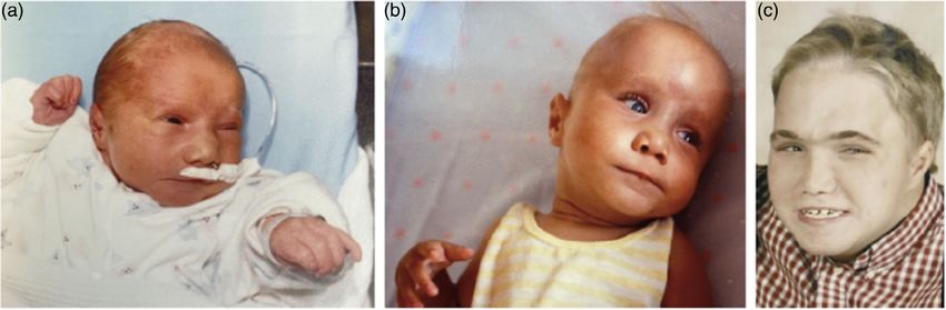
2 LEBEDOFF AND CAREY
Consent took place by telephone. A standard questionnaire about (Bugge et al., 2007; Imataka et al., 2016). While it is an important con-
medical history, surgical history, developmental history, and parents' sideration in terms of survivorship for this condition, it is outside of the
reflections on raising a child with trisomy 13 were asked over the scope of this study. We did not investigate with further chromosome
phone (available as Data S1). Families had the opportunity to submit testing in alternative tissues on any of the participants; thus, it is not
photographs of their family member with trisomy 13 (see Figures 1–7). possible to completely exclude the presence of low-level mosaicism.
Data were reviewed with the participating family member to verify Instead, we highlighted the histories of individuals with trisomy
some details of their initial report. Growth centiles were estimated 13 where initial chromosome testing did not reveal mosaicism, the fam-
based on parental reported data and use of Fenton, 2003 growth charts ilies received counseling based on these testing results and found that
for preterm and term infants. The history of one individual (Individual 3) the outcomes of their loved ones were beyond clinical expectation. We
has previously been reported. Her history reflects an update of this hope that these data will be taken into consideration by clinicians in dis-
report, which is specified in her clinical vignette below and noted in the cussing the diagnosis of trisomy 13 with families.
tables of clinical information.
Of note, this study strives to recognize and bring awareness to the
inherent uncertainty of historical and current practices in the assess- 2.1 | Participants
ment of mosaicism when making the diagnosis of trisomy 13. Previous
data have shown that while mosaicism occurs at a low rate as detected We enrolled 11 families representing 11 adult individuals with apparent
by chromosome analysis performed on cells derived from amniotic fluid, non-mosaic trisomy 13 by parental report, or verified by clinical chro-
a disproportionate number of individuals with mosaicism detected on mosome testing or clinical documentation when available. Similar stud-
chromosome testing make up the clinical reports of long-term survivors ies performed using parental reports have shown the accuracy of
giving credence to mosaicism as an important factor in terms of survival parental reporting in the distinction of testing that showed non-mosaic
F I G U R E 1 (a) Individual 2 at 1 year. (b) Individual 2 at 2 years. (c) Individual 2 as a school-aged girl [Color figure can be viewed at
wileyonlinelibrary.com]
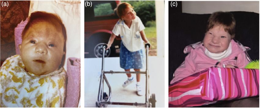
F I G U R E 2 (a) Individual 3 at 10 months old. (b) Individual 3 in kindergarten. (c) Individual 3 as an adult [Color figure can be viewed at
wileyonlinelibrary.com]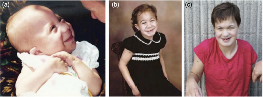
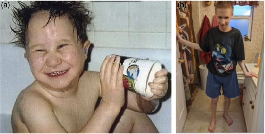
LEBEDOFF AND CAREY                                                                                                                               3

F I G U R E 3 (a) Individual 4 as a
boy. (b) Individual 4 as a young man
[Color figure can be viewed at
wileyonlinelibrary.com]

F I G U R E 4 (a) Individual 5 as an
infant. (b) Individual 5 as an adult
[Color figure can be viewed at
wileyonlinelibrary.com]

F I G U R E 5 (a) Individual 7 as a newborn. (b) Individual 7 at 12-months old. (c) Individual 7 at 18-year old [Color figure can be viewed at
wileyonlinelibrary.com]

versus mosaic trisomies, and in medical histories in general (Baty           35 years old. Three individuals are deceased and reports were given
et al., 1994; Bruns & Campbell, 2014; Hansen et al., 2000). Our study        posthumously. We were able to obtain chromosome testing reports or
included seven women and four men between the ages of 18 years and           clinical notes verifying testing results for six individuals. No co-
4                                                                                                                                 LEBEDOFF AND CAREY

F I G U R E 6 (a) Individual 8 as a newborn. (b) Individual 8 as a school-aged girl. (c) Individual 8 at 18 years of age [Color figure can be viewed at
wileyonlinelibrary.com]

F I G U R E 7 (a) Individual 11 at 1 month. (b) Individual 11 at 11 years. (c) Individual 11 at 29 years [Color figure can be viewed at
wileyonlinelibrary.com]

occurring genetic diagnoses were reported. The average age of the par-        Developmental milestones are summarized in Table 3, and Adult-onset
ticipants was 24.5 years old. Women in the study had reported adult           Conditions are listed in Table 4. We have included brief descriptions of
heights or lengths of 122 cm to 158 cm and were on average about 5.5          developmental histories below, whereas more detailed medical and
standard deviation below the mean height for adult women. Men in the          developmental information specific to the individual and the family
study had reported adult heights or lengths of 135–163 cm, which is           experiences are featured in 11 vignettes, which are included in Data S1.
about four standard deviations below the mean for adult men. The
average BMI for female participants was 23.9 and for male participants
was 19.8. The study was approved by the Institutional Review Board of         3.1     |   Cardiovascular findings
the University of Utah and consent to participate was obtained in all
families. No families withdrew participation after consent was obtained.      In this group of adults, 10 individuals have undergone an echocardio-
                                                                              gram. The reported findings included three individuals with isolated
                                                                              atrial septal defect, two individuals with isolated ventricular septal
3    |   RESULTS                                                              defect, three individuals with both an atrial septal defect and ventricu-
                                                                              lar septal defect, and one individual with dextrocardia and a ventricu-
We have summarized clinical manifestations in system-based summaries          lar septal defect (see Table 1). One person had normal cardiac
below and in Table 1. Prenatal and birth history as well as a summary of      anatomy on imaging. One individual's cardiac anatomy was unknown,
medical support in the newborn period are reported in Table 2.                but this person did not experience cardiac symptoms or complications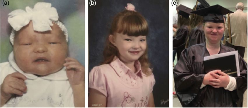
TABLE 1 Summary of clinical features
Height/
Array length
Age, characteristics, weight Central nervous Seizures, Ears, nose,
ID gender tissue BMI Cardiac system Apnea onset Gastrointestinal Renal urologic throat Eye Musculoskeletal Endocrine Dermatologic
1 35 y F Non-mosaic, 122 cm NR Brain hemorrhage Reported, Myotonic, GT, NR Hearing loss Cataracts, Natal teeth, NR Cutis aplasia,
Parental report, 32 kg, resolved Within first Reflux, Coloboma, Equinovarus Skin
Blood. (4 ft by 7– 12 months Constipation, Vision loss infections
70 lbs) 8 years of Abdominal cysts
21.5 life
2 23 y F 47,XX,+13 130 cm Small muscular NR Apnea with Myotonic, GT, VUR, Dysphagia Micropth., Scoliosis, PP, Dermal
22 cells 39 kg, VSD, seizures GTC, Umb. hernia, UTI Coloboma, Polydactyly Pancreatic sinuses
analyzed, (4 ft Low blood 21 months Malrotation, Cataracts, insufficiency
2 cells 3 in pressure Gallstones, Vision loss
karyotyped, 86 lbs) Pancreatitis
Blood. 23.1
3a 19 y F 47,XX,+13 158 cm ASD, Normal MRI, Reported Infantile Reflux, Hydronephrosis, Cleft lip/ Cataracts, Scoliosis, PP, Cutis aplasia,
50 cells 47 kg, VSD Contractures symptoms, spasms FTT, Duplicated palate, Vision loss Polydactyly Bone health Dermal
counted, (5 ft Normal sleep 12 months, Malrotation, ureter, Ear tubes, sinuses,
Metaphases 2 in study at 5 Cerebral Umb. hernia, UTI Branch. Hidradenitis
counted: 6, 103 lbs) yo palsy GT, Cleft cyst, Suppurativa
3 cells 18.8 Constipation, Hearing loss
karyotyped, “Wandering
Blood. spleen”
4 24 y M Non-mosaic, 163 cm Normal MRI: NR NR Inguinal hernia, PCK, Cleft palate, Micropth., NR NR Cysts,
Parental report, 52 kg, echocardiogram Delayed Pancreatitis, Unil. Retractile Ear tubes, Colobomas, Boils,
Parental (5 ft myelination, Malrotation, testicle Hearing loss, Cataracts, CVM
balanced 4 in Normal structure Constipation Aspiration Nystagmus,
translocation, 115 lbs) Retinal
Blood. 19.6 dysplasia,
Retinal
detachment,
Lens
displacement,
Vision loss
5 27 y F Non-mosaic, 142 cm ASD, MRI: Reported NR GT, PCK, Intubation at Micropth., Polydactyly. NR Cutis aplasia
dec. Parental report, 43 kg, VSD, Atypical position Gallstones, Duplicated birth. Cataracts, Scoliosis,
Blood. (4 ft Cardiac of cerebellum Pancreatitis ureters, Trach until Coloboma, Achilles tendon
8 in dysfunction VUR, 8 months Vision loss release
94 lbs) UTI Postnatal
21.3 stridor
6 28 y M 47,XY,+13, 135 cm ASD, NR Reported Yes Malrotation Cryptorchidism Cleft palate, Glaucoma, Polydactyly NR Cutis aplasia
Confirmed by 38 kg, HTN symptoms 8–9 years old Nasal Retinal
neonatal ICU (4 ft polyps, detachment
discharge 5 in Ear tubes,
summary, 83 lbs) Aspiration,
Blood. 20.9 Hearing loss
7 18 y M 47,XY,+13, 153 cm ASD NR Reported Myoclonic Inguinal hernias, Small genitals, Cleft palate, Blindness Polydactyly NR Cutis aplasia,
dec. Cell count not 58 kg, jerks, Malrotation VUR, Ear tag, Skin
reported. (5 ft Absence UTI Ear tubes infections
Blood. 4 in seizures
125 lbs)
24.8
(Continues)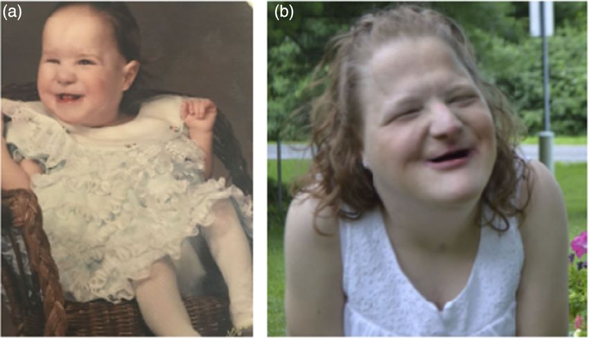
TABLE 1               (Continued)
                                               Height/
                          Array                length
             Age,         characteristics,     weight                            Central nervous                            Seizures,                                                     Ears, nose,
     ID      gender       tissue               BMI        Cardiac                system                  Apnea              onset              Gastrointestinal     Renal urologic        throat            Eye                  Musculoskeletal       Endocrine           Dermatologic

     8       20 y F       46,XX,+13,der        147 cm     ASD,                   Microcephaly            Reported,          NR                 GT,                  VUR,                  Ear tag,          Entropion,           Scoliosis,            NR                  Psoriasis
                             (13;13)(q10;      66 kg,     HTN                                            Developed at                          Slow gastric         UTI                   Aspiration,       Coloboma,            Contractures
                            q10)               (4 ft                                                        3 weeks                              emptying,                                Ear tubes         Hypertelorism
                          Confirmed by            10 in                                                                                        Eosinophilic
                             physician's       145 lbs)                                                                                           esophagitis,
                             note, Initial     30.5                                                                                            Possible
                             testing not                                                                                                         Malrotation,
                             available,                                                                                                        Gallstones
                          Blood.

     9       20 y F       Non-mosaic,          122 cm     ASD,                   NR                      NR                 GTCs,              GT/GJT,              PCK,                  Ear tubes,        Vision loss,         Polydactyly,          NR                  NR
                          Parental report,     43 kg,     VSD                                                                 myoclonic        Reflux,              UTI                   Aspiration        Myopia,              Scoliosis,
                          Amniotic fluid       (4 ft                                                                        9 months           Malrotation                                                  Lacrimal duct        Limb
                                               95 lbs)                                                                                                                                                         stenosis             contractures
                                               28.9

     10      22 y M       Non-mosaic,          139 cm     Dextrocardia           Holoprosencephaly       Reported           Myoclonic          Omphalocele,         Small genitals,       Intubation at     Coloboma,            Polydactyly,          Delayed             Cutis aplasia,
                          Parental report,     27 kg      VSD                                                                  jerks,          GT                   Cryptorchidism           birth,         Myopia               Achilles tendon          pubertal         Cyst,
                          Amniotic fluid       (4 ft 7                                                                      1-time GTC         Pancreatitis,        PCK                   Cleft lip,                               release                progression,     Psoriasis
                                                    in                                                                                         Abnormal             UTI                      palate,                                                   Bone health
                                               59 lbs)                                                                                            position of                             Ear tags
                                               14                                                                                                pancreas,
                                                                                                                                               SBO

     11      35 y F       46,XX,+13,           127 cm     VSD                    Microcephalic           NR                 GTCs,              Malrotation,         Normal imaging        Ear               Cataract,            Lower extremity       Delayed             Cutis aplasia,
             dec.         Karyotyped           38 kg,                                                                       7 months           Milk allergy                                  infections     Abnormal pupil,         contractures,         pubertal         Cysts
                            number of          (4 ft 2                                                                                         GT                                                           Vision loss             hip dysplasia         progress,
                             cells                  in                                                                                                                                                                                                 Osteoporosis
                             analyzed: 2,      84 lbs)
                          Blood                23.6

Abbreviations: ASD, atrial septal defect; CVM , capillary vascular malformation; dec., deceased; FTT , failure to thrive; GT , gastric tube; GJT , gastrojejunal tube; GTCs, generalized tonic–clonic seizures; HTN , hypertension; micropth., microphthalmia; MRI , magnetic resonance
imaging; NR , not reported or work-up not obtained; PCK , cystic kidneys; PP , precocious puberty; SBO , small bowel obstruction; umb., umbilical; unil., unilateral; UTI , urinary tract infections; VUR , vesicoureteral reflux; VSD , ventricular septal defect.
a
    Previously reported in the literature at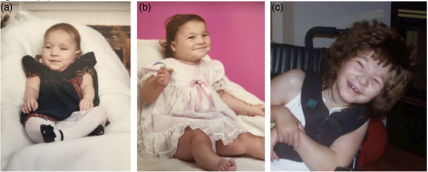
LEBEDOFF AND CAREY                                                                                                                                                  7

TABLE 2           Summary of early medical history

         Age,                                                             Known birth                                   Time until
  ID     gender       Prenatal concerns             Birth history         parameters                Early support       discharge           Support at discharge

  1      35 y F       Hyperemesis,                  42 weeks              2.72 kg                   Nasal cannula       48 hours            Room air,
                      Oligohydramnios               SVD                   (1st centile)            NG                                      Bottle feeds
  2      23 y F       IUGR                          36 weeks              2.04 kg                   Nasal cannula       10 days             Bottle feeds,
                      VUR                           Induced VD            (10th centile),          TPN                                     NG
                                                                            44.5 cm
                                                                          (20th centile)
  3a     19 y F       Reduced prenatal              38 weeks              3.4 kg                    Nasal cannula       11 days             Room air,
                        screening due to            Repeat CS             (75th centile),          NG                                      NG, Bottle feeds,
                        parental preference                               53.3 cm                                                           Palate retainer,
                                                                          (97th centile), OFC                                              Suction equipment
                                                                            34 cm (67th
                                                                            centile)
  4      24 y M       Decreased fetal movement,     39 weeks              2.9 kg                    Oxygen hood         Transferred on      Room air
                        maternal blood pressure     Spontaneous           (15th centile)                                 day 2             Bottle feeds
                        issues                        onset requiring       48.3 cm
                                                      further             (20th centile)
                                                      induction
  5      27 y F       Two vessel cord               40 weeks              2.4 kg                    Intubated           >3 months of        Tracheostomy (removed
         dec.                                       CS due to failure     (1st centile),           NG                    life                at 8 mos)
                                                      to progress         49.5 cm                                                           NG
                                                                          (35th centile)
  6      28 y M       Maternal edema                39 weeks              2.29 kg                   Room air            7 days              Room air
                                                    CS                    (1st centile)                                                    Bottle feeding
  7      18 y M       None.                         36 weeks              2.72 kg                   Nasal cannula       5 days              Nasal cannula
         dec.         Normal 1st trimester US       SVD                   (50th centile),          NG                                      NG
                                                                            53.3 cm
                                                                          (97th centile)
  8      20 y F       Breech                        Full term             3.147 kg (30th           Room air            7 days              Room air
                                                    CS                      centile), 48.3 cm       NG                                      Bottle feeds
                                                                          (15th centile)
  9      20 y F       Postaxial polydactyly,        35 weeks              1.53 kg                   Room air            3 days              Room air
                        Abnormal shape and          SVD                   (2nd centile)            Bottle feeds                            Bottle feeds
                        length of kidney
  10     22 y M       multiple congenital           39 weeks              2.5 kg (3rd %ile).      Intubated           5 days              Nasal cannula
                       anomalies found on           CS                                              IVF                                     GT
                       prenatal imaging:                                                            NG
                       holoprosencephaly,
                       omphalocele, postaxial
                       polydactyly, cleft lip and
                       palate, small genitalia,
                       dextrocardia with
                       ventricular septal defect
                       and rocker bottom feet
  11     35 y F       Limited prenatal screening    37 weeks              2.41 kg                   Room air            4 days              Room air
         dec.           due to practitioner's       SVD                   (15th centile),          NG                                      Bottle feeds
                        practice                                            43.18 cm                                                        NG
                                                                          (5th centile).

Abbreviations: Dec., deceased; GT, gastric tube; IUGR, intrauterine growth restriction; IVF, intravenous fluids; NG, nasogastric tube; SVD, spontaneous vaginal
delivery; VD, vaginal delivery.
a
 Previously reported in the literature.

later in life. Of all 11 participants, two individuals underwent cardiac              individuals. The 28-year old man (Individual 6), who had an atrial sep-
surgery (Individuals 5 and 10) for repair of septal defects. The                      tal defect, developed hypertension as an adult, which is controlled
27-year-old woman (Individual 5) was born with an atrial septal defect                with a beta-blocker. The 20-year-old woman (Individual 8), who had
and a ventricular septal defect and underwent repair at 4 months of                   an atrial septal defect, developed hypertension at about 9 years of
age. She developed a thrombus in her heart at 10–11 years old and                     age coinciding with the onset of puberty. Renal imaging performed at
was treated for cardiac dysfunction in her twenties. She succumbed                    that time was normal and laboratory studies did not provide a
to complications of heart failure. Hypertension arose in two                          diagnosis.
8
TABLE 3 Developmental milestones
Case ID Rolling over Sitting Pulling to stand Crawling Mobility Communication Fine motor
1 36 y F 12–24 mo Supported: 12–24 mo As a young child As a young With walker, Smiles, points, says “mama” Feeds herself finger foods,
child Wheelchair uses a cup
2 22 y F 12 mo Unsupported: 24 mo 24 mo NR Never walked, Smiles, cries, says “mom” Could play with toys
wheelchair
3a 19 y F 3 yo Supported: by 24 mo NR 5 yo Walked at 29 mo, Emotions expression, says Can grab toys
Unsupported: by 3 yo Walks with walker “mama”
4 24 y M 7 mo NR 2.5 yo NR Walks with guidance Had a few words, now uses Feeds himself
“mama”
5 27 y F 2.5 yo Supported: 2.5 yo 6 yo 4 yo Walks with walker Expression of emotion, Played with toys,
Unsupported: 4 yo indicating needs by Hold cups
moving through the home
6 27 y M 12–24 mo Supported:LEBEDOFF AND CAREY 9
TABLE 4 List of adult-onset conditions on noticed improvement and resolution with time. As adults, one indi-
Case ID Adult-onset medical concerns vidual will undergo sleep study to evaluate for covert apneic events at
night. She is not currently on oxygen or respiratory support for apnea
1 35yF Failure to thrive, abdominal cysts,
worsening seizures, brain hemorrhage (Individual 8). One individual had a repeat sleep study at 5 years of
2 23yF Low blood pressure, recurrent infections, age, which was normal (Individual 3). Of note, no individuals are on
insulin resistance baseline oxygen or positive pressure ventilation during the day or
3 a
19yF Low immunoglobulin levels in serum night.
4 24yM Retinal detachment, lens dislocation
5 27yF Congestive heart failure, renal failure
secondary to hypoperfusion, gallstones, 3.4 | Gastrointestinal findings
pancreatitis, urinary tract infections
6 28yM Recurrent pneumonias, hypertension Six individuals were discharged after birth with a feeding tube (five
7 18yM Recurrent pneumonias, endocarditis individuals had nasogastric tubes and one individual had gastric tube).
8 20yF Asthma, recurrent pneumonias, Low The other five individuals went home with plans to feed by bottle.
immunoglobulins, possible Surgical feeding tube placement was required in eight individuals with
hemochromatosis most surgeries taking place during childhood. One individual required
9 20yF Recurrent pneumonias, sepsis revision of a feeding tube at 18 years of age (Individual 9) and one
10 22yM Surgery to relocate pancreas, treatment of woman underwent feeding tube placement as an adult (Individual 11)
cystic skin infections, repeat occurrences (see Table 1).
of small bowel obstruction
Eight of the 11 individuals were diagnosed with malrotation. Four
11 35yF Ear infections, Clostridium difficile,
individuals had pancreatitis commonly due to gallstones. Other gastro-
Malrotation, Pneumonia, osteoporosis
intestinal findings included reflux, constipation, hernias, eosinophilic
a
Previously reported in the literature. esophagitis, omphalocele, atypical position of pancreas, pelvic spleen,
and milk allergy.
3.2 | Central nervous system findings
Abnormal brain imaging findings were reported in four individuals. 3.5 | Renal and urogenital findings
These findings included holoprosencephaly (Individual 10), atypical
position of the cerebellum (Individual 5), delayed myelination with Seven individuals experienced recurrent urinary tract infections.
otherwise normal structure (Individual 4), and brain hemorrhage with- Vesicoureteral reflux or hydronephrosis were reported in five people,
out known cause in an adult (Individual 1). Two individuals had normal and cystic kidneys were reported in four. Other findings included
brain imaging. Six families were not aware of central nervous system duplicated collecting duct, cryptorchidism or retractile testicle, and
findings or the participants had not undergone postnatal imaging. small genital size.
Seizures arose in eight individuals most with onset within the first
2 years of life. One man had seizures diagnosed at 8–9 years of age.
Types of seizures included myotonic seizures, generalized tonic–clonic 3.6 | Ears, nose, and throat findings
seizures, infantile spasms, and absence seizures.
Recurrent ear infections arose in seven individuals. Ear tubes
required placement two or more times in three people due to pre-
3.3 | Respiratory findings mature expulsion of the ear tubes (Individuals 4, 6, 8). Hearing loss
was reported in four people, cleft palate was reported in five, and
Only two individuals were intubated after birth (Individuals 5 and 10). dysphagia or aspiration was reported in five. Other findings included
Individual 10 was extubated to nasal cannula on day two of life after ear tags, branchial cleft cyst, nasal polyps and suspected
omphalocele repair. Individual 5 underwent tracheostomy, which was laryngomalacia.
later removed at 8 months of life without need for further respiratory
support. Five individuals required nasal cannula or oxygen hood in the
neonatal period. Four individuals remained on room air after birth. 3.7 | Ophthalmologic findings
Symptoms of apnea were reported in seven individuals arising
during the first months of life. One person had apnea arising outside Abnormal eye findings were reported in all participants. These
of this period coinciding with the onset of seizures. For this individual, included cataracts in six people and coloboma in seven individuals.
symptoms improved after initiation of treatment for epilepsy Visual impairment likely affected all participants. Other findings
(Individual 2). Three individuals never had findings or symptoms con- included glaucoma, retinal dysplasia, retinal detachment, lens displace-
cerning for apnea. Families who intervened for apneic episodes early ment, entropion, microphthalmia, and nystagmus.10 LEBEDOFF AND CAREY
3.8 | Musculoskeletal findings developmental history is summarized in Table 3. Individual 1 turns to
sounds and understands warnings from her loved ones if she is near a
Postaxial polydactyly was found in seven individuals and scoliosis in hazard. She smiles, makes communicative noises, and says “mama.” This
six. Six individuals had contractures affecting the upper or lower woman can crawl to her parent and grab the arm to communicate. She
extremities. Other findings included natal teeth, need for subtalar feeds herself finger foods and drinks from a cup with a straw. She
bone fusion, and hip dysplasia. enjoys music and children's television programming and stories.
3.9 | Endocrine findings 3.12.2 | Individual 2 (Figure 1a–c)
Issues with bone health due to osteoporosis is reported in two women The individual is a 23-year-old woman born late preterm, who was
(Individuals 3 and 11). Two individuals underwent investigations for diagnosed by prenatal genetic testing and confirmed by postnatal
delayed onset of puberty. In one of these individuals (Individual 10), a chromosome analysis. Her developmental history is summarized in
brain MRI to assess the pituitary was normal. He was treated initially Table 3. When she was hospitalized for pancreatitis at 8 years of age,
with testosterone, which his parents chose not to continue. He now she lost many skills including her ability to sit up, pull to stand, and
receives zoledronic acid to support bone health. Individual 11 was also manipulate objects with her hands. Her parent notes that as a child
evaluated by an endocrine team. Imaging of her uterus was normal. she was able to do summersaults. She enjoys attention, snuggling,
Ovaries were not found on imaging, though parent did not know if this feeling hands run through her hair, and sunshine.
was due to difficulty of exam or confirmed absence of these structures.
She had not been on hormonal therapies, but received zoledronic acid,
vitamin D and calcium. Precocious puberty was diagnosed by an endo- 3.12.3 | Individual 3 (Figure 2a–c)
crine team in two individuals (Individuals 2 and 3).
This individual is a 19.5-year-old woman born at term and diagnosed
by postnatal chromosome analysis. Her developmental history is sum-
3.10 | Dermatologic findings marized in Table 3. She can sit and stand on her own, uses a walker
several hours per day and can take a few steps on her own. She
Seven individuals had cutis aplasia. Seven individuals had recurrent swipes to grab objects. She understands simple commands, such as
skin infections involving dermal sinuses, skin abscesses, or cysts. “stand,” “sit” and can assist her parent with dressing herself. Her par-
Other findings included psoriasis in two individuals, and a capillary ent reports that she functions at the level of a toddler. She is a gener-
vascular malformation. ally happy and loving woman. She enjoys shows and movies with
music. She loves to go outside, use her walker and an adaptive tricycle
or tandem bicycle with her parent.
3.11 | Early history
Please see Table 2 for details of prenatal, birth history, growth param- 3.12.4 | Individual 4 (Figure 3a,b)
eters, and early support required in the nursery or neonatal intensive
care unit for each person described below. Of note, eight individuals The individual is a 24-year-old man born at term with trisomy
were born after 36 weeks gestation. 13, translocation type, diagnosed by postnatal chromosome analysis.
His developmental history is summarized in Table 3. It is notable that
Individual 4 was able to walk independently at 3.5 years. He used a
3.12 | Vignettes stander for a short period of time when he was acquiring new skills.
He can now walk independently in his home, but requires guidance
We provide more detailed accounts of the early history, medical outside of the home due to vision loss. He requires a wide-handled
course, and developmental milestones in the Data S1. Below we high- spoon for self-feeding. He was able to make some signs at 2 years of
light the developmental histories because few data are available on age. He said “mama,” and used to have a few more words, but no lon-
persons with trisomy 13 over the age of 5 years. Figures 1–7 show ger uses them. He uses a communication device (Tec-top) and can ask
photographs at different ages of Individuals 2, 3, 4, 5, 7, and 11. to go to the bathroom and understands simple commands. He can
unzip and pull up his pants. He uses a pull-up during the day and a dia-
per at night. He demonstrates an awareness of his weekly schedule
3.12.1 | Individual 1 and likes routine. He can feed himself and eats soft foods, such as
pizza and ham sandwiches. The man is currently able to help with
This individual is a 35-year-old woman born at term with trisomy bathing, toileting, and dressing himself. He enjoys music, shows,
13 diagnosed by postnatal chromosome testing in blood. Her swimming at the beach, and pizza.LEBEDOFF AND CAREY 11
3.12.5 | Individual 5 (Figure 4a,b) walking independently at 29 months of age. She has an abnormal gait,
but does not require additional support to ambulate. She can use
This individual was a woman, who died at 27 years of age. She was utensils to feed herself, colors and scribbles, and plays with toys and
born at term and was diagnosed by postnatal chromosome analysis. cards. She can go to the kitchen and get a drink, though she spills
Developmental history is outlined in Table 3. It is notable for playing when she pours. She babbled until 5–6 years of age. She has situa-
with toys with her hands at 3 years of age. She would hold cups and tional phrases, such as “I love you,” “here's your towel,” “take a
put her arms in the sleeves while dressing. After removal of tracheos- shower,” can name her favorite foods, can say “momma,” “dada,” and
tomy, she started babbling. She was nonverbal and would indicate her uses words for yes and no, among some others. She uses a communi-
needs and desires through expressions and moving through the house cation device at school and can use some signs.
to show what she needed by her position. She enjoyed bright, light-up She enjoys socializing, eating out, music and motorcycle rides.
toys, and swimming with her parent's support. As an adult, she used a She receives support with feeding herself, but manages mostly on her
walker. She was able to feed herself, and carry her cup. She helped own in this arena. Individual 8 can get in and out of the bath, help
with bathing, but required support for toileting and diapering. clean herself, and assists with toileting. She otherwise requires diaper-
ing. She can use a zipper and can start to put on articles of clothing by
herself. She washes her own hands.
3.12.6 | Individual 6
This individual is a 28-year-old man born at term and diagnosed by 3.12.9 | Individual 9
postnatal chromosome analysis. Developmental history is summarized
in Table 3. In addition, he sat supported before 12 months and This individual is a 20-year-old woman born preterm and diagnosed
unsupported at 3–4 years of age. Currently, he uses a wheelchair. He by prenatal genetic testing obtained via amniocentesis. Amniocentesis
plays with toys and can grab a spoon. He started babbling at about 3– was performed at 7 months gestation and chromosome testing
4 years of age and can say “dada.” He is otherwise nonverbal, and showed apparent non-mosaic trisomy 13. Her developmental history
communicates with sounds and expressions. He requires support for is summarized in Table 3. It is notable for sitting with support after
all activities of daily living. 12 months of age and standing supported at 3 years of age. She now
gets around with a wheelchair. She developed contractures in her
limbs and no longer uses her hands for grabbing. Her family notes that
3.12.7 | Individual 7 (Figure 5a–c) she was able to pick up objects starting at about 24 months. Verbally
she can utter sounds indicating “no,” and is otherwise nonverbal. She
Individual 7 is a man, who passed away during his eighteenth year of enjoys music, watching birds, car rides, beach sounds, and bright car-
life. He was born late preterm and the diagnosis of apparent non- toons. She requires support for all activities of daily living.
mosaic trisomy 13 was made with postnatal chromosome analysis. His
developmental history is summarized in Table 3. It is notable for sit-
ting supported at about three to three and half years of age. At this 3.12.10 | Individual 10
time, he started to stand with support, but was not able to stand on
his own. He used a wheelchair, and could guide his wheelchair by Individual 10 is a 22-year-old man born at term and diagnosed with
8 years of age. He was able to hold a bottle within his first year of life non-mosaic trisomy 13 by amniocentesis. Developmental milestones
and started using a cup and spoon when he was in elementary school. are summarized in Table 3. Of note he sat on his own at about one
He made a few signs at 11–12 months of age, indicating hunger, thirst and a half years and was able to get around by sitting up and rolling to
and satiation and was able to mimic other's words at 2–3 years of a new position. He began crawling at 2 years of age. He currently uses
age. For the most part, he would make nonverbal sounds to convey a wheelchair, which he self-propels with his hands, and a stander. He
his emotions. He enjoyed milkshakes, bright toys, riding in the car in uses his hands to pick options that his parent gives him. He is nonver-
the carwash, swimming with parental support, swinging, and rides on bal but communicates through expression of emotions and tapping on
an adapted bicycle and tricycle. He received some support with feed- an object. His parent notes that he can follow simple instructions. He
ing. He used a shower chair and support from his parents to bathe. He enjoys movies, music, laughter, watching children play, and riding in
needed assistance with diapering/toileting and dressing. cars or boats.
3.12.8 | Individual 8. (Figure 6a–c) 3.12.11 | Individual 11 (Figure 7a–c)
This individual is a 20-year-old woman born at term and diagnosed by Individual 11 is a woman, who passed away during her 35th year of
karyotype with apparent non-mosaic trisomy 13 in the setting of a life. She was born at term and was diagnosed with apparent non-
Robertsonian translocation [46,XX,+13der(13;13)(q10;q10)]. Her mosaic trisomy 13 by karyotype. Her developmental milestone history
developmental milestones are summarized in Table 3. It is notable for is summarized in Table 3. It is notable for rolling over at 13 months.12 LEBEDOFF AND CAREY
She did not learn to crawl, but would scoot on her back with her legs. Feeding by nasogastric tube was common in the neonatal period
She sat unsupported at 9 months, stood supported by 24 months and and varied by parental preference and the infant's immediate need.
walked with a walker by 5 years of age. She would grab objects by Eight individuals required permanent placement of a feeding tube for
6 months old. She did not have full control of her hands, but could use nutrition or medication management after the neonatal period. Dys-
her hand to rock and open a doorknob and feed herself with a spoon. phagia was common in individuals who take nutrition by mouth. Other
She would say “mama,” and express herself through emotions and by complications that have been reported in individuals with trisomy
nodding “yes” and “no.” She had some signs that she used to commu- 13, such as reflux, gallstones, pancreatitis, malrotation were common
nicate, such as waving to indicate desire to have the lights turned in this group. Notably, three individuals had diagnosis of gallstones,
on. Her parent notes that she was at the developmental level of a with an additional two who had recurrent pancreatitis without this
“wise toddler.” She enjoyed interacting with others, exploring, church diagnosis (one individual had the pancreas located beneath the dia-
music, band music, and swimming. She received support with all activ- phragm that required surgical repositioning). Eight individuals were
ities of daily living, but she was able to help with feeding and diagnosed with intestinal malrotation. This finding suggests that intes-
undressing. tinal malrotation should be considered in all patients with trisomy
13 presenting at any age with symptoms of gastrointestinal
obstruction.
4 | DISCUSSION Seizures were reported in 8 of 11 participants with onset pre-
dominantly within the first years of life, though one individual devel-
This series of adults with trisomy 13 is the largest published compila- oped seizures at 8–9 years of age. Seizure types included myoclonic
tion of adult individuals with the syndrome. Individuals 1 (currently seizures, generalized tonic–clonic seizures and infantile spasms. Imag-
35 years) and 11 (lived to 35 years) represent the oldest persons with ing of the brain was reported in five people. Abnormal brain imaging
trisomy 13 documented in the medical literature. A summary of the findings included holoprosencephaly, delayed myelination, abnormal
clinical features of each individual is compiled in Table 1. The constel- position of the cerebellum, and brain hemorrhage. Normal anatomy
lation of findings is typical of classical trisomy 13 syndrome was found in one individual. In the persons in whom echocardiogram
suggesting that this series is representative of the syndrome and was obtained (10 individuals), septal defects were common and com-
reflects individuals who survived despite the high mortality rate. At plex cardiac malformations were rare. One individual had normal car-
this time, there are limited data about risk factors that affect mortality. diac anatomy on echocardiogram.
Some studies have suggested increased survival in infants without Some degree of vision loss likely affected nearly all participants.
heart defects (Rasmussen et al., 2003) and higher mortality in infants Six individuals had cataracts. Glaucoma arose in one individual and
with central apnea (Wyllie et al., 1994). According to a multistate pop- retinal detachment occurred in two. Five individuals had cleft palate
ulation study from 2016, the strongest predictor of mortality in tri- and three had hearing loss. Cystic kidneys were found in five people,
somy 13 was prematurity. Increased survival rate was associated with and six individuals required prophylactic management to prevent uri-
female gender, term birth and residing in a metropolitan area. The nary tract infections. Recurrent skin infections or cystic lesions of the
presence of heart defects and omphalocele showed a small decreased skin were common. Precocious puberty was reported in two people,
rate of survival, which was not clinically significant. Association with while two others were evaluated for delayed pubertal onset.
apneas was not reported (Meyer et al., 2016). While this remains a Adult onset conditions included intra-abdominal cysts, spontane-
small group, notable features include a majority of term births (8 of ous brain hemorrhage, hypertension, retinal detachment, glaucoma,
11) and increased ratio of women to men (seven to four). In addition, congestive heart failure, recurrent pneumonias, endocarditis, gall-
only two individuals underwent cardiac surgery. Surprisingly, 7 of stones and pancreatitis, small bowel obstruction, asthma, low immu-
10 individuals had symptoms of apnea in the neonatal period that noglobulins, possible hemochromatosis, cystic skin lesions and other
improved with time. Reflecting on the methods of this study, it is skin infections. Please see Table 4 for summary of these conditions
important to highlight that apneic symptoms described by parents with respect to reported histories. The finding of low immunoglobu-
could represent other etiologies aside from central apnea, such as lins deserves further study of the immune status of older infants and
obstructive apnea, cyanosis, or seizures. We were unable to verify the children with Patau syndrome.
etiology of these episodes with clinical documentation. Adult growth parameters were notable for an average height or
Other interesting findings include the extent of respiratory sup- length of 134.6 cm in women and 149.8 cm in men. Average weight
port used at birth. Two individuals were intubated. In one of these for this group of women was 43.9 kg and for men was 43.3 kg.
individuals, the infant needed a surgery and was able to be extubated Milestone attainment is summarized in Table 3 and varied greatly
to nasal cannula and weaned to room air when the surgery was com- among the participants. All families reported significant delays in
pleted (Individual 10). The other person underwent tracheostomy as acquisition of milestones and cognitive delays in their adult children.
an infant, but was able to have it removed before 1 year of life Five adults use a walker or walk independently, while six individuals
(Individual 5). (These two individuals are also the only participants use wheelchairs. Independent ambulation in the two individuals, while
where cardiac surgery was performed for repair of septal defects.) All uncommon in the syndrome, has been reported previously (Baty
other postnatal care was limited to oxygen supplementation by nasal et al., 1994) and indicates that when counseling families of newborns,
cannula or the infant remained on room air. stating that a child will “never walk” is inaccurate.LEBEDOFF AND CAREY 13
Communication varied with most individuals expressing them- however, this is becoming a more commonly used method for collect-
selves through emotions, pointing, or another use of spatial relation- ing information about patient outcomes and is supported by the use
ship to convey a need or desire. Five individuals could identify their of such methodologies in previous studies, and routine clinical prac-
parent with a name. Some individuals used limited signs, a communi- tice. Furthermore, as discussed previously, standard chromosome
cation device or situational phrases. analysis cannot completely exclude mosaicism.
Most individuals could partially assist with activities of daily living, In conclusion, we describe 11 adults with Patau syndrome and
either by walking, using a wheelchair, feeding him- or herself, or par- characterize their medical and developmental histories. Two individ-
tially assisting with dressing, bathing, or toileting. uals were able to walk independently, and most showed some skills
All 11 adults were reported to have apparent non-mosaic trisomy beyond those typically obtained during the first year of life. The two
13. As we noted previously, mosaicism was not completely excluded adults who reached their 35th birthday are the oldest known individ-
with clinical testing in these 11 individuals. The standard chromosome uals with trisomy 13 reported in the medical literature.
analysis used to diagnose trisomy 13 remains one of the best methods
to detect low-level mosaicism, though it is now surpassed by SNP ACKNOWLEDG MENTS
microarray and FISH analysis (Robberecht et al., 2010). Chromosome We appreciate the support and commitment to the project given to
analysis, however, remains the standard for confirming this diagnosis in us by ThereseAnn Siegle (coordinator of Living with Trisomy 13); this
clinical practice, which was the case for all of our participants. These work could not have occurred without her leadership. We thank the
individuals' diagnostic stories remain relevant to current practices today. families that shared their stories and wisdom. Lastly, we thank Carrie
We asked the parent of each individual to comment on what they Bailey for administrative support.
would like a family to know who has a new diagnosis of trisomy 13 in
their infant. Quotes from their experiences are listed in Table 5 in no CONFLIC T OF INT ER E ST
particular order. Comments that identified the family with clinical The authors declare none.
information were left out of this list.
DATA AVAILABILITY STAT EMEN T
Exempt, n/a.
4.1 | Study limitations
OR CID
Amy N. Lebedoff https://orcid.org/0000-0001-6918-9145
The study has several limitations. As acknowledged previously, data
John C. Carey https://orcid.org/0000-0002-6007-8518
come from parent report rather than review of medical records;
TABLE 5 Comments from family members RE FE RE NCE S
Parent comments Baty, B. J., Blackburn, B. L., & Carey, J. C. (1994). Natural history of trisomy
18 and trisomy 13: I. Growth, physical assessment, medical histories, survival,
“There is definitely hope.” “Don't settle for what the books say.” “If I
and recurrence risk. American Journal of Medical Genetics, 49(2), 175–188.
had done what they had told me, she would definitely not be alive.”
Bruns, D. A., & Campbell, E. (2014). Nine children over the age of one year
“You cannot fully listen to what doctors say.” “They always give you with full trisomy 13: A case series describing medical conditions. Amer-
the worst-case scenario.” “Take it day by day.” “She reminds me of ican Journal of Medical Genetics, 164A(12), 2987–2995. https://doi.
a toddler; she wants attention and is easy to please.” org/10.1002/ajmg.a.36689
“First year it is pretty scary.” “Living in rural America, there are not Bugge, M., Collins, A., Hertz, J. M., et al. (2007). Non-disjunction of chro-
too many kids like him around.” “I can help—live his life to the best mosome 13. Human Molecular Genetics, 6(16), 2004–2010.
of his ability.” Fenton, T. R. (2003). A new growth chart for preterm babies: Babson and
Benda's chart updated with recent data and a new format. BMC Pedi-
“It's so overwhelming at first, but don't be afraid. Parents are their
atrics, 3(1), 13. http://dx.doi.org/10.1186/1471-2431-3-13.
child's best advocate.” “Don't be afraid to take 1 day at a time.”
Hansen, B., Barnes, A., Fergestad, M., Tani, L. Y., & Carey, J. C. (2000). An
“They're still fun. It's worth the adventure.” analysis of heart surgery in children with trisomy 18, 13. Journal of
“I wish we had a day program to be out in public more.” “If you Investigative Medicine, 48, 47A.
encourage things, they can happen.” “Things have gotten a lot Imataka, G., Hagisawa, S., Nitta, A., Hirabayashi, H., Suzumura, H., &
easier for us, we've kind of grown up together.” Arisaka, O. (2016). Long-term survival of full trisomy 13 in a 14 year
old male: A case report. European Review for Medical and Pharmacologi-
“She definitely fits in to our family.” “It is hard if you're in a small area.
cal Sciences, 20(5), 919–922.
There are more opportunities in urban areas.”
Meyer, R. E., Liu, G., Gilboa, S. M., Ethen, M. K., Aylsworth, A. S.,
“Fear of the unknown is so powerful, but you can grow with your Powell, C. M., Flood, T. J., Mai, C. T., Wang, Y., Canfield, M. A., &
children.” “Even during harder times, you learn to deal with it, you National Birth Defects Prevention Network. (2016). Survival of children
learn to adjust.” with trisomy 13 and trisomy 18: A multi-state population-based study.
“It was difficult to navigate therapies before he got in to school. It American Journal of Medical Genetics. Part A, 170A(4), 825–837.
took a lot of my time.” “There's a grieving period for the child you Nelson, K. E., Rosella, L. C., Mahant, S., & Guttmann, A. (2016). Survival
thought you would have. How can we move forward and make him and surgical interventions for children with trisomy 13 and 18. Journal
a part of our family?” “We were his biggest advocate[s]. We of the American Medical Association, 316(4), 420–428.
brought people on board.” Peterson, R., Calamur, N., Fiore, A., Huddleston, C., & Spence, K. (2018).
Factors influencing outcomes after cardiac intervention in infants with
“They know love.”
trisomy 13 and 18. Pediatric Cardiology, 39(1), 140–147.14 LEBEDOFF AND CAREY
Rasmussen, S. A., Wong, L. Y., Yang, Q., May, K. M., & Friedman, J. M. SUPPORTING INF ORMATION
(2003). Population-based analyses of mortality in trisomy 13 and tri- Additional supporting information may be found online in the
somy 18. Pediatrics, 111(4 Pt 1), 777–784.
Supporting Information section at the end of this article.
Robberecht, C., Fryns, J., & Vermeesch, J. R. (2010). Piecing together the
problems in diagnosing low-level chromosomal mosaicism. Genome
Medicine, 2(7), 47.
Wu, J., Springett, A., & Morris, J. K. (2013). Survival of trisomy 18 (Edwards How to cite this article: Lebedoff AN, Carey JC. Parent-
syndrome) and trisomy 13 (Patau syndrome) in England and Wales: reported histories of adults with trisomy 13 syndrome. Am
2004-2011. American Journal of Medical Genetics, 161A(10), 2512– J Med Genet Part A. 2021;1–14. https://doi.org/10.1002/
2518. https://doi.org/10.1002/ajmg.a.36127
ajmg.a.62165
Wyllie, J. P., Wright, M. J., Burn, J., & Hunter, S. (1994). Natural history of
trisomy 13. Archives of Disease in Childhood, 71(4), 343–345.You can also read