Permanent jugular catheterization in miniature pig: treatment, clinical and pathological observations
←
→
Page content transcription
If your browser does not render page correctly, please read the page content below
Veterinarni Medicina, 53, 2008 (7): 365–372                                                          Original Paper

Permanent jugular catheterization in miniature pig:
treatment, clinical and pathological observations
D. Usvald1, J. Hlucilova1, J. Strnadel1, R. Prochazka1, J. Motlik1,
M. Marsala2
1
  Institute of Animal Physiology and Genetics of the Academy of Sciences of the Czech Republic,
  Libechov, Czech Republic
2
  University of California, San Diego, La Jolla, California, USA

ABSTRACT: The aim of present study was the installation of permanent jugular catheter to miniature pigs, which
are frequently used as animal model for the experiments closely related to human medicine. We describe here
in many details surgical interventions leading to the localization and fixation of Seldinger needle in v. jugularis
externa and its use for extended period of time. Eight animals were included in these experiments and their heath
status was currently monitored and no visible problems were recorded. After two months they were euthanased
and potential function of catheters was carefully inspected. Only in two instances we found pathological changes
resulting in the obstruction of catheter and trombophlebitis of v. jugularis externa sin and v. cava cranialis. In six
remaining animals, it was easy to inject any time the solutions with drugs to blood system or to take safely blood
samples. During whole post operation period the animals were maintained in conventional conditions, without
any special care.

Keywords: miniature pig; central venous catheter; Seldinger’s method

  Miniature pig is currently often used as a unique         et al., 1988; Gardner and Johnson, 1988; Smith et
model in biomedicine. The significance of the mini-         al., 1990; Mullen et al., 1992; Brown and Terris,
pig, as an experimental animal, is constantly in-           1996). Thus, the minipig model may help to create
creasing, even though breeding minipigs is time             optimal pre-clinical protocol that will be safe, surgi-
and space consuming and costly process (Swindle,            cally manageable and that will enable to predict all
1983, 1992, 1998; Stanton and Mersmann, 1986;               complications associated with surgical operations
Tumbleson, 1986; Swindle and Adams, 1988;                   and convalescence in humans.
Tumbleson and Schook, 1996). For use in biomedi-              Access to blood-vessel system represents an
cine, the minipigs have considerable advantages             essential factor limiting success of a surgical ex-
over other experimental animals. Anatomical simi-           periment. Provision of a safe and reliable entry to
larity of some organs with humans and also suffi-           venous system enables application of injection so-
cient anatomical size of organs and tissues enable          lutions to experimental animals, collection of blood
performance of different surgical interventions and         samples for diagnostics purposes, measurement
executions in an extent comparable with humans              of central venous blood pressure or performance
(Swindle, 1983, 1998; Swindle et al., 1986; Bolton          of mini-invasive surgical interventions through

Supported by the Institutional Research Plan of the Institute of Animal Physiology and Genetics, Academy of
Sciences of the Czech Republic (Grant No. AVOZ 50450515), Grant Agency of the Academy of Sciences of the Czech
Republic (Grant No. IAA 600450601), Ministry of Education, Youth and Sports of the Czech Republic (Grant No.
1M 0538), National Institutes of Health, USA (Grant No. NS 11149).

                                                                                                                  365
Original Paper Veterinarni Medicina, 53, 2008 (7): 365–372 large-bore vessels. In many cases, peripheral veins complications of this procedure are puncture of can be used for these purposes, mostly auricular trachea or a. carotis. veins in pigs. The advantages of such peripheral An alternative to the peripheral access to venous system are represented by an easy introduction into system is a permanent catheterization of v. jugu- well-identified vein, low risk of serious complica- laris externa. This is a medium-bore vein; its di- tions and last, but not least an easy treatment and ameter reaches in minipigs weighing 25 kg about maintenance. However, the peripheral system is 3–5 mm. Vena jugularis externa branches out of not suitable for long-term experiments or inten- vena cava cranialis in front of apertura thoracis sive post-operation treatments. Such system often cranialis and courses in sulcus jugularis under fails for obstruction of the intravenous cannula, lamina superficialis fasciae cervicis. In the region local infection in the insertion site or insufficient of the third neck vertebra is v. jugularis externa fixation to skin basis. For these reasons, the use of limited by m. cleidomastoideus from dorsal side a central intravenous catheter is unambiguously and by m. sternomastoideus from ventral side. superior to the peripheral system for animals deter- Musculus cutaneus colli then limits the vein from mined for long-term experiments. An implantation lateral side. of the central catheter is more time-consuming and systematic treatment is more demanding, but this system eliminates complications associated with Material and Methods the peripheral cannulations and ensures convenient access to the central venous system. Animals Seldinger’s method is method for introducing a catheter into a vessel via a needle puncture. The For the study, eight miniature male pigs, six vessel is located with a special needle that con- months of age and 25 kg of weight were used. All tains a wire; the needle is removed. The catheter animals were clinically healthy. The pigs were is threaded into the vein while being guided by kept individually in stalls and fed twice a day by a the wire over which it is moving. The wire is then complete feeding mixture A2 (ZZN Melnik, Czech removed from the needle. This method is used in Republic). Access to water was ad libitum. angiography, cardiac catheterization, and cannu- lation of the central venous system. The Seldinger method spread fast, and in the late 1950’s was a Catheter routine procedure (Seldinger, 1953, 1984). Miniature pigs have a very limited number of ac- A set for catheterization of central veins by cessible surface veins. Ear veins (v. auricularis cau- Seldinger ’s method – Certofix Mono V 330 dalis or v. auricularis intermedia) are suitable for (B. Braun, Melsungen, Germany) was used in our intravenous administration of drugs or collection experiments. The set includes Seldinger needle of small-volume blood samples. Course, branching (18 G, 70 mm of length), guide wire with lengths and size of ear veins differ from animal to animal. marking and a flexible J-tip, in dispenser (wire of Moreover, these veins are liable to formation of 0.89 mm in diameter, 30 cm of length) and opaque haematoms that complicate their repeated punc- polyurethane catheter with a soft tip (16 G, 30 cm ture by a needle. In small animals, up to 15 kg of of length). weight, intravenous administration can be accom- plished after proper animal fixation into v. saphena, localized on medial surface of thigh. The last al- Anesthesia ternative for collection of small amount of blood is v. epigastrica superficialis cranialis. Collection of The animals had not been fed 12 h before sur- large-volume blood samples is possible from v. cava gery. They were pre-medicated with 0.02 mg/kg cranialis. However, collection of blood from this of atropine (Hoechst-Biotika, Martin, Slovakia) deeply located central vein is associated with sever- followed 10 min later by 2.0 mg/kg of azaperone al risks, since it occurs without exact visual control. (Stresnil, Janssen Pharmaceutica, Beerse, Belgium). An irritation of left-side localized nervus vagus may As soon as sedation had occurred, anesthesia was lead to excessive vagotonic stimulation of heart and induced by intravenous administration of 10 mg/kg consequently to a cardiac arrest. Another possible of ketamine (Narkamon, Spofa, Prague, Czech 366
Veterinarni Medicina, 53, 2008 (7): 365–372                                                    Original Paper

Republic) into an ear vein. The animals were fixed       part of the catheter was directed caudo-dorsally
in back position and intubated by an endotracheal        and through a tunnel in subcutaneous tissue was
cannula of 7.0 mm in diameter. Deep level of an-         led out on surface in regio parotidea sin., about
esthesia was maintained by inhalation of halothane       3 cm behind ear base (Figure 1B). The catheter
(Narcotan 0.5%, Zentiva, Prague, Czech Republic)         was checked for breaks and its function was test-
with medicinal oxygen (25 ml/kg/min). SPO2, ECO2         ed by aspiration and application of venous blood.
and three-lead ECG were recorded during opera-           Consequently, the operation wound was sutured by
tion of the experimental animals by the use of a         layers, in the following order: musculus cutaneus
patient monitor MMED 6000 DP (Beijing Choice             colli, subcutaneum (Vicryl) and skin (3M silk). The
Electronic Technology Co., Ltd.).                        skin around the exterior part of the catheter was
                                                         firmly sutured by 3M silk and the outlet itself was
                                                         fixed to skin by a simple suture. Surgical wounds
Catheter implantation                                    were treated by fluid bandage (Novikov sol.). The
                                                         animals were given antibiotics amoxicilin (Zentiva,
  An incision was made in caudal part of sulcus          Prague, Czech Republic; 10 mg/kg) for seven day.
jugularis sin. and subcutaneous tissues were blunt-      To increase welfare of animals, analgesia was in-
preparated up to musculus cutaneus colli. The mus-       duced for the first four days after operation by
cle was cut through and two lower located muscles        i.m. administration of metamizol (20 mg/kg/day,
(m. brachiocephalicus and m. sternocephalicus)           Vetalgin, Intervet).
were separated. A retractor was inserted into the
operation wound and v.jugularis externa was pre-
parated. Two fixation ligatures (4M silk) were set       Catheter maintenance
around the vein, proximally and distally from the
point of cannulation, about 5 cm apart, and the vein       The operation wounds were regularly checked
was drawn into the operation wound. To prevent va-       and treated by sol. Novikov. Surroundings of the
soconstriction, the vein was moistened by 2% lido-       catheter outlet was daily washed by Betadine sol.
cain. The vein was punctured with Seldinger needle       (iodpovidonum) and then overlaid by antibiotic and
connected with a syringe. Penetration of the needle      disinfection ointment (Framykoin ung., Bactroban
into lumen of the vein was checked by aspiration of      ung., Dermazin ung., Betadine ung.). The mini-
blood. Afterwards, the guide wire with dispenser         pigs well tolerated this treatment without necessity
was hooked to the needle and inserted into the           of a fixation. An administration of drugs through
vein. When the wire was in the desired position, the     catheter was preceded by aspiration of blood, to
dispenser and the needle were removed, retaining         make sure that the catheter is vent and to remove
the position of the guide wire. The distal part of the   a possible coagulum from lumen of the catheter.
catheter was advanced over the extracorporeal part       Following administration, the catheter was washed
of the guide wire up to the puncture site and, rotat-    through by application of 20 ml of a sterile saline
ing it slightly, moved through the site to the desired   solution. Finally, 1 ml of the saline with 200 IU/kg of
position. The tip of the central venous catheter was     heparin was injected in the catheter and its orifice
introduced directly into v. cava cranialis. Intravasal   was closed by a screw cap. The cap was changed
position of the introduced catheter was checked          every seven days of the experiment.
by length markings (Figure 1A). The depth of im-
plantation of the catheter is determined by the size
of the minipig and was controlled by ECG curve           Monitoring of animals, autopsy
on the patient monitor. Implantation of the cath-
eter into right atrium was excluded by auscultation        Health and behavior of animals, status of opera-
of heart. Following the implantation of the cath-        tion wounds and patency of the catheter were mon-
eter, the site of insertion was covered by fascia of     itored during the experiment. Next, we assessed
m. brachiocephalicus and m. sternocephalicus and         occurrence of respiratory diseases, complications
both muscles were sutured together. The catheter         after cannulation, and ascendent inflammatory
was then turned into a loop, to prevent its breaks       processes originating from the catheter. The ani-
in the wound, and fixed to surrounding muscles           mals were euthanased in two months after opera-
by two positioning sutures (Vicryl). The proximal        tion by i.v. application of barbiturates (Thiopental,
                                                                                                            367
Original Paper Veterinarni Medicina, 53, 2008 (7): 365–372 368
Veterinarni Medicina, 53, 2008 (7): 365–372 Original Paper
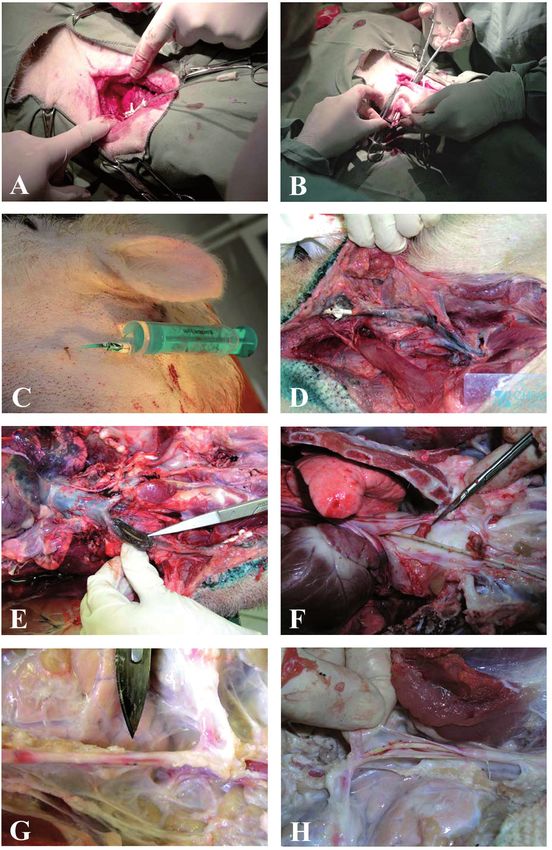
Figure 1. Implantation of the catheter and pathological findings in the experimental animals.
A – introduction of the catheter into v. jugularis externa sin.; B – preparation of subcutaneous tunnel for catheter outlet;
C – outlet of the catheter in regio parotidea; D – trombophlebitis of v. jugularis externa sin. found in the minipig E105;
E – trombophlebitis in v. jugularis externa sin.; F – wall-thrombi in v. cava cranialis found in the minipig E 103; G – v. jugularis
externa sin. without pathological findings; H – v. jugularis externa sin. – catheter inside, without pathological findings
VUAB Pharma, Prague, Czech Republic; 170 mg/kg) tion, using puncture of v. jugularis externa by the
followed by a myorelaxans (Pipecuronii bromidum Seldinger needle. The vein is well accessible for a
– 0.08 mg/kg; Arduan, Chemical Works, Budapest, safe surgical preparation and is the first choice for
Hungary) and subjected to an anatomical autopsy. clinics without monitoring technique since posi-
The autopsy was primarily concentrated on assess- tion of the implanted catheter can be reliably de-
ment of pathological changes in the site of can- termined. Pigs are very liable to vasocontriction
nulation and in the course of the catheter in vein, during surgical manipulation with vessels (Swindle
pathological changes in heart and other organs. and Adams, 1988; Swindle et al., 1998). The va-
soconstriction complicates manipulation with the
vessel and the diminished lumen prevents insertion
Results and Discussion of the catheter into the vein. Such a vein is also
more liable to damage or occlusion during manipu-
Implantation of the catheter and lation, for example during drawing the vein into op-
complications of catheterization eration wound by the use of ligatures. This should
be considered during preparation of the cannulated
The long-term cannulation of v. jugularis exter- vein that must always be done carefully by the blunt
na has been performed in pigs by surgical method method. A topical application of lidocain or pa-
(Schmitd et al., 1988; Swindle and Adams, 1988; paverin proved to prevent spasm of vessels (Swindle
Swindle et al., 1998; Stukelj et al., 2005) or trans- and Adams, 1988). We have topically applied 5 ml
cutaneously (Carrol et al., 1999; Matte, 1999; Fudge of 2% lidocain directly into operation wound and let
et al., 2002) and proved convenient for application is work for 5–7 min. After this period of time, the
of medicaments or collection of blood. We have vein did not exhibit tendency to vasoconstriction
approached to the surgical method of catheteriza- and manipulation with it was safe and comfortable.
Table 1. Pathological findings
Pathological finding E66 E67 E103 E104 E105 E107 E108 E110
Granulom in operation wound + – – – – – – –
Inflammation in subcutum – – – – – – – –
Serom – – – – – – – –
Haematoma – – – – – – – –
Trombophlebitis of v. jugularis externa sin. – – – – + – – –
Trombophlebitis of v. cava cranialis – – + – + – – –
Endocarditis parietalis cordis – – – – – – – –
Endocarditis valvularis cordis – – – – + – – –
Pneumonia catarrhalis acuta – – – – + – – –
Pneumonia intersticialis chronica – – – – – – – –
thrombus
Other – – – – – – –
at catheter
+ = presence of pathological finding
– = absence of pathological finding
369Original Paper Veterinarni Medicina, 53, 2008 (7): 365–372
A small incision in the vein wall has been used in The local swelling was cured by local application
previous studies to facilitate insertion of catheter of Heparoid ung. and disappeared completely in a
into vessels (Yoshikuni et al., 1984; Moritz et al., week. Obstruction of the catheter persisted until
1989; Bain et al., 1991). We used a direct penetra- the end of the experiment, application in the distal
tion of the vein wall by the sharp Seldinger needle. direction worked without problems. Due to indi-
This approach restricts manipulation with the vein, vidual stalling of the pigs, the catheters were well
excludes bleeding into the operation wound and maintained and no withdrawal or dislocation of
prevents the vein wall form an extensive damage. them was observed.
Following implantation of the catheter and removal
of the dispenser, the vein wall firmly embraced the
catheter, which safely prevented leakage of blood, Pathological assessment of animals
but did not limit mobility of the catheter in the
vein. To prevent bleeding, soaking and descendent All experimental animals were euthanased in two
infections at the site of cannulation, we carried out months after catheter implantation. The course of
a tight suture of a part of fascia from m. brachio- the catheter was anatomically preparated and path-
cephalicus and m. sternocephalicus and also sutured ological changes were recorded. No macroscopi-
both muscles together. The loop in the course of the cally visible signs of inflammation were noticed
catheter well prevented the breaks but also an ac- around the outlet of the catheters (Figure 1C). All
cidental removal of the catheter from lumen of the catheters were correctly introduced into v. jugu-
vein. Next, the catheter was anchored to surround- laris externa sin. No inflammation, haematoma
ing muscle tissue by suture through the moveable or blood soaking from the point of implantation
fixation wings. Thus, the catheter was reliably fixed was observed in operation wound. No breaks or
on the site of implantation. obstructing clots were found in the catheters. All
Implantation of the catheter was carried out catheters were implanted into the upper third of v.
without complications in all experimental animals. cava cranialis. We did not observed haemothorax
Preparation of v. jugularis sin. and its catheteriza- or pneumothorax in any of the experimental ani-
tion by the Seldinger method proved to be a safe mals. Afterward, a general autopsy of all animals
and well manageable procedure. No heart arrest was carried out. The results of the autopsies are
or arrhythmias were recorded immediately after summarized in Table 1.
catheterization. A trombophlebitis of v. jugularis ext. sin. was
found in the minipig E105. The trombophlebitis
afflicted the hole course of v. jugularis up to joining
Maintenance of the catheter, its function with v. cava cranialis that was also severely afflicted
and complications by the inflammation (Figure 1D). Clinical symp-
toms of a respiratory disease had been recorded in
Several complications have been reported to oc- this animal and these were confirmed during the
cur during or soon after the central catheterization: autopsy by finding pneumonia catarrhalis acuta in
Catheter displacement, a wrong implantation into both diaphragm lung lobes. However, this finding is
artery, haemotorax, haematoma, catheter embo- frequent in conventional breeding. No other clini-
lism, arrhythmias, air embolism, damage or per- cal symptoms of a disease were observed in this ani-
foration of vein wall. Some complications occur mal, v. jugularis externa dx. was not affected by the
later after catheterization, like thrombosis, throm- inflammation. Catheter was functional for applica-
bophlebitis, thromboembolism, local or system in- tion as well as aspiration. The thrombophlebitis
fection and sepsis (Sznajder et al., 1986; Swindle displayed during autopsy by darkening and thick-
and Adams, 1988; Gonzales et al., 1991; Doierau et ness of the vein wall (Figure 1E) and crepitation in
al., 1993; Yilmazlar et al., 1997; Swindle et al., 1998; the lumen. A thrombus was found in the right heart
Mickley, 2002; Badge et al., 2003; Yoshida, 2003). ventricle on cuspis septalis valvae tricuspidalis,
We have noticed only a local tempered swelling of which argues for a descendent progress of inflam-
subcutum, localized in the site of catheterization at mation from the proximal parts of venous system.
the third day after operation in the minipig E66, and Infection probably originated at the site of catheter
an obstruction of the catheter in proximal direction outlet at the skin surface, even though no symptoms
in the minipig E103 at the Day 20 of experiment. of inflammation were observed there. The autopsy
370Veterinarni Medicina, 53, 2008 (7): 365–372 Original Paper
of the minipig E103 demonstrated presence of two duced Animal Models of Human Disease. Lippincott,
wall-thrombi in v. cava cranialis (Figure 1F). No Williams & Wilkins, Baltimore. 1–9.
other thrombi were found in tissues and this animal Brown D.R., Terris J.M. (1996): Swine in physiological
did not show any clinical symptoms of a disease. and pathophysiological research. In: Tumbleson M.E.,
An interesting finding in this animal was a throm- Schook L.B. (eds.): Advances in Swine in Biomedical
bus attached to the distal tip of the catheter that Research. Vol. 1. Plenum Press, New York. 5–6.
was probably the cause of the failure of aspiration Carroll J.A., Daniel J.A., Keisler D.H., Matteri R.L. (1999):
described above. An amplification of subcutane- Non surgical catheterization of the jugular vein in
ous tissue was observed in the animal E66 and was young pigs. Laboratory Animals, 33, 129–134.
diagnosed as a granulom of the size 3.8 × 5 cm. The Doireau V., Daoud P., Pasche J., Le Biodos J. (1993): Right
granulom was freely located in subcutum, transi- intraventricular thrombus. Rare complication of caval
tion of inflammation to the catheterized vein was catheterization. Archives Francaises de Pediatrie, 50,
not observed. In the other catheterized animals, no 887–889.
symptoms of thrombophlebitis or endocarditis was Fudge M., Coleman R.E., Parker S.B. (2002): A minimally
noticed (Figure 1G, H). No macroscopic pathologi- invasive percutaneous technique for jugular vein cath-
cal changes were observed in other organs. eterization in pigs. Contemporary Topics in Labora-
In conclusion, we have demonstrated in the tory Animal Science, 41, 38–42.
present study that central venous catheterization by Gardner T.J., Johnson D.L. (1988): Cardiovascular sys-
Seldinger method is suitable for experimental ani- tem. In: Swindle M.M., Adams R.J. (eds.): Experimen-
mals that must be repeatedly given large volumes tal Surgery and Physiology: Induced Animal Models
of solutions into venous system or in which blood of Human Disease. 74–124.
samples are often collected. The Seldinger method Gonzales A., Rahimtoola S.H., Kulick D.L. (1991): Tech-
of catheter implantation eliminates the complica- nique of vascular access. In: Kulick D.L., Rahimtoola
tions associated with the central venous cathetri- S.H. (eds.): Techniques and Applications in Interven-
zazation. Even though the experimental animals tional Cardiology. Mosby, St. Louis. 1–16.
were kept in conventional breeding conditions in Matte J.J. (1999): A rapid and non-surgical procedure
this study, the autopsy showed complications in for jugular catheterization of pigs. Laboratory Ani-
only two of eight animals. mals, 33, 258–264.
Mickley V. (2002): Central venous catheters: many ques-
tions, few answers. Nephrology, Dialysis, Transplan-
Acknowledgement tation, 17, 1368–1373.
Moritz W.M., Dawe J.E., Holliday F.J., Elliot S., Mattei A.J.,
The authors would like to thank Ms. Jirina Zelen- Thomas L.A. (1989): Chronic central vein catheteriza-
kova for competent technical assistance. Ms. Jitka tion for intraoperative and long-term venous access in
Cervena and Ms. Lenka Cizkova for excellent care swine. Laboratory Animal Science, 39, 153–155.
of experimental animals. Mullen Y., Taura Y., Nagata M., Miyazawa K., Stein E.
(1992): Swine as a model for pancreatic betacell trans-
plantation. In: Swindle M.M. (ed.): Swine as Models
References in Biomedical Research. Iowa State University Press,
Ames, I.A. 16–34.
Badge V.S., Ninan B., Rajan S., Cherian K.M. (2003): Schmitd W., Dehn A., Hutter J.F. (1988): A central venous
Kinked triple-lumen central venous catheter due to catheter for long-term studies on drug effect and phar-
superior vena caval cannula: An unusual complica- macokinetics in Munich minipigs. European Journal
tion. Journal of Cardiothoracic and Vascular Anesthe- of Drug Metabolism and Pharmacokinetics, 13, 143–
sia, 17, 145–146. 147.
Bain A.F.S., Ting J., Simeonovic J.C.H., Wilson D.J. Seldinger S.I. (1953): Catheter replacement of the needle
(1991): Technique of venous catheterization for se- in percutaneous arteriography – a new technique. Acta
quential blood sampling from the pig. Journal of In- Radiologica, 39, 368–376.
vestigative Surgery, 4, 103–107. Seldinger S.I. (1984): The Seldinger technique – cath-
Bolton L.L., Pines E., Rovee D.T. (1988): Wound healing eter replacement of the needle in percutaneous arte-
and integumentary system. In: Swindle M.M., Adams riography – a new technique (reprinted). American
R.J. (eds.): Experimental Surgery and Physiology: In- Journal of Roentgenology, 142, 5–7.
371Original Paper Veterinarni Medicina, 53, 2008 (7): 365–372 Smith A.C., Spinale F.G., Swindle M.M. (1990): Cardiac Swindle M.M., Smith A.C., Goodrich J.A. (1998): Chronic function and morphology of Hanford miniature swine cannulation and fistulization procedures in swine: A and Yucatan miniature and micro swine. Laboratory review and recommendations. Journal of Investigative Animal Science, 40, 47–50. Surgery, 11, 7–20. Stanton H.C., Mersmann H.J. (1986): Swine in Cardio- Sznajder J.I., Zveibil F.R., Bitterman H. (1986): Central vascular Research. Vol. 1–2. CRC Press, Inc., Boca vein catheterization: Failure and complication rates Raton. by three percutaneous approaches. Archives of Inter- Stukelj M., Mihelcic D., Butinar J., Nemec A., Pecar J. nal Medicine, 146, 259–261. (2005): Surgical intravenous catheterisation of pig. Tumbleson M.E. (1986): Swine in Biomedical Research. Slovenian Veterinary Research, 42, 43–48. Vol. 1–3. Plenum Press, New York, N.Y. 698 pp. Swindle M.M. (1983): Basic Surgical Exercises Using Tumbleson M.E., Schook L.B. (eds.) (1996): Advances in Swine. Praeger Publishers, New York. 237 pp. Swine in Biomedical Research. Vol. 1–2. Plenum Press, Swindle M.M. (1992): Swine as Models in Biomedical Re- New York, N.Y. 462 pp. search. Iowa State University Press, Ames I.A.462 pp. Yilmazlar A., Bilgin H., Korfali G., Eren A., Ozkan U. Swindle M.M. (1998): Surgery, Anesthesia and Experi- (1997): Complications of 1303 central venous cannu- mental Techniques in Swine. Iowa State University lations. Journal of the Royal Society of Medicine, 90, Press, Ames, I.A. 329 pp. 319–321. Swindle M.M., Adams R.J. (1988): Experimental Surgery Yoshida S. (2003): A lethal complication of central ve- and Physiology. In: Induced Animal Models of Human nous catheterization. The Lancet, 362, 569. Disease. Lippincott Williams & Wilkins, Baltimore, Yoshikuni T., Yukio Y., Nobuo A., Hiroyasu O., Nahoko M.D. 350 pp. K., Akira E. (1984): Chronic blood vessel catheteriza- Swindle M.M., Horneffer P.J., Gardner T.J., Gott V.L., tion in Gottingen miniature pigs and application to a Hall T.S., Sturat R.S., Baumgartner W.A., Borkon A.M., preliminary bioavailability study of nalidixic acid. Che- Galloway E., Reitz B.A. (1986): Anatomic and anes- mical & Pharmaceutical Bulletin, 32, 2851–2854. thetic considerations in experimental cardiopulmo- nary surgery in swine. Laboratory Animal Science, 36, Received: 2007–09–03 357–361. Accepted after corrections: 2008–06–06 Corresponding Author: Dusan Usvald, DVM, Institute of Animal Physiology and Genetics of the Academy of Sciences of the Czech Republic, v.v.i., Rumburska 89, 277 21 Libechov, Czech Republic Tel. +420 315 639 565, fax +420 315 639 510, e-mail: usvald@iapg.cas.cz 372
You can also read