POLYMERIC DRUG DELIVERY TECHNIQUES Translating Polymer Science for Drug Delivery - Sigma-Aldrich
←
→
Page content transcription
If your browser does not render page correctly, please read the page content below
POLYMERIC DRUG DELIVERY TECHNIQUES Translating Polymer Science for Drug Delivery
aldrich.com/matsci
Preface Solubility Enhancement
Low drug solubility and stability often reduce the effectiveness of an
Rapid advances in medicine and otherwise promising therapeutic candidate. Drug delivery systems
biotechnology have driven the field of drug can be formulated to improve the in vivo solubility of lipophilic and
discovery and led to the development of hydrophobic drugs by encapsulation in a drug delivery carrier or by
many new highly potent and target-specific conjugation with a polymer.
drug candidates. Despite the fast pace of
research and early-stage discovery, many drug
candidates fail during preclinical evaluation Selecting a Polymeric Drug Delivery System
due to poor efficacy, limited bioavailability,
There are three main categories of polymeric drug delivery systems;
and other challenges associated with effective Nicolynn Davis, Ph.D.
colloidal carriers (micro, nanoparticles, micelles, micro/nanogels),
drug delivery. Small molecule drugs can Aldrich Materials Science
implantable networks or hydrogels, and polymer drug conjugates.
suffer from low solubility, poor stability, short Sigma-Aldrich, Milwaukee, WI USA
Email: nicolynn.davis@sial.com Unfortunately, there is no “silver bullet” for effective delivery of
circulation time, and non-specific toxicity
broad classes of therapeutics. Rather, selection of a drug delivery
limiting their therapeutic efficacy. Biopharmaceuticals such as nucleic
system must be driven by the nature of the drug and the inherent
acids, peptides, and proteins are often limited by poor stability and
properties of the drug delivery system (Figure 1). Drug properties,
rapid clearance from the body. These challenges, coupled with the
including chemistry, solubility, potency, site of action, and clearance
complexity and diversity of new pharmaceuticals, are fueling the
rate, each impact the proper selection of a drug delivery system that
evolution of novel drug delivery systems that overcome bioavailability
can achieve the desired outcomes. In addition, the choice of drug
and delivery obstacles. However, despite the growing importance of
delivery system determines the drug loading capacity, longevity of
polymer drug delivery methodologies, the materials and methods of
release, and the route best suited for administration. Furthermore,
drug delivery are not widely available to those outside the polymer
characteristics of the drug delivery system (size, surface charge and
synthesis field.
hydrophobicity, shape, flexibility, inclusion of targeting moieties) will
The objective of effective drug delivery is improving the affect performance and distribution in the body. Each drug delivery
pharmacokinetics and pharmacodynamics of each therapeutic to system has inherent advantages and limitations (Table 1). It should be
enable drug delivery to the right place, at the right time and in the noted that drug release from any carrier is determined by a complex
right amount. Delivery systems apply three main strategies to enable interaction between the drug properties, polymer characteristics, and
improved drug efficacy. environmental/in vivo conditions.
Controlled Release Drug
Drug efficacy can be enhanced by maintaining the concentration
• Drug properties
within the therapeutic window (effective dose). Polymer carriers loaded (solubility, stability)
with therapeutics enable controlled temporal and spatial release • Desired site of Drug Delivery System Formulation
action
of a drug by controlling drug diffusion, the rate of dissolution, or • Desired release rate
• Loading capacity
degradation of the carrier. • Delivery challenge
• Route of
administration Polymer Selection
associated with
drug • Compatibility with
Targeted Delivery drug
Drug efficacy can be enhanced and toxicity minimized by localization • Desired release
kinetics, including
at the organ, tissue, cellular, or organelle level. Targeting can be degradation rate
achieved by coating or conjugating the carrier with affinity reagents
such as nucleic acids, peptides, antibodies, or others that bind specific
cell receptor proteins, nucleic acids, or polysaccharides. Figure 1. Drug delivery formulation selection process.
Table 1. Advantages and limitations of drug delivery systems.
Drug Delivery System and
Polymer Types Advantages Limitations
Microparticles
• Biodegradable polymers • Encapsulate a variety of drugs • Burst release possible, may lead to local toxicity
• Natural polymers • Sustained release can be achieved
Nanoparticles
• Biodegradable polymers • Stable delivery system • Non-specific uptake in RES
• Natural polymers • Small size enables enhanced retention and permeation into tissue and tumor
Micelles
• Amphiphilic block copolymers • Enhanced solubility for hydrophobic drugs • Less stable, may require additional crosslinking
• Facile synthesis
Drug Conjugates
• Hydrophilic polymers • E xtended circulation half-life, reduced clearance due to increased drug • Activity of drug can decrease due to conjugation
• Dendrimers hydrodynamic radius • Approach provides sustained but not controlled release
• Decreased drug immunogenicity and degradation • Low loading capacity of drug
Hydrogels or Implants
• Hydrophilic polymers • Broad range of release timeframes (weeks to months) • Drug solubility may limit utility
• Biodegradable polymers • Useful for localized delivery • Limitation to route of delivery achievable
• Natural polymers • Improved patient compliance due to infrequent dosing • Delivery may require incision or larger gauge needle
• Risk of local dose dumping
TO ORDER: Contact your local Sigma-Aldrich office or visit aldrich.com/matsci.
Drug Delivery Material Choices                                                                  Table of Contents
Polymer selection greatly influences the performance of the drug
delivery system. Careful polymer selection is essential to control the                            Articles
encapsulation efficiency, release rate, and duration of release. Many
polymers can be formulated into various drug delivery systems to                                  Colloidal Carriers for Drug Delivery
address the three key drug delivery strategies to enable improved                                 Polymer Micelles for Drug Delivery                                       3
drug efficacy (Table 2). The diversity of polymer building blocks                                 Alice Du, Martina Stenzel
can further complicate formulation decisions. As discussed by Du
and Stenzel (in this publication), the most critical factor in polymer                            Biodegradable Colloidal Carriers in Drug Delivery Applications           8
selection is considering the interaction of the drug and polymer.                                 Bin Wu, Theresa Logan
Polymer selection will determine the mechanism for drug release (bulk                             Lipid-polymer Hybrid Nanoparticles for Drug Delivery Applications        14
erosion, system degradation), and the choice of polymer properties                                Sangeetha Krishnamurthy, Juliana M. Chan
(molecular weight, surface charge) will influence release rate and
impact pharmacokinetics. Further fine-tuning of release from drug                                 Crosslinked Chitosan Nanoparticles and Chemical Modifications for        18
delivery systems can be achieved by using multiple types of polymers                              Drug Delivery Applications
or including additives.                                                                           Shady Farah, Joshua Doloff, Daniel Anderson, Robert Langer
Table 2. Polymer categories and the drug delivery strategies they enable.                         Poly(N-isopropylacrylamide)-based Stimuli-responsive Materials           22
                                                                                                  Ganga Panambur, Nicolynn Davis
                                          Controlled
                                           Release
                                                             Targeted
                                                             Delivery
                                                                             Solubility
                                                                            Enhancement
                                                                                                  Shape Change Poly(N-isopropylacrylamide) Microstructures                 28
                                                                                                  for Drug Delivery
                                                                                                  Tanvi Shroff, ChangKyu Yoon, David H. Gracias
Biodegradable nanoparticles                   ✔                 ✔

Biodegradable micelles                                                          ✔                 Hydrogels for Drug Delivery
Responsive polymers                           ✔                 ✔                                 Formulation of Poly(ethylene glycol) Hydrogels for Drug Delivery         31
Polymeric hydrogels                           ✔                 ✔                                 Tyler Lieberthal, W. John Kao
PEG conjugation                                                                 ✔

Polyoxazoline polymers                                                          ✔
                                                                                                  Drug Conjugates
Dendrimers                                                      ✔               ✔                 Protein PEGylation                                                       36
                                                                                                  Steve Brocchini

About This Guide                                                                                  Poly(2-oxazoline)s for Drug Delivery
                                                                                                  Rainer Jordan, Robert Luxenhofer, Alexander V. Kabanov
                                                                                                                                                                           42

This guide is intended to provide an overview of polymeric drug                                   Polyoxazolines: An Alternative to Polyethylene Glycol                    46
delivery systems as well as provide the corresponding example                                     Nicolynn Davis
formulation protocols and product information required to utilize these
techniques in the laboratory. The publication has been developed                                  Dendritic Polyester Scaffolds: Functional and Biocompatible Precision    47
to enable those without a polymer chemistry background to use                                     Polymers for Drug Delivery Applications
polymers to solve their drug delivery research challenges; but, we have                           Sandra García-Gallego, Michael Malkoch
also kept the expert in mind by including a number of cutting-edge
methodologies. This guide is arranged according to drug delivery
                                                                                                  RAFT Polymeric Carriers for Antibody Drug Conjugates of Biologic Drugs   52
                                                                                                  Patrick S. Stayton, Anthony Convertine, Geoffrey Berguig
strategies, and these strategies are noted within each method. We
hope this publication will enable chemists, engineers, pharmaceutical                             Linear and Branched PEIs as Nonviral Vectors for Gene Delivery           57
scientists, and biologists to explore different drug delivery techniques                          Philip Dimitrov, Nicolynn Davis
to facilitate translational research.
                                                                                                  Featured Products
                                                                                                  Diblock Copolymers                                                        6
                                                                                                  Poly(lactide-co-glycolide) Copolymers                                    11
                                                                                                  End-functionalized Poly(l-lactide)s                                      12
                                                                                                  PNIPAM and End-functionalized PNIPAM                                     26
                                                                                                  Bifunctional and Multi-arm PEGs                                          33
                                                                                                  Poly(oxazoline)s                                                         45
                                                                                                  bis-MPA Dendrimers and Hyperbranched PEG Dendrimers                      50

                                                                                                  Indexes
                                                                                                  Method                                                                   60
                                                                                                  Trademark                                                                60

                                                  For questions, product data, or new product suggestions, contact us at matsci@sial.com.                                   1
NANOMATERIALS FOR
  DRUG DELIVERY AND
THERANOSTICS
Appropriate surface modification allows the conjugation of
nanoparticles to a wide range of biomolecules, enabling their
delivery and preferential accumulation at the site of action. This
leads to enhanced therapeutic efficacy and reduced cytotoxicity.
Aldrich® Materials Science continues to expand its nanomaterials
product portfolio, with a wide selection of nanomaterials of
varying dimensions and surface functionalization for biomedical
applications, including:
 Gold nanoparticles
yy
 Silver nanoparticles
yy
 Iron oxide nanoparticles
yy
 Carbon nanotubes
yy
 Fluorescent nanodiamonds
yy
 Silica nanobeads
yy

To access the complete portfolio, visit
aldrich.com/nanobiomed
Polymer Micelles for Drug Delivery
                                                                                                                                          Controlled     Targeted      Solubility
                                                                                                                                           Release       Delivery     Enhancement

POLYMER MICELLES FOR DRUG DELIVERY
                                                                                          Since many drugs have a strong tendency to crystallize, theoretical
                                                                                          models of the polymer–drug interactions treat this like a solution
                                                                                          where the presence of the homogenous mixture is determined by the
                                                                                          miscibility curve of its phase diagram on the molecular level. Moreover,
                                                                                          the models discussing the thermodynamic stability of a binary system
                                                                                          are based on a fast equilibrium. This may not always be the case since
                                                                                          polymers with high Tg values may trap the drug in the matrix, resulting
                                                                                          in a kinetically stable system. Readers who are interested in the
Alice Du, Martina Stenzel*                                                                underpinning thermodynamic principles are referred to an excellent
Centre for Advanced Macromolecular Design                                                 review article.8
School of Chemistry
University of New South Wales, Australia                                                  How, then, can one choose the right polymer for the right drug to
*
 Email: M.Stenzel@unsw.edu.au
                                                                                          achieve good loading and high stability? The assumption “like dissolves
                                                                                          like” is a good starting point. This rule of thumb is based on the Flory–
Introduction                                                                              Huggins parameter χ in Equation 1:
Polymeric micelles obtained from the self-assembly of amphiphilic                                                               (     −   )2
                                                                                                                            =                                           (1)
block copolymers are probably one of the most common drug
delivery carriers among polymeric nanoparticles.1–4 The rise of highly
                                                                                          where δs and δp are the Scatchard–Hildebrand solubility parameter
controlled polymerization techniques, especially processes such as
                                                                                          of the solute and the polymer, respectively.7 In short, polymers that
ATRP5 and RAFT,6 has led to an extraordinary surge of new types of
                                                                                          are chemically similar to the drug should enable the highest loading
block copolymers fit for biomedical applications. Facile control over
                                                                                          capacity. A good example is doxorubixin conjugated to a polymer.9
the polymer structure has also meant access to a large array of self-
                                                                                          While the drug attached to the polymer was found to be inactive,
assembled morphologies including micelles, cylindrical micelles, and
                                                                                          polymer micelles constructed with the polymer-drug conjugate
polymersomes. Micelles in particular are at the center of attention
                                                                                          created an environment that had the highest compatibility possible
as potential drug carriers due to a core-shell structure that is highly
                                                                                          with free doxorubicin leading to an increased loading capacity.
water soluble while still maintaining a hydrophobic core suitable for
                                                                                          Decorating the polymer with the same drug to be loaded is an
hydrophobic drugs. This is crucial for many drugs since they are often
                                                                                          effective but cost-prohibitive option. Alternatively, subtle changes
rendered insoluble in water, and loading them into drug carriers can
                                                                                          to the interior polymeric structure by altering the substitution of the
increase their solubility by several orders of magnitude.4,7
                                                                                          polymer can maximize loading. For example, a PEO-b-PCL polymer
                                                                                          was modified with benzyl, carboxyl, stearyl, palmitoyl, and cholesteryl
Choice of Block Copolymers                                                                functional groups with the aim of varying the hydrophobicity to tailor
                                                                                          the polymer matrix toward the highest possible loading capacity of the
The choice of drug carriers can be daunting. In addition to a range                       chosen drug.10
of commercially available block copolymers, there is basically no                         However, not every lab has synthetic chemists capable of carefully
limit to the design of amphiphilic structures thanks to advances in                       tailoring a drug carrier to the drug. A tool is needed to help predict
polymer design. Block copolymers can be further complemented                              the best possible polymer structure for the drug. This is not easy, but
by other amphiphilic polymers (such as miktoarm starpolymers,                             an initial estimate can be obtained using the group contribution
multiblock copolymers, and star polymers) to enable the formation                         method to determine approximate partial solubility parameters. In
of compartmentalized micelles. Whatever architecture is chosen, the                       this approach, the polymer and drug are essentially dissected into
primary consideration should be the compatibility between the drug                        their different functional groups, which then are used to determine
and the polymers.7 The polymer–drug interaction plays an important                        dispersion forces, dipole-dipole interactions, and hydrogen bonding of
role in the drug-loading capacity of a carrier and the stability of the                   polymer and drug.11 This approach frequently has been employed to
drug in the matrix, which ultimately affects the shelf-life of the carrier.               predict the most suitable polymeric drug carrier,12–13 but one also needs
The miscibility of a drug with the polymeric matrix can be described                      to exercise caution since many aspects are not taken into account
by the Flory–Huggins theory. This contains both entropy and enthalpy                      resulting in unsuitable predictions. More refined approaches are based
components, expressed by the Flory–Huggins interaction parameter χ,                       on molecular dynamics simulation,14 which can reveal the critical role of
that describe the interaction between the polymer and the drug.                           H-bonding, an interaction that is often more crucial than hydrophobic
In other words, the Flory–Huggins parameter χ is a measure of                             forces to achieve high drug loading.15 Further theories, based on a free
compatibility between polymer and drug.

                                            For questions, product data, or new product suggestions, contact us at matsci@sial.com.                                                 3
aldrich.com/matsci

energy model, describe the solubilization of low molecular weight                             Solvent Evaporation
compounds in micelles. Based on this information, one can derive                              In the solvent evaporation technique, polymer and drug are dissolved
conclusions on the distribution of the drug in the micelle. aggregation                       in an organic solvent with a low boiling point, followed by evaporation
number, the size of the micelle, the effect on micelle stability, and                         and subsequent dehydration.19 The chosen organic solvent is selective
the maximum extent of solubilization.16 A summary of various                                  toward one block, which results in the formation of micelles in non-
computational approaches can be found in a review article by Allen                            aqueous solutions. The outcome is usually determined by the type
and co-workers.17                                                                             of solvent, the concentration of polymer and drug, and the rate of
                                                                                              evaporation. The limitation of this approach lies in the limited choice of
Methods: Micelle Drug Loading                                                                 solvents, and there is no guarantee the resulting particles will be well-
                                                                                              defined core-shell particles that can be easily redissolved in water.
Once a suitable polymer has been identified, the drug must be loaded
into the micelles.7,18 Direct mixing of the hydrophobic drug and the                          Example Method
micelle in water, although suitable for some selected systems, is rarely                      1. Dissolve 2 mg of drug and 20 mg of polymer in methanol (or any
capable of dissolving both the drug and polymer. Therefore, other                                other low-boiling solvent that can dissolve both components).
techniques must be used to ensure solubilization of both the drug                             2. Evaporate solvent under vacuum.
and the polymer. A common solvent is capable of fully dissolving                              3. Add distilled water, incubate at 40 °C for 10 min, and vortex to
both the drug and the block copolymer into the unimeric state (single                            obtain a clear solution.
block copolymers). A clear solution can serve as an initial indication
the polymer has dissolved, but it is advisable to test for the absence                        Co-solvent Evaporation
of micelles or other aggregates using light scattering techniques to                          Co-solvent evaporation proceeds by adding water directly to the
ensure full solvation. Examples of commons drug-loading techniques                            organic solvent to cause the self-assembly of the micelle and
are described in Figure 1.                                                                    encapsulation of the drug. The outcome is controlled by the type of
                                                                                              solvent, the ratio between organic solvent and water, the concentration
    Solvent Evaporation                                                                       of water and drug, rate of solvent evaporation, and the order and
                                                                                              rate of mixing.20 This approach is limited by the choice of solvent, but
                                                                                              usually results in higher drug encapsulation efficiencies.

                                                                                              Example Method
                                                                                              1. Dissolve 20 mg of polymer and 2 mg of drug in 1 mL of acetone,
      Drug and ABC in             Evaporate organic             Redissolve in water              THF, or acetonitrile.
      organic solvent             solvent under vacuum                                        2. Add 2 mL of water dropwise to the organic solvent (or vice versa).
                                                                                              3. Mix for 4 h, followed by the evaporation of the organic solvent.
    Co-solvent Evaporation
                                                                                              Dialysis
                                                                                              Dialysis is probably the most versatile and most common technique
                                                                                              used for drug encapsulation since it allows the use of high-boiling
                                                                                              solvents such as DMSO, which is removed by dialysis and replaced
                                                                                              with water. Although this approach is applicable to many solvent
      Drug and ABC in       Addition of organic                Evaporation of                 systems, the drug loading efficiency is usually lower than the
      organic solvent       phase into water                   organic solvent                co-solvent evaporation and the technique can be time-consuming.
                            phase or vice versa
                                                                                              The slow process can aid the formation of thermodynamically stable
 Dialysis                                                                                     morphologies. A final dialysis step to remove solvent and free drug
                                                                                              is often crucial to obtain a product free of organic solvent while
                                                                                              maintaining maximum drug loading. However, while extensive dialysis
                                                                                              can assist in the thorough purification of the product, it also can cause
                                                                                              the release of the already encapsulated drug and result in low drug
                                                                                              encapsulation efficiencies.
         Slow addition of water to drug             Removal of organic
         and ABC in organic solvent                 solvent by dialysis                       Example Method
                                                                                              1. Dissolve 20 mg of polymer and 2 mg of drug in 1 mL of DMF.
    Flash Nanoprecipitation                                                                   2. Add 5 mL of water slowly, with the help of a syringe pump, if
                                                                                                 possible, to control the rate of water addition.
                               H2O
                                                                                              3. Dialyze against water using a tubular cellulose membrane (Sigma
                               Fast mixing                                                       Prod. No. Z726176).
                               of both
                               phases
                               using
                               confined
                               impinging                   Removal of organic
                               jet mixer                   solvent by dialysis

Figure 1. Techniques for drug loading of micelles

4                                                    TO ORDER: Contact your local Sigma-Aldrich office or visit aldrich.com/matsci.
Polymer Micelles for Drug Delivery

Flash Nanoprecipitation                                                              producing a family of relatively uniform spheroids without the use of
Flash nanoprecipitation is a relatively new technique that offers a                  specialized equipment.
more rapid solution than other time-consuming methods. Fast mixing
and precipitation into a non-solvent for the drug and one polymer
block results in a kinetically trapped structure. Although the resulting             Method: Characterization of Toxicity
structures do not have well-defined internal phase boundaries,
as would be the case in thermodynamically stable structures, the                     with Spheroids
approach provides an alternative to achieve a fast throughput.21                     Typical steps for culturing spheroids in this manner include:
Example Method                                                                       1. Pre-determine the seeding number of cells required for each
1. Prepare a solution of 40 mg of polymer and 20 mg of drug in                          spheroid (usually 1,000–2,000 cells/spheroid).
   1 mL of THF.                                                                      2. Prepare a cell suspension with a per mL concentration 100× that of
2. Use a confined impinging jet mixer to mix the solution with                          the initial seeding cell number where the suspension is prepared in
   1 mL of water.                                                                       the growth medium used to culture the cells.
3. Introduce the exit stream into 8 mL of water:THF (9:1 v/v%).                      3. Place 10 µL of the well-mixed cell suspension on the inside surface
4. Dialyze against water using a tubular cellulose membrane.                            of the lid of a petri dish.
                                                                                     4. Repeat until the required number of seeding droplets are placed.

Characterization                                                                     5. Flip the petri dish lid upside down so that the seeding droplets
                                                                                        are “hanging” and place on top of a petri dish filled with 15 mL of
Independent of the technique, the characterization of drug-loaded                       sterile PBS.
micelles is similar to other nanoparticles. Parameters of interest are the           6. Incubate at 37 °C and 5% CO2 for a minimum of 3 days without
drug encapsulation efficiency (EE) and the drug-loading capacity (LC)                   disturbing the petri dish.
(see Equation 2):                                                                    7. Check the spheroids daily until the desired size has been reached
                                                                                        (usually 300–400 µm in diameter).
                                %=                                                   8. Once culturing is complete, move the spheroids into a 96-well plate
                                                                     (2)                filled with 200 µL of medium/well.
                                %=                                                   9. Culture the spheroids for one more day at 37 °C and 5% CO2 with
                                                                                        slow rotation of the plate on a 4-way mixer during that time.
where WL is the weight of loaded drug, W0 the quantity of drug initially             Once the spheroids have been cultured and are of the desired shape
added, and WN the weight of the nanoparticle.                                        and size, the micelle formulation can be loaded for testing:
The International Organization for Standardization (ISO) published a                 1. Remove 170 µL of medium from each well in the 96-well plate
catalog of properties for the full characterization of nanoparticles. The               containing the spheroids.
Guidance on Physico-chemical Characterization of Engineered Nanoscale                2. Add 100 µL of twice-concentrated culture medium and 100 µL of
Materials for Toxicologic Assessment (ISO/TR 13014:2012) includes                       the twice-concentrated micellar solution to each well.
dimensions, shape, specific surface area, surface charge, composition,
                                                                                     3. Incubate for the desired amount of time at 37 °C and 5% CO2.
and purity, among others. While most of these points apply to micelles
                                                                                     4. At the desired time points, move the spheroids to be tested into a
as well, the evaluation of the stability of the micelle can be considered
                                                                                        new plate and wash with PBS.
a crucial aspect when trying to evaluate micelles for drug delivery
purposes. It has been shown the dynamic behavior of micelles can                     5. Measure the viability of the spheroids via several methods,
affect their cellular uptake22 and their rate of exocytosis.23 The physico-             including determination of DNA amount or measuring acid
chemical characterization of nanoparticles is then complemented by                      phosphatase activity.32
the Compilation and Description of Toxicological Screening Methods for               Additional tips for culturing spheroids include:
Manufactured Nanomaterials (ISO/TR 16197:2014), which contains a list
of recommended experiments to understand toxicity, accumulation,                     1. Use the surface tension of the droplet to naturally maintain its
and other factors. The reader is referred to comprehensive                               shape when initially placing it on the petri dish lid.
publications on this topic, which describe background and also give                  2. Place droplets at least 1 cm apart to give room for the droplets to
practical advice.24,25                                                                   spread slightly upon culturing.
                                                                                     3. Prepare at least 25% more spheroids than required in case of
Multicelluar spheroids are an established drug discovery technique that                  undesired spheroid shape or size.
is making its way into the nanomedicine area, including the testing
                                                                                     Examples of spheroids are provided in Figure 2. Confocal microscopy
of drug-loaded micelles.26–28 Interestingly, while some drug-loaded
                                                                                     can be used to visualize micelle penetration if fluorescence is
micelles can have poor performance in monolayer cell models, the
                                                                                     incorporated into the polymeric structure.
results in a three-dimensional (3D) cell culture experiment can differ
noticeably.26 The key to this behavior is the differences in penetration
of drugs and micelles into the multicellular spheroid. While this aspect
only plays a minor role in two-dimensional (2D) models, it becomes
one of the main parameters in understanding enhanced delivery in
a 3D environment.29 These 3D models can be further combined with
2D models to create sophisticated systems that mimic the tumor
microenvironment to simulate the behavior of micelles in vivo.30
A typical procedure to test the toxicity of drug-loaded micelles is
outlined below. Although the researcher can choose from a range                      Figure 2. A) Penetration of uncrosslinked and fluorescent micelles into prostate cancer
                                                                                     (LNCaP) spheroids as visualized by confocal microscopy at a depth of 90 µm (yellow scale
of ways to culture spheroids,31 only the “hanging drop” method is                    bar = 100 µm); B) LNCaP spheroids after 4 days culture (black scale bar = 300 µm); C) LNCaP
discussed here. The hanging drop method is a convenient method of                    spheroids after 14 days treatment with the uncrosslinked micelles which have been
                                                                                     conjugated with paclitaxel.

                                       For questions, product data, or new product suggestions, contact us at matsci@sial.com.                                                     5
aldrich.com/matsci
Conclusions
(10) Falamarzian, A.; Lavasanifar, A. Macromol. Biosci. 2010, 10, 648–656.
(11) J. Liu, Y. X., C. Allen. J. Pharm. Sci 2004, 93, 132–143.
(12) Kim, Y.; Liemmawa, E. D.; Pourgholami, M. H.; Morris, D. L.; Stenzel, M. H. Macromolecules
The delivery of drugs using polymeric micelles has now matured into 2012, 45, 5451–5462.
(13) Sharma, A.; Soliman, G. M.; Al-Hajaj, N.; Sharma, R.; Maysinger, D.; Kakkar, A.
a well-established field. Compatibility between the drug and polymer Biomacromolecules 2012, 13, 239–252.
is the key to success in obtaining maximum loading efficiency. The (14) Patel, S. K.; Lavasanifar, A.; Choi, P. Biomacromolecules 2009, 10, 2584–2591.
scientist can choose from a range of tools to load the drug. Although (15) Schulz, A.; Jaksch, S.; Schubel, R.; Wegener, E.; Di, Z.; Han, Y.; Meister, A.; Kressler, J.; Kabanov,
many drugs can be loaded using the above techniques, it should be A. V.; Luxenhofer, R.; Papadakis, C. M.; Jordan, R. ACS Nano 2014, 8, 2686–2696.
(16) Nagarajan, R.; Ganesh, K. Macromolecules 1989, 22, 4312–4325.
noted that a range of drugs—such as drugs that have a low drug- (17) Huynh, L.; Neale, C.; Pomès, R.; Allen, C. Nanomedicine: Nanotechnology, Biology and
loading efficiency—are best delivered by conjugating them to the Medicine 2012, 8, 20–36.
block copolymer directly, instead of relying on physical attraction alone. (18) Gaucher, G.; Dufresne, M.-H.; Sant, V. P.; Kang, N.; Maysinger, D.; Leroux, J.-C. J. Controlled
Release 2005, 109, 169–188.
It must be noted that this article has not touched upon the benefits
(19) Lavasanifar, A.; Samuel, J.; Kwon, G. S. J. Controlled Release 2001, 77, 155–160.
of crosslinking micelles.2,33 As briefly discussed here, the dynamic (20) Aliabadi, H. M.; Elhasi, S.; Mahmud, A.; Gulamhusein, R.; Mahdipoor, P.; Lavasanifar, A. Inter.
properties of the micelles and the potential disassembly may affect J. Pharm. 2007, 329, 158–165.
the interaction with biological media, and crosslinking may circumvent (21) York, A. W.; Zablocki, K. R.; Lewis, D. R.; Gu, L.; Uhrich, K. E.; Prud’homme, R. K.; Moghe, P. V.
Adv. Mater. 2012, 24, 733–739.
the issue as it may enhance characteristics such as cellular uptake,22,23 (22) Kim, Y.; Pourgholami, M. H.; Morris, D. L.; Stenzel, M. H. Biomacromolecules 2012, 13,
movement in multicellular tumors,29 and in vivo circulation.33 In 814–825.
summary, polymeric micelles provide limitless avenues of modification (23) Kim, Y.; Pourgholami, M. H.; Morris, D. L.; Lu, H.; Stenzel, M. H. Biomater Sci 2013, 1, 265–275.
possibilities and represent a versatile method of delivering a wide (24) Hall, J. B.; Dobrovolskaia, M. A.; Patri, A. K.; McNeil, S. E. Nanomedicine (Lond) 2007, 2,
789–803.
range of drugs. (25) McNeil, S. Characterization of Nanoparticles Intended for Drug Delivery. Humana Press:
2011; Vol. 697.
References (26) Du, A. W.; Lu, H.; Stenzel, M. H. Biomacromolecules 2015, 16, 1470–1479.
(1) Xiong, X.-B.; Falamarzian, A.; Garg, S. M.; Lavasanifar, A. J. Controlled Release 2011, 155, (27) Sarisozen, C.; Abouzeid, A. H.; Torchilin, V. P. Eur J Pharm Biopharm 2014, 88, 539–50.
248–261. (28) Jiang, Y.; Lu, H.; Khine, Y. Y.; Dag, A.; Stenzel, M. H. Biomacromolecules 2014, 15, 4195–4205.
(2) Elsabahy, M.; Wooley, K. L. Chem. Soc. Rev. 2012, 41, 2545–2561. (29) Lu, H.; Utama, R. H.; Kitiyotsawat, U.; Babiuch, K.; Jiang, Y.; Stenzel, M. H. Biomaterials
(3) Cabral, H.; Kataoka, K. J. Controlled Release 2014, 190, 465–476. Science 2015.
(4) Lu, Y.; Park, K. Int. J. Pharm. 2013, 453, 198–214. (30) Gao, H.; Yang, Z.; Zhang, S.; Pang, Z.; Liu, Q.; Jiang, X. Acta Biomater 2014, 10, 858–67.
(5) Siegwart, D. J.; Oh, J. K.; Matyjaszewski, K. Prog. Polym. Sci. 2012, 37, 18–37. (31) Hickman, J. A.; Graeser, R.; de Hoogt, R.; Vidic, S.; Brito, C.; Gutekunst, M.; van der Kuip, H.
Biotechnol J 2014, 9, 1115–28.
(6) Gregory, A.; Stenzel, M. H. Prog. Polym. Sci. 2012, 37, 38–105.
(32) Friedrich, J.; Seidel, C.; Ebner, R.; Kunz-Schughart, L. A. Nature Protocols 2009, 4, 309–324.
(7) Kowalczuk, A.; Trzcinska, R.; Trzebicka, B.; Müller, A. H. E.; Dworak, A.; Tsvetanov, C. B. Prog.
Polym. Sci. 2014, 39, 43–86. (33) van Nostrum, C. F. Soft Matter 2011, 7, 3246–3259.
(8) Qian, F.; Huang, J.; Hussain, M. A. J. Pharm. Sci. 2010, 99, 2941–7.
(9) Yokoyama, M.; Fukushima, S.; Uehara, R.; Okamoto, K.; Kataoka*, K.; Sakurai, Y.; Okano, T. J.
Controlled Release 1998, 50, 79–92.
Diblock Copolymers
For more information on these products, visit aldrich.com/block.
Name Structure Molecular Weight/Viscosity Degradation Time Prod. No.
Poly(L-lactide-block-acrylic acid) OH PAA average Mn 18,000 - 805718-1G
O CH3 PLA average Mn 4,500
S H 3C CN
O H PAA average Mn 18,000 - 799246-250MG
H3C(C10H20)H2C S y O
S x PLA average Mn 10,000
O
Poly(D,L-lactide-block-acrylic acid) OH PAA average Mn 18,000 - 798126-1G
O H3C
S CH3 PLA average Mn 5,000
CN
CH3(CH2)10CH2 O H
S S O
n m
O
Poly(DL-lactide-co-caprolactone) O inherent viscosity 0.7-0.9 dL/g in - 457639-5G
chloroform, DL 86%
O
O inherent viscosity 0.7-0.9 dL/g in - 457647-5G
CH3 O chloroform, DL 40%
x y
Poly(L-lactide)-block-poly(ethylene O H3C PEG average Mn 5,000 - 570281-250MG
glycol)methyl ether O O PLA average Mn 5,000 570281-1G
H3C O OH
m CH3 O n
Poly(ethylene glycol)-block- O PEG average Mn 350 - 659665-1G
polylactide methyl ether O OH PLA average Mn 1,000
H3C O
PEG average Mn 750 - 659657-1G
x H3C x
PLA average Mn 1,000
Poly(ethylene glycol) methyl ether- O PEG average Mn 2,000 2-4 weeks 764779-1G
block-poly(D,L lactide) O O PLA average Mn 2,200
H O CH3 average Mn 4,000 (total)
n
H3C m
6 TO ORDER: Contact your local Sigma-Aldrich office or visit aldrich.com/matsci.
Polymer Micelles for Drug Delivery
Name Structure Molecular Weight/Viscosity Degradation Time Prod. No.
Poly(ethylene glycol) methyl ether- PEG Mn 2,000 1-4 weeks 764825-1G
block-poly(lactide-co-glycolide) CH3 O PLGA Mn 4,000
H O O average Mn 6,000 (total)
O O CH3
O n PEG average Mn 5,000 1-4 weeks 765139-1G
x y PLGA Mn 10,000
m average Mn 15,000 (total)
PEG average Mn 2,000 1-4 weeks 764760-1G
PLGA average Mn 15,000
average Mn 17,000 (total)
PEG average Mn 5,000 1-4 weeks 764752-1G
PLGA Mn 55,000
average Mn 60,000 (total)
Poly(ethylene glycol) methyl ether- PEG average Mn 2,000 2-5 weeks 764736-1G
O
block-poly(D,L lactide)-block-decane PLA average Mn 2,000
O O CH2(CH2)8CH3 average Mn 4,000 (total)
H3C O
n
H3C O
m
Poly(ethylene glycol)-block-poly(ε− O PCL average Mn 5,000 >12 months 570303-250MG
caprolactone) methyl ether O OH PEG average Mn 5,000 570303-1G
H3C O average Mn 10,000 (total)
m n
PCL average Mn 13,000 >12 months 570311-250MG
PEG average Mn 5,000 570311-1G
average Mn 18,000 (total)
PCL average Mn 32,000 >12 months 570338-250MG
PEG average Mn 5,000 570338-1G
average Mn 37,000 (total)
Triblock Copolymers
For more information on these products, visit aldrich.com/block.
Name Structure Molecular Weight/Viscosity Degradation Time Prod. No.
Polylactide-block-poly(ethylene CH3 O PEG average Mn 900
aldrich.com/matsci
Controlled Targeted Solubility
Release Delivery Enhancement
BIODEGRADABLE COLLOIDAL CARRIERS
IN DRUG DELIVERY APPLICATIONS
acetate microspheres used for the treatment of prostate cancer and
endometriosis, can be subcutaneously administered at 1-month,
3-month, or 6-month intervals. When drug-loaded PLGA microspheres
are administered, the PLGA polymer starts to degrade in vivo, and
as it degrades the drug molecules are gradually released from the
microspheres. The drug release rate can be modulated by the selection
of the type of PLGA polymer and by adjusting the encapsulation
Bin Wu, Theresa Logan
process. For example, the following parameters can affect the drug
Phosphorex, Inc. release profile:
Hopkinton, MA USA
bin.wu@phosphorex.com and tlogan@phosphorex.com yy The ratio of lactide to glycolide (L/G ratio) in the PLGA polymers;
e.g., PLGA with an L/G ratio of 50:50 have the fastest drug release.
Introduction yy The molecular weight or inherent viscosity of the PLGA polymer,
where higher molecular weight provides slower drug release.
Colloidal carriers are particles or vesicles of nanometer to micron
size that facilitate drug delivery. Common colloidal carrier systems yy The terminal group of the PLGA polymer, where carboxyl-
include liposomes, polymeric microspheres and nanoparticles, terminated PLGA polymers offer faster drug release compared to
nanocrystals, and microemulsions. Colloidal carriers can be used to ester-terminated PLGA.
improve the therapeutic index of APIs by transporting loaded drugs
to the target site and modifying their distribution within the body.
Furthermore, colloidal carriers can alter the pharmacokinetics of drug
molecules, increase efficacy, reduce toxicity, and provide controlled and
sustained release.
Polymeric Microspheres
Polymeric microsphere drug carriers are spherical particles in the size
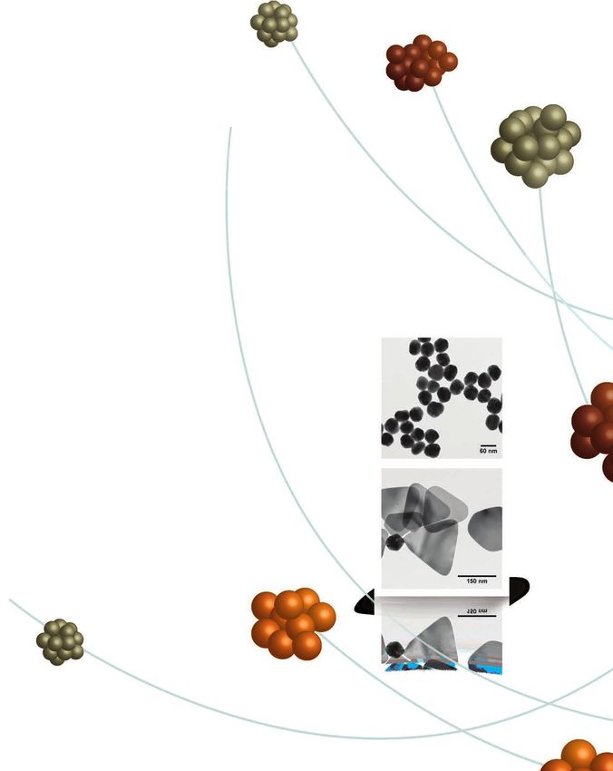
range of several to hundreds of microns that can protect unstable Figure 1. Image of API-loaded PLGA microspheres.
drugs pre- and post-administration. Microspheres have the ability to
release a drug continuously over time,1 thereby providing a prolonged
therapeutic effect and reducing the dosing frequency. In addition to Polymeric Nanoparticles
controlled release, microspheres allow for the targeted drug delivery of
Polymeric nanoparticles (NP), either plain or drug loaded, are
potent drugs at reduced concentrations, thereby minimizing systemic
typically less than 1 micron in size. The use of API-loaded polymeric
exposure and adverse side effects. Finally, polymeric microspheres
nanoparticles for intravenous administration is a promising approach
facilitate manipulation of in vivo behavior, pharmacokinetic profile,
for achieving the controlled release and site-specific delivery of
tissue distribution, and cellular interaction of the drug.2
drugs. The nanoparticle delivery system can be designed to maintain
Microspheres are typically comprised of biodegradable polymers such appropriate therapeutic concentration in the bloodstream (controlled
as poly(lactide-co-glycolide) (PLGA), polylactic acid (PAA), polylactide release) or to target a specific cell type (e.g., bone marrow, blood cells).
(PLA), and polycaprolactone (PCL). These polymers degrade in vivo Various types of APIs, including small molecule drugs and biologic
by hydrolysis of their ester backbone into non-toxic products, which compounds, can be incorporated into PLGA polymer nanoparticles by
are excreted by the kidneys or eliminated as CO2 and water through either microencapsulation or surface conjugation. Nanoencapsulation
biochemical pathways. PLGA microspheres have been widely used can protect the API from early degradation, facilitate cell entry, and
to encapsulate drug molecules and have been used as long-acting, increase solubility and bioavailability.
sustained-release pharmaceutical formulations. There are several
The surface properties of intravenously injected particles are important
drug-loaded PLGA microspheres approved by the FDA and marketed
factors determining in vivo organ distribution and fate. Furthermore,
for clinical use. For example, depot products, such as luprolide
surface modification can be an effective approach to targeting specific
8 TO ORDER: Contact your local Sigma-Aldrich office or visit aldrich.com/matsci.Biodegradable Colloidal Carriers in Drug Delivery Applications
tissues. Surface modification of nanoparticles with polyethylene Natural polymers such as chitosan and alginate have been studied
glycol (PEG) can be used to prolong the in vivo circulation lifetime extensively for the preparation of hydrogel nanoparticles. Hydrogel
of drug-loaded nanoparticles. PEGylation of the nanoparticle can nanoparticles based on synthetic polymers including poly (vinyl
be accomplished by adding a copolymer containing PEG chains alcohol) (PVA), PEG, poly (ethyleneimine) (PEI), poly vinyl pyrrolidone,
during the nanoparticle fabrication process. For example, the and poly-N-isopropylacrylamide have also been used for drug delivery.
addition of an ethylene glycol monomer during lactide and glycolide
Hydrogel systems have various applications including oral, transdermal,
copolymerization can lead to a PEGylated PLGA polymer. PEGylation
nasal, rectal, and ocular drug delivery. Hydrogel membranes facilitate
can increase nanoparticle hydrophilicity and improve degradation rate
transdermal drug delivery through the skin at a predetermined and
and crystallization.3 In addition to being biocompatible, PEG is resistant
controlled rate. They are advantageous as they prevent the first pass
to immunological recognition. PEG units on the NP surface prevent
metabolism effect.12 Additionally, hydrogel delivery systems can
opsonin-NP binding, thus preventing the nanoparticles from being
increase the bioavailability of ophthalmic drugs by increasing the
recognized by monocytes and macrophages and, therefore, increasing
contact time of the drugs with cornea.13
circulation time in the body.4 In some circumstances nanoparticles
have been shown to remain in circulation 40× longer when coated Embolization therapy is another application of hydrogel microspheres.
with PEG compared to uncoated nanoparticles.5 Other advantages of It is typically used to prevent the growth of solid tumors by blocking
PEGylated nanoparticles include increased drug loading of hydrophilic the blood supply to the feeding artery. For example, trans-arterial
drugs, reduced initial burst and improved bioavailability.6 PEGylated chemical embolization is an application where anticancer drug loaded
nanoparticles have been used as carriers for vaccine and protein APIs particles are injected into the feeding artery of cancer tumors. In
and are particularly useful in both sustained/controlled release and addition to blocking the blood supply to the tumor, they release the
targeted drug delivery systems. Currently, there are more than 35 U.S. anticancer drugs at high concentrations inside the tumor.14
FDA-approved products utilizing PEG in their biomedical applications.4
Nanoparticles can also facilitate the crossing of the blood brain barrier
(BBB). Surfactants such as Polysorbate 80 and Poloxamer 188 have been
Methods: Colloidal Carrier Fabrication
shown to facilitate the BBB crossing of drug molecules encapsulated in Colloidal carriers can be fabricated using a variety of techniques. The
polybutyl cyanoacrylate nanoparticles or solid lipid nanoparticles.7 method for synthesis should be selected based on the type and nature
In some nanoparticle formulations, the API is attached to the surface of the drug to be encapsulated as well as the desired particle size,
instead of being encapsulated inside the particle. For example, a delivery route, and release characteristics for the final formulation.
peptide for ocular delivery (POD) and a human immunodeficiency virus
transactivator were conjugated to the surface of PLGA nanoparticles, Nanoprecipitation
and the conjugate was found to improve ocular drug bioavailability.8 Nanoprecipitation is a facile and low energy process for the preparation
The conjugation can be done by reacting the terminal functional group of polymeric nanoparticles. It is based on interfacial deposition due
(e.g., terminal COOH) on the PLGA molecule with a reactive group to the displacement of a solvent with the non-solvent. Miscibility
(e.g., amino) on the peptide, API or protein. Finally, in some cases, of the solvents and the dilute polymer solutions are required for
PLGA nanoparticles themselves can have therapeutic effects against nanopreciptation.13 For drug delivery applications, nanoprecipitation
certain diseases.2 is often used for small-scale preparation of nanoparticles of polylactic
acid, PLGA, and polycaprolactone. It is well-suited for hydrophobic APIs.
Microgels and Nanogels In a typical nanoprecipitation process, a polymer (e.g., PLGA)
(and hydrophobic drug, if desired) is dissolved in a solvent that is
Hydrogel particles, including microgels and nanogels, consist of miscible with water (e.g., acetone). The polymer/drug solution is
crosslinked networks of hydrophilic polymer chains that form added dropwise to an aqueous solution under continuous stirring.
colloidal gels. Hydrogel particles are made from natural or synthetic Surfactants or polymer stabilizers may be added to the aqueous
polymeric networks, are highly absorbent, and can contain over 90% solution to stabilize the nanoparticles during formation. Common
water. Crosslinking between polymer chains is either chemically or surfactants and stabilizers include TWEEN® 20, sodium dodecyl sulfate
physically induced and prevents the dissolution of these networks (SDS), PVA, hydroxymethylcellulose (HPMC), and Pluronic® F-68. The
in water.11 The release of the loaded API from the hydrogel particles organic solvent is removed by evaporation or repetitive washing.
may occur through diffusion, hydrogel matrix swelling, or chemical Additional washing steps may be performed to remove surfactant
reactivity of the drug/matrix. The physical properties of microgels and and unincorporated API. The purified nanoparticles can be lyophilized
nanogels (i.e., swelling, permeation, mechanical strength, and surface for storage.
characteristics) can be optimized by structural modification.11 Many The following is a typical protocol for preparing PLGA nanoparticles, of
hydrogel particles are also environmentally sensitive and have the approximately 200 nm, using a one-step nanoprecipitation.
ability to respond to changes of pH, temperature, or the concentration
of metabolites, and release their load as a response to these stimuli for 1. PLGA polymer with an L/G ratio of 50/50, COOH terminated, and
controlled drug release. inherent viscosity of 0.55–0.75 dL/g (additional PLGA polymers may
be substituted, such as Aldrich Prod. No. 719900) is dissolved in
In drug delivery, microgels and nanogels fill a unique niche because acetone (Aldrich Prod. No. 179124).
they can encapsulate water-soluble, small molecule APIs that are 2. Prepare a polyvinyl alcohol (PVA) solution by dissolving the
difficult to encapsulate using traditional biodegradable polymeric appropriate amount of PVA (Mw 85,000–124,000, 87–89%
particles comprised of PLGA and PCL. As a result, these gels are hydrolyzed, Aldrich Prod. No. 363081) in DI water. A typical
particularly suitable for the sustained release of water-soluble drugs concentration is 1%.
or proteins. Other advantages of hydrogel particles include their high 3. Transfer 100 mL of the 1% PVA solution prepared in Step 2 to a
drug loading and activity, biocompatibility, and biodegradability. 500 mL beaker equipped with a magnetic stir bar. Stir the PVA
Additionally, the manufacturing of colloidal gels does not require solution at 400 rpm.
organic solvents, eliminating toxicity risks and the potential for
protein denaturation.
For questions, product data, or new product suggestions, contact us at matsci@sial.com. 9aldrich.com/matsci
4. Using a disposable pipette, slowly and dropwise add 10 mL of the In double emulsions, achieving high drug loading of hydrophilic
PLGA solution into the stirring PVA solution. The nanoparticles form APIs can be challenging since the drug partitions away from the
on contact when the PLGA solution is added. hydrophobic polymer solution into the aqueous surfactant solution.
5. After the PLGA solution is completely added to the PVA solution, Macromolecular drugs such as proteins and antibodies have all been
continue stirring in a fume hood for 3 h to allow acetone encapsulated into polymeric microspheres and nanoparticles using a
evaporation. double-emulsion processes.
6. Wash the nanoparticles three times using refrigerated However, protein aggregation or denaturing may occur during
centrifugation and follow with lyophilization. formation. During the microencapsulation process, proteins are
7. Store lyophilized PLGA nanoparticles dry at –20 °C. constantly exposed to cavitation, heat, solvents, and high shear force,
which could lead to aggregation and denaturation.17,18–20 Alternative
Emulsification Process techniques have been pursued to encapsulate protein drugs into
Emulsification can be used to prepare plain or drug-loaded polymeric microspheres and nanoparticles and are reviewed elsewhere.19–29
microspheres and nanoparticles. Depending on the type of drug to be
loaded, either a single emulsion or double emulsion may be used. The following is a typical protocol for preparing API-loaded PLGA
nanoparticles using a double-emulsion process:
Single Emulsion 1. Prepare a 1% bovine serum albumin (BSA) solution by dissolving
In a single emulsion, the polymer (e.g., PLGA, PCL) is dissolved in a BSA (lyophilized powder, Sigma Prod. No. 05470) in DI water.
solvent that is not miscible with water. If a drug is to be encapsulated, 2. Prepare a 5% PLGA solution by dissolving PLGA polymer with an
it is preferably hydrophobic and solvent soluble. The hydrophobic L/G ratio of 50/50, COOH terminated, and inherent viscosity of
drug is dissolved in the same solution as the polymer. The polymer/ 0.55–0.75 dL/g (additional PLGA polymers may be substituted,
drug solution is emulsified in an aqueous solution containing a such as Aldrich Prod. No. 719900) in methylene chloride. Prepare
surfactant or a polymeric stabilizer (as noted earlier). Emulsification a 1% PVA (Mw 85,000–124,000, 87–89% hydrolyzed, Aldrich Prod.
can be completed by sonication, magnetic or mechanical stirrer, rotor No. 363081) solution in DI water.
stator, high-pressure homogenizer, or microfluidizer. After the oil-in-
3. Mix 0.5 mL of the BSA solution prepared in Step 1 and 5 mL of the
water emulsion is formed, the solvent is removed by evaporation or
5% PLGA solution prepared in Step 3 in a 15 mL glass vial.
extraction. The particles can be washed to remove the surfactant and
possible unincorporated drug molecules, and then lyophilized. 4. Homogenize the PLGA and BSA solution using an IKA Ultra Turrax
High Speed Homogenizer at 20,000 RPM for 25 seconds.
The single emulsion process can also be used to prepare nanocrystals 5. Mix the resulting emulsion with 100 mL of the 1% PVA solution
of poorly soluble compounds. For example, a single emulsion process prepared in Step 3 in a 500 mL beaker.
was used to prepare albumin bound paclitaxel nanoparticles.14–16 6. Homogenize the mixture of the first emulsion and the PVA solution
The following is a typical protocol for preparing PLGA nanoparticles using an IKA Ultra Turrax High Speed Homogenizer generator at
using a single-emulsion process: 8,000 rpm for 2 min.
7. Stir the resulting double emulsion on a stir plate at 400 rpm in a
1. Prepare a 5% PLGA solution by dissolving PLGA polymer with an
fume hood for 3 h to allow the methylene chloride to evaporate.
L/G ratio of 50/50, COOH terminated, and inherent viscosity of
0.55–0.75 dL/g (additional PLGA polymers may be substituted, such 8. Wash the nanoparticles three times using refrigerated
as Aldrich Prod. No. 719900) in methylene chloride (Sigma Prod. centrifugation. Follow with nanoparticle lyophilization.
No. 443484). 9. Store lyophilized BSA-PLGA nanoparticles dry at –20 °C.
2. Prepare a 1% solution of polyvinyl alcohol (PVA) (Mw 85,000–124,000, Spray Drying
87–89% hydrolyzed, Aldrich Prod. No. 363081) solution in DI water.
In spray drying, the feed (a solution, emulsion, or suspension
3. Mix 5 mL of the 5% PLGA solution prepared in Step 1 with 100 mL of containing the API and the matrix material) is atomized into hot
the 1% PVA solution prepared in Step 2 in a 500 mL beaker. nitrogen, leading to rapid drying and particle formation. The particles
4. Homogenize the mixture of the PLGA and PVA solutions by using an are then separated in a cyclone and/or filter bag.
IKA Ultra Turrax High Speed Homogenizer at 18,000 rpm for 2 min.
5. Stir the resulting emulsion at 400 rpm on a stir plate in a fume hood Spray drying has been used extensively by the pharmaceutical industry
for 3 h to allow the methylene chloride to evaporate. to formulate solid dispersions to overcome solubility limitations.
Crystalline APIs can be encapsulated within polymer microspheres. For
6. Wash the nanoparticles three times using refrigerated
example, enteric polymers can be used to protect a drug from harsh
centrifugation and lyophilize.
gastric conditions or for enhanced delivery to the site of maximum
7. Store lyophilized PLGA nanoparticles dry at –20 °C. absorption. Similar approaches can be used for taste masking and
Double Emulsion protecting the drug from physical environments such as light or
For hydrophilic API encapsulation, a double-emulsion technique is moisture. These drug-loaded particles are normally in the micron to
necessary to prepare the polymeric microspheres and nanoparticles. millimeter range, although the spray-drying process is capable of
The double emulsion is predominately a water-in-oil-in-water producing sub-micron or nanometer sized particles.
emulsion, although in some cases it can be a reverse double emulsion, In addition to enhancing the oral bioavailability of poorly water-soluble
or an oil-in-water-in-oil. In a typical double-emulsion process, the compounds, spray drying offers the ability to control particle size,
hydrophilic API is dissolved in an aqueous media and emulsified in the morphology, and other properties with direct effect in Fine Particle
polymer solution to form the first emulsion. The first emulsion is again Fraction (FPF) and lung deposition. It also allows for a reproducible,
emulsified in an aqueous solution containing appropriate surfactants or controllable, and scalable manufacturing process. Spray drying is the
polymer stabilizers to form the second emulsion, or double emulsion. method of choice where abrasion or shearing needs to be avoided,
The solvent is then removed by evaporation or extraction processes. as in the case of biologic compounds. Inhaled insulin formulations
The equipment used in single-emulsion processes can be used to of Affrezza (MannKind) and Exubera (Pfizer) are manufactured by
generate double emulsions. spray drying.
10 TO ORDER: Contact your local Sigma-Aldrich office or visit aldrich.com/matsci.Biodegradable Colloidal Carriers in Drug Delivery Applications
The following is a typical spray-drying protocol for the preparation (10) Giri, T. K.; Thakur, A.; Alexander, A.; Ajazuddin, H.; Badwaik, H.; Tripathi, D. K.; Acta
Pharmaceutica Sinica B 2012, 2, 439.
of chitosan microspheres. This method uses a Mini Spray Dryer B-290 (11) Genta, I.; Conti, B.; Perugini, P.; Pavanetto, F.; Spadaro, A.; Puglisi, G. J. Pharm. Pharmacol.
(BUCHI Corporation, New Castle, DE, USA) with a 0.7 mm standard 1997, 49, 737–742.
nozzle. (12) Saralidze, K.; Koole, L. H.; Knetsch, M. L. W. Materials 2010, 3 (6), 3537–3564.
(13) Hornig, S.; Heinze, T.; Becer, C. R.; Schubert, U. S. J. Mater.Chem. 2009, 3838–3840.
1. Dissolve an appropriate amount of chitosan (medium Mw, (14) Green, M. R.; Manikhas, G. M.; Orlov, S.; Afanasyev, B.; Makhson, A. M.; Bhar, P.; Hawkins, M. J.
Aldrich Prod. No. 448877) in 1.0% v/v acetic acid (Aldrich Prod. Annals of Oncology 2006, 17 (8), 1263–1268.
(15) Miele, E.; Spinelli, G. P.; Miele, E.; Tomao, F.; Tomao, S. Int. J Nanomed 2009, 4, 99–105.
No. 695092) solution to prepare a 2.5% (w/v) chitosan solution in
(16) Stinchcombe, T. E. Nanomedicine 2007, 2 (4), 415–423.
a 500 mL Erlenmeyer flask. (17) Cleland, J.; Jones, A. Pharm Res 1996, 13, 1464–1475.
2. Use the suction mode as the method of operation. Set the flow rate (18) Morlocka, M.; Kollb, H.; Winterb, G.; Kissel, T. Eur J Pharm Biopharm 1997, 43, 29–36.
of the compressed air at 600 L/h, the inlet temperature at 160 °C, (19) Pérez, C.; Castellanos, I. J.; Costantino, H. R.; Al-Azzam, W.; Griebenow. K. J Pharm Pharmacol
2002, 54, 301–303.
and sample flow rate at 700 mL/h. (20) Boury, F.; Ivanova, T.; Panaieotov, I.; Proust, J. E.; Bois, A.; Richou, J. Langmuir 1995, 11,
3. Start the operation. The chitosan solution is fed to the spray dryer, 1636–1644.
atomized by the force of the compressed air, and blown by heated (21) Carrasquilloa, K. G.; Stanleya, A. M.; Aponte-Carroa, J. C.; Jésusa, P. D.; Costantinob, H. R.;
Bosquesc, C. J.; Griebenow, K. J. Controlled Release 2001 76, 199–208.
air to the drying chamber. (22) Sáncheza, A.; Villamayora, B.; Guob, Y.; McIverb, J.; Alonso, M. J. Int J Pharm 1999, 185,
4. Collect the dried microspheres. The mean residence time is 255–266.
1–1.5 seconds. (23) Sturesson, C.; Carlfors, J. J. Controlled Release 2000, 67, 171–178.
(24) Sah, H. J. Controlled Release 1999, 58, 143–151.
References (25) Péan, J-M.; Boury, F.; Venier-Julienne, M-C.; Menei, P.; Proust, J-E.; Benoit, J-P. Pharm Res
1999, 16, 1294–1299.
(1) Sahil, K.; Akanksha, M.; Premjeet, S.; Bilandi, A.; Kapoor, B. Int. J. Res.Pharm. Chem. 2011, 1,
(26) Benoita, J-P.; Faisanta, N.; Venier-Juliennea, M-C.; Menei, P. J. Controlled Release 2000, 65,
1184.
285–296.
(2) Ramteke, K.; Jadhav, V. B.; Dhole, S. N. IOSR J. of Pharm. 2012, 2, 44.
(27) Jain, R. A. Biomaterials 2000, 21, 2475–2490.
(3) Xiao, R. Z.; Zeng, Z. W.; Zhou, G. L.; Wang, J. J.; Li, F. Z.; Wang, A. M. Int J Nanomedicine 2010,
(28) Iwata, M.; McGinity, J. W. J Microencapsul 1992, 9, 201–214.
5, 1057.
(29) O’donnell, P. B.; Iwata, M.; McGinity, J. W. J Microencapsul 1995, 12, 155–163.
(4) Moffatt, S. MOJ Proteomics Bioinform 2015, 2, 00037.
(5) van Vlerken, L. E.; Vyas, T. K.; Amiji, M. M. Pharm Res 2007, 24, 1405–1414.
(6) Xiao, R. Z.; Zeng, Z. W.; Zhou, G. L.; Wang, J. J.; Li, F. Z.; Wang, A. M. Int J Nanomedicine 2010,
5, 1057.
(7) Sharma, H. S. Progress in Brain Research in Nanoneuroscience and
Nanoneuropharmacology, 2009, page 198 and reference therein.
(8) Vasconcelos, A.; Vega, E.; Pérez, Y.; Gómara, M. J.; García, M. L.; Haro, I. Int. J. Nanomed. 2015,
10, 609.
(9) Ahmed, E. M. J Adv. Res. 2015, 7, 105.
Poly(lactide-co-glycolide) Copolymers
For more information on these products, visit aldrich.com/biodegradable.
Name Feed Ratio End Group Molecular Weight Degradation Time Prod. No.
Resomer® RG 502 H, Poly(D,L-lactide-co-glycolide) lactide:glycolide 50:50 acid terminated Mw 7,000‑17,000You can also read