PROSTATE GLAND SEGMENTATION IN HISTOLOGY IMAGES VIA RESIDUAL
←
→
Page content transcription
If your browser does not render page correctly, please read the page content below
PROSTATE GLAND SEGMENTATION IN
                                            HISTOLOGY IMAGES VIA RESIDUAL AND
                                                 MULTI-RESOLUTION U-NET?

                                           Julio Silva-Rodrı́guez1 , Elena Payá-Bosch2 , Gabriel Garcı́a2 , Adrián Colomer2 ,
                                                                          and Valery Naranjo2
arXiv:2105.10556v1 [eess.IV] 21 May 2021

                                               1
                                                Institute of Transport and Territory, Universitat Politècnica de València, Spain
                                                                              jjsilva@upv.es
                                           2
                                               Institute of Research and Innovation in Bioengineering, Universitat Politècnica de
                                                                               València, Spain

                                                    Abstract. Prostate cancer is one of the most prevalent cancers world-
                                                    wide. One of the key factors in reducing its mortality is based on early
                                                    detection. The computer-aided diagnosis systems for this task are based
                                                    on the glandular structural analysis in histology images. Hence, accurate
                                                    gland detection and segmentation is crucial for a successful prediction.
                                                    The methodological basis of this work is a prostate gland segmentation
                                                    based on U-Net convolutional neural network architectures modified with
                                                    residual and multi-resolution blocks, trained using data augmentation
                                                    techniques. The residual configuration outperforms in the test subset
                                                    the previous state-of-the-art approaches in an image-level comparison,
                                                    reaching an average Dice Index of 0.77.

                                           Keywords: Prostate Cancer, Histology, Gland Segmentation, U-Net, Residual.

                                           1       Introduction
                                           Prostate cancer was the second most prevalent cancer worldwide in 2018, accord-
                                           ing to the Global Cancer Observatory [1]. The final diagnosis of prostate cancer
                                           is based on the visual inspection of histological biopsies performed by expert
                                           pathologists. Morphological patterns and the distribution of glands in the tis-
                                           sue are analyzed and classified according to the Gleason scale [2]. The Gleason
                                           patterns range from 3 to 5, inversely correlating with the degree of glandular
                                           differentiation. In recent years, the development of computer-assisted diagnostic
                                           systems has increased in order to raise the level of objectivity and support the
                                           work of pathologists.
                                               One of the ways to reduce mortality in prostate cancer is through its early
                                           detection [3]. For this reason, several works have focused on the first stage of
                                           ?
                                               This work was supported by the Spanish Ministry of Economy and Competitiveness
                                               through project DPI2016-77869. The Titan V used for this research was donated by
                                               the NVIDIA Corporation. Preprint accepted for publication on 21st International
                                               Conference on Intelligent Data Engineering and Automated Learning - IDEAL 2020.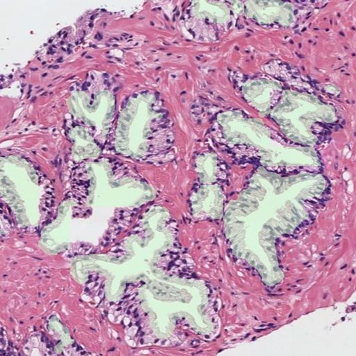
2 J. Silva-Rodrı́guez et al.
prostate cancer detection by differentiating between benign and Gleason pattern
3 glands [4–6]. The benign glands differentiate from Gleason pattern 3 structures
in the size, morphology, and density in the tissue (see Fig. 1). In order to au-
tomatically detect the early stage of prostate cancer, the main methodology
used in the literature is based on detecting and segmenting glands and then,
classifying each individual gland. For the classification of cancerous glands, both
classic approaches based on hand-driven feature extraction [7] and modern deep-
learning techniques [5] have been used. Nevertheless, those results are limited by
a correct detection and delimitation of the glands in the image. This encourages
the development of accurate systems able to detect and segment the glandular
regions.
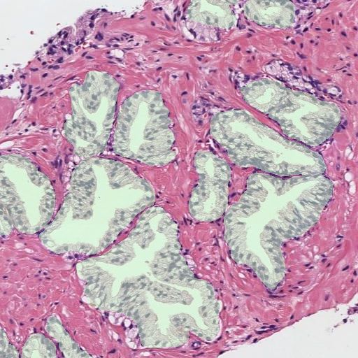
(a) (b) (c) (d)
Fig. 1: Histology regions of prostate biopsies. Examples (a) and (b) present be-
nign glands, including dilated and fusiform patterns. Images (c) and (d) contain
patterns of Gleason grade 3, with small sized and atrophic glands.
For the prostate gland segmentation, different approaches have been carried
out. In the work of Nguyen et al. [4,7–9] this procedure is based on the unsuper-
vised clustering of the elements in the tissue, i.e. lumen, nuclei, cytoplasm, and
stroma. Then, for each detected lumen, a gland is associated if enough nuclei are
found in a region surrounding the lumen’s contour. In the research carried out
by Garcı́a et al. [5, 6, 10] the components in the image are clustered by working
in different color spaces, and then a Local Constrained Watershed Transform
algorithm is fitted using the lumens and nuclei as internal and external mark-
ers respectively. As final step, the both aforementioned methodologies require a
supervised model to differentiate between artifacts or glands. To the best of the
authors’ knowledge, the best performing state-of-the-art techniques for semantic
segmentation, based on convolutional neural networks, have not been studied yet
for prostate gland segmentation. In particular, one of the most used techniques
in recent years for semantic segmentation is the U-Net architecture, proposed
for medical applications in [11].
In this work, we present an U-Net-based model that aims to segment the
glandular structures in histology prostate images. To the extent of our knowl-
edge, this is the first time that an automatic feature-learning method is used for
this task. One of the main contributions of this work is an extensive validation
about different convolutional block modifications and regularization techniques
on the basic U-Net architecture. The proposed convolutional block configura-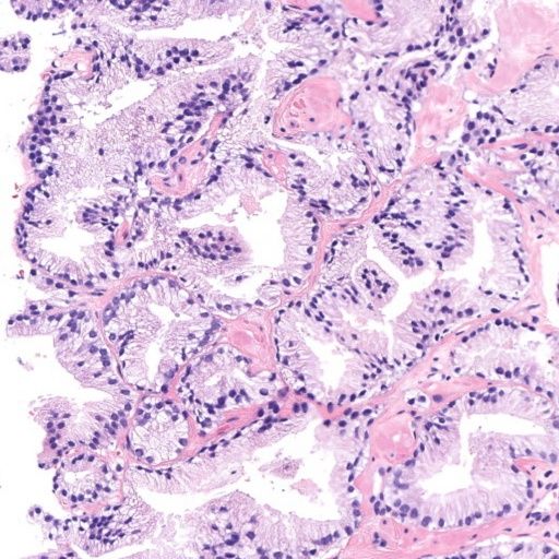
PROSTATE GLAND SEGMENTATION VIA U-NET                      3

tions are based on well-known CNN architectures of the literature (i.e. residual
and Inception-based blocks). Furthermore, we study the impact of regularization
approaches during the training stage based on data augmentation and using the
gland contour as an independent class. Finally, we perform, as novelty, an image-
level comparison of the most relevant methods in the literature for prostate gland
segmentation under the same database. Using our proposed modified U-Net with
residual blocks, we outperform in the test subset previous approaches.

2     Materials and methods
2.1   Materials
The database used in this work consists of 47 whole slide images (WSIs, histol-
ogy prostate tissue slices digitised in high-resolution images) from 27 different
patients. The ground truth used in this work was prepared by means of a pixel-
level annotation of the glandular structures in the tissue. In order to work with
the high dimensional WSIs, they were sampled to 10× resolution and divided
into patches with size 10242 and overlap of 50% among them. For each image, a
mask was extracted from the annotations containing the glandular tissue. The
resulting database includes 982 patches with its respective glandular masks.
2.2   U-Net architecture
The gland segmentation in the prostate histology images process is carried out
by means of the U-Net convolutional neural network architecture [11] (see Fig.
2). As input, the images of dimensions 10242 are resized to 2562 to avoid memory
problems during the training stage. The U-Net configuration is based on a sym-
metric encoder-decoder path. In the encoder part, a feature extraction process
is carried out based on convolutional blocks and dimensional reduction through
max-pooling layers. Each block increases the number of filters in a factor of 2×,
starting from 64 filters up to 1024. After each block, the max-pooling operation
reduces the activation maps dimension in a factor of 2x. The basic convolutional
block (hereafter referred to as basic) consist of two stacked convolutional layers
with filters of size 3 × 3 and ReLU activation. Then, the decoder path builds the
segmentation maps, recovering the original dimensions of the image. The recon-
struction process is based on deconvolutional layers with filters of size 3 × 3 and
ReLU activation. These increase the spatial dimensions of the activation volume
in a factor of 2× while reducing the number of filters in a half. The encoder
features from a specific level are joined with the resulting activation maps of the
same decoder level by a concatenation operation, feeding a convolutional block
that combines them. Finally, once the original image dimensions are recovered,
a convolutional layer with as many filters as classes to segment and soft-max
activation creates the segmentation probability maps.
2.3   Loss function
The loss function defined for the training process is the categorical Dice. This
measure takes as input the reference glands and background masks (y) and the
predicted probability maps (by ) and is defined as follows:
4      J. Silva-Rodrı́guez et al.

          Fig. 2: U-Net architecture for prostate gland segmentation.

                                           C   P
                                        1 X 2 ybc ◦ yc
                          Dice(y, yb) =       P 2                             (1)
                                        C c=1   ybc + yc2

where y is the pixel-level one hot encoding of the reference mask for each class
c and yb is the predicted probability map volume.
   Using a categorical average among the different classes brings robustness
against class imbalance during the training process.

2.4   Introducing residual and multi-resolution blocks to the U-Net

To increase the performance of the U-Net model, different convolutional blocks
are used to substitute the basic configuration. In particular, residual and multi-
resolution Inception-based blocks are used during the encoder and decoder stages.
    The residual block [12] (from now on RB) is a configuration of convolutional
layers that have shown good performance in deep neural networks optimisation.
The residual block proposed to modify the basic U-Net consist of three convo-
lutional layers with size 3 × 3 and ReLU activation. The first layer is in charge
of normalizing the activation maps to the output’ amount of filters for that
block. The resultant activation maps from this layer are combined in a shortcut
connection with the results of two-stacked convolutional layers via an adding
operation.
    Regarding the multi-resolution block (referred to in this work as M RB), it
was recently introduced in [13] as a modification of the U-Net with gains of ac-
curacy on different medical applications. This configuration, based on Inception
blocks [14], combines features at different resolutions by concatenating the out-
put of three consecutive convolutional layers of size 3×3 (see Fig. 3). The number
of activation maps in the output volume (Fout ) is progressively distributed in
the three blocks ( Fout Fout      Fout
                    4 , 4 , and 2 respectively). Furthermore, a residual con-
nection is established with the input activation maps, normalizing the number
of maps with a convolutional layer of size 1 × 1.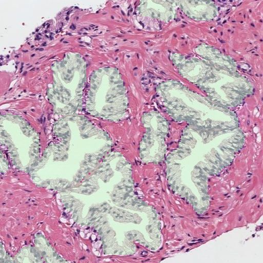
PROSTATE GLAND SEGMENTATION VIA U-NET                     5

Fig. 3: Multi-resolution block. Fin : activation maps in the input volume. Fout :
number of activation maps in the output volume. D: activation map dimensions.

2.5   Regularization techniques
To improve the training process, two regularization techniques are proposed:
data augmentation and the addition of the gland border as an independent class.
Data augmentation (DA) is applied during the training process making random
translations, rotations, and mirroring are applied to the input images. Regarding
the use of the gland contour as an independent class (BC), this strategy has
shown to increase the performance in other histology image challenges such as
nuclei segmentation [15]. The idea is to highlight more (during the training
stage) the most important region for obtaining an accurate segmentation: the
boundary between the object and the background. Thus, the reference masks and
the output of the U-Net are modified with an additional class in this approach.

3     Experiments and Results
To validate the different U-Net configurations and the regularization techniques,
the database was divided in a patient-based 4 groups cross-validation strategy.
As figure of merit, the image-level average Dice Index (DI) for both gland and
background was computed. The Dice Index for certain class c is obtained from
the Dice function (see Equation 1) such that: DI = 1 − Dice. The metric ranges
0 to 1, from null to perfect agreement.
    The different U-Net architectures, composed of basic (basic), residual (RB)
and multi-resolution (M RB) blocks were trained with the proposed regularisa-
tion techniques, data augmentation (DA) and the inclusion of the border class
(BC), in the cross-validation groups. The training was performed in mini-batches
of 8 images, using NADAM optimiser. Regarding the learning rates, those were
empirically optimised at values 5 ∗ 10−4 for the basic and RB configurations and
to 1 ∗ 10−4 for the M RB one. All models were trained during 250 epochs. The
results obtained in the cross-validation groups are presented in Table 1.
    Analysing the use of different U-Net configurations, the best performing
method was the U-Net modified with residual blocks (RB + DA), reaching an
average DI of 0.75, and outperforming the basic architecture in 0.06 points.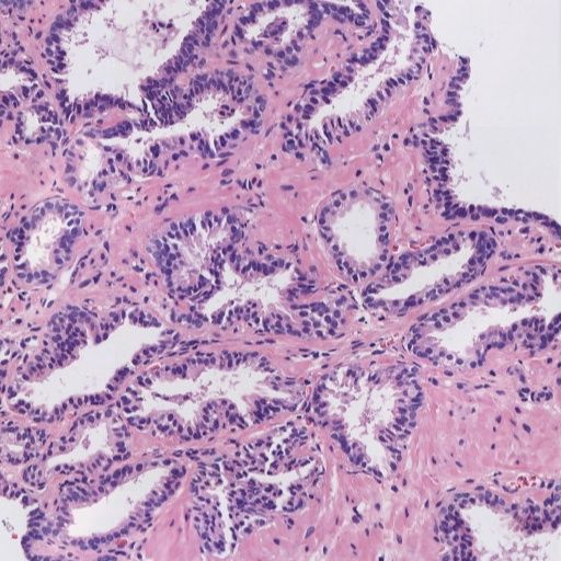
6      J. Silva-Rodrı́guez et al.

Table 1: Results in the validation set for gland and background segmentation.
The average Dice Index is presented for the different configurations. DA: data
augmentation, BC : border class, RB : residual and MRB : multi-resolution blocks.
                     Method             DIgland       DIbackground
                basic               0.5809(0.2377)   0.9766(0.0240)
                basic + DA          0.6941(0.2515)   0.9845(0.01664)
                basic + DA + BC     0.6945(0.2615)   0.9842(0.0168)
                RB                  0.5633(0.2340)   0.9759(0.0255)
                RB + DA             0.7527(0.2075)   0.9862(0.0148)
                RB + DA + BC        0.7292(0.2395)   0.9854(0.0161)
                M RB                0.5710(0.2378)   0.9765(0.0253)
                M RB + DA           0.7294(0.2173)   0.9843(0.0161)
                M RB + DA + BC      0.7305(0.2247)   0.9846(0.0163)

Regarding the use of the multi-resolution blocks (M RB), an increase in the
performance was not registered. Concerning the use of the gland profile as an
independent class (BC), the results obtained were similar to the ones with just
two classes.
    The best performing strategy, RB + DA, was evaluated in the test subset.
A comparison of the results obtained in this cohort with the previous methods
presented in the literature is challenging. While previous works report object-
based metrics, we consider more important to perform an image-level comparison
of the predicted segmentation maps with the ground truth, in order to take into
account false negatives in object detection. For this reason, and in order to
establish fair comparisons, we computed the segmentation results in our test
cohort applying the main two approaches found in the literature: the work of
Nguyen et al. [4] and the research developed by Garcı́a et al. [10]. This is, to
the best of the authors’ knowledge, the first time in the literature that the main
methods for prostate gland segmentation are compared at image level in the
same database. The figures of merit obtained in the test set are presented in the
Table 2. Representative examples of segmented images for all the approaches are
shown in Fig. 4.

Table 2: Results in the test set for gland and background segmentation. The
average Dice Index for both classes is presented for the state-of-the-art methods
and our proposed model. DA: data augmentation and RB : residual blocks.
                    Method              DIgland       DIbackground
                Nguyen et al. [4] 0.5152(0.2201)     0.9661(0.0168)
                Garcı́a et al. [10] 0.5953(0.2052)   0.9845(0.01664)
                U-Net + RB + DA 0.7708(0.2093)       0.9918(0.0075)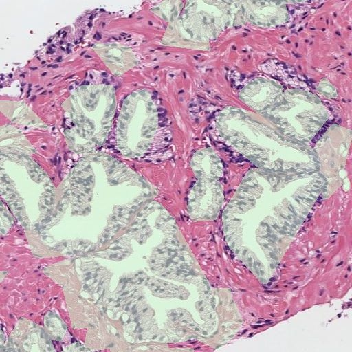
PROSTATE GLAND SEGMENTATION VIA U-NET 7
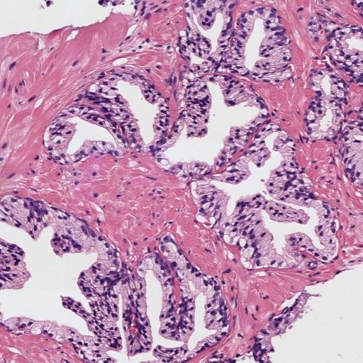
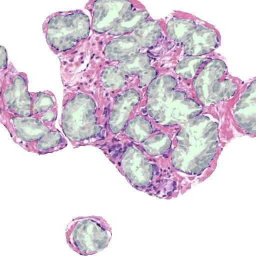
(a) (b) (c) (d)
Fig. 4: Semantic gland segmentation in regions of images from the test set. (a):
reference, (b): Nguyen et al., (c): Garcı́a et al., and (d): proposed U-Net.
Our model outperformed previous methods in the test cohort, with an average
DI of 0.77 for the gland class. The method proposed by Nguyen et al. and the
one of Garcı́a et al. obtained 0.51 and 0.59 respectively. The main differences
were observed in glands with closed lumens (see first and second row in Fig. 4).
The previous methods, based on lumen detection, did not segment properly those
glands, while our proposed U-Net obtains promising results. Our approach also
shows a better similarity in the contour of the glands with respect the reference
annotations (see third row in Fig. 4).
4 Conclusions
In this work, we have presented modified U-Net models with residual and multi-
resolution blocks able to segment glandular structures in histology prostate im-
ages. The U-Net with residual blocks outperforms in an image-level comparison
previous approaches in the literature, reaching an average Dice Index of 0.77 in
the test subset. Our proposed model shows better performance in both glands
with closed lumens and in its shape definition. Further research will focus on
studying the gains in accuracy in the first-stage cancer identification with a
better gland segmentation based on our proposed U-Net.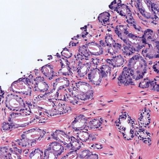
8       J. Silva-Rodrı́guez et al.

References
 1. World Health Organization, “Global cancer observatory,” 2019.
 2. Donald Gleason, “Histologic grading of prostate cancer: A perspective, human
    pathology,” 1992.
 3. Andrew J. Vickers and Hans Lilja, “Predicting prostate cancer many years before
    diagnosis: How and why?,” World Journal of Urology, vol. 30, no. 2, pp. 131–135,
    2012.
 4. Kien Nguyen, Bikash Sabata, and Anil K. Jain, “Prostate cancer grading: Gland
    segmentation and structural features,” Pattern Recognition Letters, vol. 33, no. 7,
    pp. 951–961, 2012.
 5. Gabriel Garcı́a, Adrián Colomer, and Valery Naranjo, “First-stage prostate cancer
    identification on histopathological images: Hand-driven versus automatic learning,”
    Entropy, vol. 21, no. 4, 2019.
 6. José Gabriel Garcı́a, Adrián Colomer, Fernando López-Mir, José M. Mossi, and
    Valery Naranjo, “Computer aid-system to identify the first stage of prostate cancer
    through deep-learning techniques,” European Signal Processing Conference, pp. 1–
    5, 2019.
 7. Kien Nguyen, Anindya Sarkar, and Anil K. Jain, “Prostate cancer grading: Use
    of graph cut and spatial arrangement of nuclei,” IEEE Transactions on Medical
    Imaging, 2014.
 8. Kien Nguyen, Anil K. Jain, and Ronald L. Allen, “Automated gland segmentation
    and classification for gleason grading of prostate tissue images,” International
    Conference on Pattern Recognition, pp. 1497–1500, 2010.
 9. Kien Nguyen, Anindya Sarkar, and Anil K. Jain, “Structure and context in pro-
    static gland segmentation and classification,” MICCAI 2012, vol. 7510, pp. 115–
    123, 2012.
10. Jose Gabriel Garcı́a, Adrián Colomer, Valery Naranjo, Francisco Peñaranda, and
    M. A. Sales, “Identification of Individual Glandular Regions Using LCWT and
    Machine Learning Techniques,” IDEAL, vol. 3, pp. 374–384, 2018.
11. Olaf Ronneberger, Philipp Fischer, and Thomas Brox, “U-net: Convolutional net-
    works for biomedical image segmentation,” Lecture Notes in Computer Science
    (including subseries Lecture Notes in Artificial Intelligence and Lecture Notes in
    Bioinformatics), vol. 9351, pp. 234–241, 2015.
12. Kaiming He, Xiangyu Zhang, Shaoqing Ren, and Jian Sun, “Deep residual learning
    for image recognition,” Proceedings of the IEEE Computer Society Conference on
    Computer Vision and Pattern Recognition, vol. 2016-Decem, pp. 770–778, 2016.
13. Nabil Ibtehaz and M. Sohel Rahman, “MultiResUNet: Rethinking the U-Net ar-
    chitecture for multimodal biomedical image segmentation,” Neural Networks, vol.
    121, pp. 74–87, 2020.
14. Christian Szegedy, Wei Liu, Yangqing Jia, Pierre Sermanet, Scott Reed, Dragomir
    Anguelov, Dumitru Erhan, Vincent Vanhoucke, and Andrew Rabinovich, “Going
    deeper with convolutions,” Proceedings of the IEEE Computer Society Conference
    on Computer Vision and Pattern Recognition, vol. 07-12-June, pp. 1–9, 2015.
15. Neeraj Kumar, Ruchika Verma, Sanuj Sharma, Surabhi Bhargava, Abhishek Va-
    hadane, and Amit Sethi, “A Dataset and a Technique for Generalized Nuclear Seg-
    mentation for Computational Pathology,” IEEE Transactions on Medical Imaging,
    vol. 36, no. 7, pp. 1550–1560, 2017.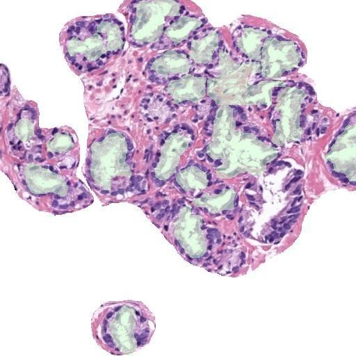
You can also read