PSO-KNN BASED EFFECTIVE OPTIC DISC SEGMENTATION AND CLASSIFICATION IN FUNDUS IMAGES
←
→
Page content transcription
If your browser does not render page correctly, please read the page content below
European Journal of Molecular & Clinical Medicine
                                                       ISSN 2515-8260           Volume 7, Issue 4, 2020

      PSO-KNN BASED EFFECTIVE OPTIC DISC
     SEGMENTATION AND CLASSIFICATION IN
               FUNDUS IMAGES
                                   B.Sakthi Karthi Durai1, J.Benadict Raja2
 1
   Assistant Professor, Department of Computer Science and Engineering, PSNA College of Engineering and
                                            Technology, India
 2
   Assistant Professor, Department of Computer Science and Engineering, PSNA College of Engineering and
                                            Technology, India

ABSTRACT
Glaucoma is the most common sources of the retinal disease that leads to permanent impaired vision
worldwide. An automatic Optic Disc (OD) findings in retinal images utilized to diagnosis eye-related
diseases like diabetic retinopathy. Numerous methods are offered to detect OD in low-resolution retinal
images. This work presents an automatic glaucoma diagnosis using an image processing technique from
the digital fundus image. In this work, a novel Particle Swarm Optimization (PSO) optimized KNN used for
glaucoma disease classification. PSO is a naturally inspired optimization algorithm, utilized to find
optimization parameters of KNN to improve classification accuracy. The proposed algorithm divided into
three stages. Preprocessing stage includes noise removal, contrast enhancement using histogram
equalization. For OD detection FCM has been used. Finally, PSO-KNN classifier used for categorizing
healthy and non-healthy images of Optic Disc. The proposed technique has been coded in MATLAB and
tested in the standard database of DRIVE and STARE fundus image. From the result observed that
compared to other algorithms proposed approach improves accuracy considerably.

1.INTRODUCTION

        Medical imaging is a development of producing images of inner portions of the human body for
clinical l diagnosing purpose. The inner parts of the human body can be effortlessly envisaged by the used
image processing. Various imaging techniques have been introduced to produce images such as X-ray, CT
and MRI techniques. It is used to improve medical science, particularly to Ophthalmology. Ophthalmology
[1] is the main part of the medical division used to diagnosis eye-related treatment and disorder. There are
various diseases related to retinal, such as cataracts, glaucoma and diabetic retinopathy, etc. From the
earlier years, Ophthalmology techniques improved by introducing numerous automatic detection methods but
these approaches need further development[2][3].

Glaucoma is a collection of disease-related with the human eye that leads blindness without any indications
and cautions. Firstly, glaucoma increases Intraocular Pressure (IOP) and if not identified at an early stage, it
spontaneously abolishes the optic nerve and ultimately leads to impaired vision.

                                                                                                           2015
European Journal of Molecular & Clinical Medicine
ISSN 2515-8260 Volume 7, Issue 4, 2020
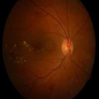
Position of Optic Disc (OD) is an important portion of fundus which is used to decide the harshness of
glaucoma. It is normally the bright portions in fundus images and optic disc centre is the origin of blood
vessels. OD can be classified by shape, the pattern of vessels and sharpness of margins.
By the structure of OD, it can be segmented with shape regression method, template matching [16]
and Snake based Contour Refinement. OD can be divided into two parts such as the central bright zone named
Optic Cup (OC) and a peripheral region as the neuroretinal rim. Cup identification can be done by threshold
level set approach, r-bends information [6], convex hull [8], boundary segmentation method and shape
regression method.
The contribution of the proposed method is listed as follows:
• Using an improved pre-processing method - RGB to Grayscale conversion, Green channel separation.
Normalization, adaptive median filter, Noise removal and contrast enhancement using histogram equalization.
The optic disc is detected by using FCM, that’s Fuzzy C-means clustering approach.
The optic disc is segmented by using the OTSU scheduling approach.
Finally, we used PSO-KNN classifier for categorizing healthy and non-healthy images.
. The structure of this work is as follows. In section 2, presented a related work in the area of glaucoma
diagnosis. Section 3 describes the proposed diagnosis system. Section 4 comprises of the implementation.
Section 5 presents the conclusion of this work.
2.RELATED WORK
In the literature, many researchers have worked on OD segmentation and classification in their particular
application domain.
Jasem Almotiri 2017 et al have presented an automated technique for the segmentation of the OD region in
retinal images. The proposed preprocessing technique tested in various data sets of DRIVE, DRISHTI-GS and
DiaRetDB1 datasets. Jun Cheng et al 2013 have proposed OD and optic cup segmentation using superpixel
classification method. In OD segmentation, histograms, and centre edging statistics are utilized to categorize
each superpixel. The proposed method attains higher areas under the curve than other methods. Shijian Lu et
al 2011 have presented an OD detection and segmentation technique using circular transformation.
Implementation results show that OD detection accuracies of 99.15%, 96.3%, and 97.66% are achieved for
the STARE dataset. Ting Yu et al 2015 have presented a fully automatic localization and segmentation of OD
in fundus images. The border of the OD is identified by using distance regularized narrowband level set
evolution (DRLSE) method. The proposed method achieves a high success rate of 99.52% than the existing
one. Junjie Bai et al 2014 have proposed a graph-based optimal segmentation method to concurrently segment
multiple star-shaped surfaces. Further, the segmented surfaces are confirmed to be smooth by integrating
smoothness limits. Huajun Ying et al have proposed an algorithm to detect OD location in retinal images
using simple local histogram analysis. In a high fractal dimension of blood vessel environment, OD can be
separated from other bright portions such as hard exudates and artifacts. Compared to other methods, the
proposed method has a lower computational cost and is more robust. Canan Çelik et al 2016 have proposed an
OD detection method for diagnosing eye diseases. In this work, the red channel is utilized to remove blood
vessels in the retina images applied in MESSIDOR database. Then, the Graph Cut algorithm is used for
segmentation.
2016European Journal of Molecular & Clinical Medicine
ISSN 2515-8260 Volume 7, Issue 4, 2020
Huazhu Fu et al 2018 have proposed a deep learning architecture based OD segmentation named M-
Net, which consider the OD and OC segmentation concurrently. The proposed M-Net contains input and
output layers with U-shape convolutional network. The U-shape network is used to learn the complex
hierarchical.
Ana Salazar-Gonzalez et al 2014 have presented a Markov random field (MRF) image reconstruction method
based segmentation for blood vessels and optic disk in the fundus retinal images. The first step of extraction
of the retina vascular tree using the graph cut technique. Then, blood vessel data is then utilized to calculate
the location of the OD. The proposed technique is validated on three public datasets, DIARETDB1, DRIVE,
and STARE.
Feng Pan et al 2020 have used the GrabCut technique to create the coarse foreground segmentation in retinal
images. The proposed method train the network based on an improved U-net model with the produced
foreground map. Validation performed on the RIM-ONE benchmarks to show the effectiveness of our
algorithm.
3. PROPOSED METHODOLOGY
The proposed system mainly consists of the following steps,
Step1: Image Acquisition: Fundus image mainly collected from datasets DRIVE and STARE. For prime
research taken 40 from each dataset (40 healthy, 40 non-healthy).
Step 2: Pre-processing: Main steps are the following,
1. RGB to gray
2. Green channel separation.
3. ROI extracted using morphological operations.
4. A used adaptive median filter for noise reduction. For contrast enhancement used histogram
equalization.
Step3: Healthy image’s consists of the optic disc, Fovea, Macula and blood vessels. In this work, an
algorithm only detected and localized an optic disc.
Step 4: Optic disc is detected by using FCM, that’s Fuzzy C-means clustering approach[14].
FCM works by allocating membership values to each pixel corresponding to each cluster centre on the
basis of the distance between the cluster centre and the pixel. More the pixel is close to the cluster center
more is its membership towards the specific cluster centre.
Step 5: Optic disc is segmented by using the OTSU scheduling approach[15].
Otsu method was introduced by Scholar Otsu in 1979. Which is extensively used because it is simple and
effective. The Otsu method needs computing a grey level histogram before running
Step 6: Finally we used PSO-KNN classifier for categorizing healthy and non-healthy images. If (Number
of classes==2), we received comparatively good accuracy than more than two classes.
Step 7: Calculated Accuracy in terms of usually used measures.
2017European Journal of Molecular & Clinical Medicine
ISSN 2515-8260 Volume 7, Issue 4, 2020
Fundus FCM OTSU
image
OD OD
PREPROCESSING
DETECTION SEGMENTATION
Normal
CLASSIFIER
Severe
Figure 1: Proposed System
The overall accuracy and efficiency of KNN based on the method used to find K nearest neighbours. The
identification of K nearest neighbours and the similarity metric of KNN classifier considered as an
optimization problem. In this work Particle Swarm Optimization (PSO) utilized to find optimization
parameters of KNN like attribute selection, voting power of neighbours, value for K. weight vector and
instance selection.
PSO is a well-improved optimization model created by Kennedy and Eberhart [13]. A Particle Swarm
Optimization (PSO) is an occupant based stochastic optimization calculation model by the recreation of the
regular conduct of bird gatherings. Swarm Intelligence (SI) is a novel scattered intellectual model for working
out of optimization inconveniences that at absolute originally took its brainwave from the biological
delineations by swarming, gathering and coordinating occasions invertebrates. PSO incorporates swarming
deeds which are seen in herds of birds, trains of fish, or runs of honey bees, and furthermore in human shared
conduct, from which the proposition is developed. Consequently, the PSO is a populace based optimization
approach, which could be utilized and furthermore applied easily to determine an assortment of optimization
challenges.
The proposed PSO strategy characterizes each particle as an expected key to an emergency in D-
dimensional space. Every last one of the particles knows about its best an incentive up to now (pbest) and its
spot. Moreover, every particle knows the best expense as of recently in the gathering (gbest) among pbests.
This data is the connection of information for how the further particles in the district have been performed.
Every single particle attempts to modify its area and destinations utilizing the accompanying in succession:
• The separation among both present position and pbest
• The separation among both position and gbest
This change can be represented by the idea of speed velocity. This speed Velocity of every single
arbiter can be adjusted by the underneath condition (1) in the Inactivity Weight Approach (IWA).
2018European Journal of Molecular & Clinical Medicine
ISSN 2515-8260 Volume 7, Issue 4, 2020
VK+1 = W * VK + C1 * Rand1 * (PK – XK) + C2 * Rand2 * (GK – XK) (1)
where, W – non-negative inactivity factor, VK – speed velocity of article, XK - present situation of
particle, C1 - the psychological segment for relative impact, C2-decide the public segment for relative impact
of the, PK - pbest of particle , GK - gbest of the particle, Rand1 , Rand2 - arbitrary numbers which are utilized
to save the scope of the populace, and are reliably appropriated in the stretch [0,1].From the condition (1), a
particle settles on a choice where to move straightaway, thinking about its own insight, which is the
remembering memory of its best point of reference position, and the ability of its most triumphant particle in
the swarm. In the particle swarm strategy, the particle searches for the arrangements in the emergency
opening with a range [−s, s]. Every thing refreshes its area as indicated by condition (2).
Xk+1 = Xk + Vk+1 (2)
After the PSO fitness applied, the local maximum fitness value is estimated and compared with the
global maximum. The cluster points will be changed corresponding to the particle having a global maximum.
Otherwise, the next iteration is continued with the same old population. The KNN classification algorithm
finds the test sample’s category according to the K training samples which are the close neighbors to the test
sample, and judge it to that category which has the largest category probability.
4.EXPERIMENTAL RESULTS
The proposed method implemented using MATLAB tool and experiments have been conducted in a
database of DRIVE and STARE fundus images and results of the experiments are presented below.
Figure .2 Output images of the proposed method
Image Fundus Image Green Histogram Optic Disc Optic Disc
No Channel Equalization Segmentation Mapped to
Retinal Image
diaretdb
1_imag
e001
diaretdb
1_imag
e002
diaretdb
1_imag
e03
2019European Journal of Molecular & Clinical Medicine
ISSN 2515-8260 Volume 7, Issue 4, 2020
diaretdb
1_imag
e04
diaretdb
1_imag
e05
diaretdb
1_imag
e06
The MATLAB implementation results on datasets is assessed. The pre-processing and final segmentation
result is shown in Figure 2.The performance of the PSO-KNN is compared with the standard KNN, SVM and
GO (Genetic optimization) algorithms. It must be considered that the overall average classification accuracy
of the PSO-KNN is 98.3 %. Figure .3 shows a bar chart demonstration for the comparison of the proposed
method with standard classifiers.
Table .1 Accuracy comparison
Method Accuracy-%
KNN 95
SVM 94.1
GO-KNN 96.7
PSO-KNN 98.3
Accuracy-%
100
98
98.3
96 95 96.7
94 94.1
92
KNN Accuracy-%
SVM
GO-KNN
PSO-KNN
Figure 3 Performance analysis
2020European Journal of Molecular & Clinical Medicine
ISSN 2515-8260 Volume 7, Issue 4, 2020
5.CONCLUSION
Glaucoma is types of eye disease which causes permanent injury to the optic nerve and leads to
impairment. In the earlier stage, it experiences no symptoms but finally leads to blindness when untreated. To
find the occurrence of glaucoma at the starting stage, this work proposes a new PSO optimized KNN method
for automatic classification of glaucoma disease. The proposed PSO-KNN classifier shows higher accuracy
compared to other conventional algorithms. Thus the proposed technique automatically classify the glaucoma
disease of the human being which supports for medical treatment according to the classification result.
REFERENCES
1. Pan, F., Lu, Z., Chen, D., & Xue, D. (2020, August). An optic disk semantic segmentation method
based on weakly supervised learning. In 2020 Chinese Control And Decision Conference (CCDC) (pp.
4791-4794). IEEE.
2. Agarwal, A., Issac, A., Singh, A., & Dutta, M. K. (2016, August). Automatic imaging method for
optic disc segmentation using morphological techniques and active contour fitting. In 2016 Ninth
International Conference on Contemporary Computing (IC3) (pp. 1-5). IEEE.
3. Salazar-Gonzalez, A., Kaba, D., Li, Y., & Liu, X. (2014). Segmentation of the blood vessels and optic
disk in retinal images. IEEE journal of biomedical and health informatics, 18(6), 1874-1886.
4. Fu, H., Cheng, J., Xu, Y., Wong, D. W. K., Liu, J., & Cao, X. (2018). Joint optic disc and cup
segmentation based on multi-label deep network and polar transformation. IEEE transactions on
medical imaging, 37(7), 1597-1605.
5. Çelik, C., & Erdoğmuş, P. (2016, May). A new approach in human retina optic disc segmentation
using Graph Cut. In 2016 24th Signal Processing and Communication Application Conference
(SIU) (pp. 1517-1520). IEEE.
6. Sedai, S., Roy, P. K., Mahapatra, D., & Garnavi, R. (2016, August). Segmentation of optic disc and
optic cup in retinal fundus images using shape regression. In 2016 38th Annual International
Conference of the IEEE Engineering in Medicine and Biology Society (EMBC) (pp. 3260-3264).
IEEE.
7. Ying, H., Zhang, M., & Liu, J. C. (2007, August). Fractal-based automatic localization and
segmentation of optic disc in retinal images. In 2007 29th Annual International Conference of the
IEEE Engineering in Medicine and Biology Society (pp. 4139-4141). IEEE.
8. Bai, J., Miri, M. S., Liu, Y., Saha, P., Garvin, M., & Wu, X. (2014, April). Graph-based optimal multi-
surface segmentation with a star-shaped prior: Application to the segmentation of the optic disc and
cup. In 2014 IEEE 11th International Symposium on Biomedical Imaging (ISBI) (pp. 525-528). IEEE.
9. Yu, T., Ma, Y., & Li, W. (2015, March). Automatic localization and segmentation of optic disc in
fundus image using morphology and level set. In 2015 9th International Symposium on Medical
Information and Communication Technology (ISMICT) (pp. 195-199). IEEE.
10. Cheng, J., Liu, J., Xu, Y., Yin, F., Wong, D. W. K., Tan, N. M., ... & Wong, T. Y. (2013). Superpixel
classification based optic disc and optic cup segmentation for glaucoma screening. IEEE transactions
on medical imaging, 32(6), 1019-1032.
11. Lu, S. (2011). Accurate and efficient optic disc detection and segmentation by a circular
transformation. IEEE Transactions on medical imaging, 30(12), 2126-2133.
12. Almotiri, J., Elleithy, K., & Elleithy, A. (2018, May). An automated region-of-interest segmentation
for optic disc extraction. In 2018 IEEE Long Island Systems, Applications and Technology Conference
(LISAT) (pp. 1-6). IEEE.
2021You can also read